Id-1 and Id-2 genes and products as markers of epithelial cancer
a technology of epithelial cancer and id-2, which is applied in the field of epithelial cancer diagnosis, prognosis and treatment, can solve the problems of no marker available to detect or determine the metastatic aggressivity of cancer cells, serious undesirable side effects, tumorigenesis and metastasis, etc., and achieve the effect of enhancing the id-2 protein action
- Summary
- Abstract
- Description
- Claims
- Application Information
AI Technical Summary
Problems solved by technology
Method used
Image
Examples
example 1
Production of pBabe-Id-1 Retroviral Vector and Virus
[0295] This example describes production of pBabe-Id-1 retroviral vector and virus.
[0296] The full-length human Id-1 cDNA was excised from CMV-Id-1 and cloned into pBabe-puro, a gift from Dr. Hartmut Land, ICRF, London, United Kingdom. Clones in which the Id-1 cDNA was inserted in the sense orientation (pBabe-Id-1) were selected for use.
[0297] pBabe-Id-1 was transfected into the TSA54 packaging cell line (Cell Genesis; Foster City, Calif.) using calcium phosphate. Twenty-four hours after transfection, culture medium containing infectious virus was harvested twice at 4 hour intervals and was frozen at −80 C. Viral titers were determined by reverse-transcriptase activity. Briefly, thawed aliquots of harvested media were incubated with poly(A) (20 ng / μl), oligo dT (10 ng / μl), and [3H]TTP (0.1 μCi / μl) in reaction buffer (50 mM Tris-HCl, 75 mM Kcl, 0.5 mM EGTA, and 5 mM MgCl2) for 30 minutes at 37° C. The reaction mixture was spotted...
example 2
Cell Culture and Retroviral Infection
[0298] This example describes cell lines, cell culture conditions and retroviral infection.
[0299] Human breast cancer cell lines MCF7, T47D, and MDA-MB-231 were purchased from the American Tissue Culture Collection (ATCC). Metastatic MDA-MB-435 cells from ATCC were selected for a highly aggressive phenotype by passage in immunodeficient mice. Briefly, cells were injected into nude mice and fast growing tumors were harvested 3-4 weeks later and processed for in vitro cultivation. Fibroblasts were eliminated from the culture by differential trypsinization, and the tumor cells were expanded and cryopreserved for future use.
[0300] Breast cancer cell lines were grown in DMEM or RPMI 1640 obtained from University of California, San Francisco, containing 10% fetal bovine serum and insulin (5 μg / ml, Sigma). For experiments using serum-free medium, fetal bovine serum was omitted.
[0301] Approximately eight RT-units of either pBabe-puro or pBabe-Id-1 re...
example 3
RNA Isolation and Northern Analysis
[0302] This example describes conditions used for RNA isolation and Northern analysis.
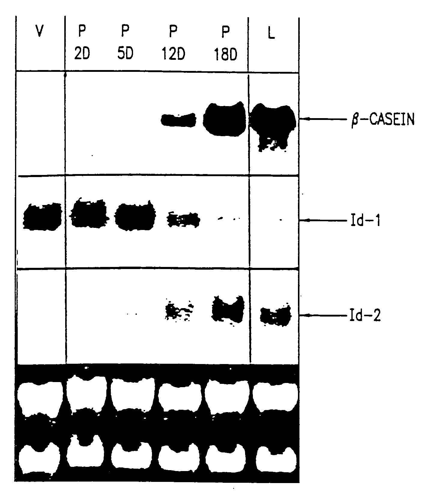
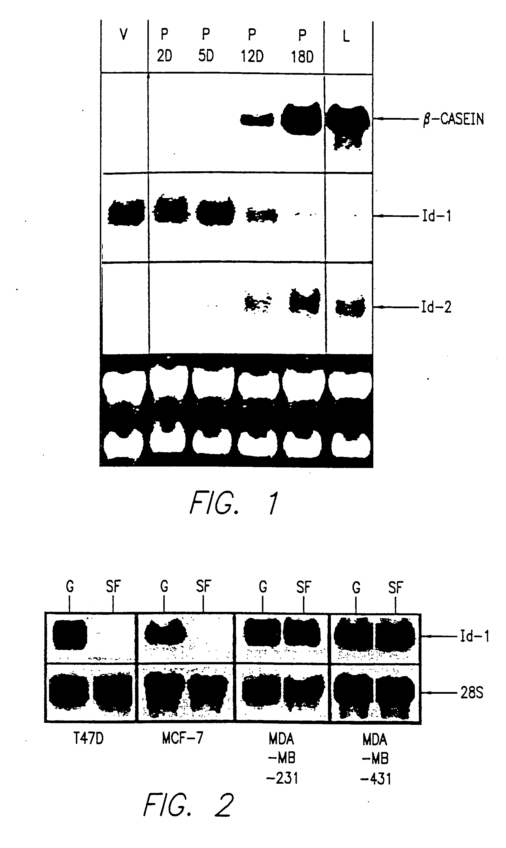
[0303] Total cellular RNA was isolated and purified as described in Anal. Biochem., 162:156-159 (1987). Twenty μg were separated by electrophoresis through formaldehyde-agarose gels and transferred to a nylon membrane (Hybond N; Amersham). The membrane was hybridized to a 32P-labeled human Id-1 cDNA or Id-2 or.-casein probe according to J. Biol. Chem., 269:2139-2145 (1994) and was washed and exposed to XAR-5 film for autoradiography. The same blot was hybridized to a 28S rRNA probe to control for RNA integrity and quantitation.
PUM
| Property | Measurement | Unit |
|---|---|---|
| temperature | aaaaa | aaaaa |
| pH | aaaaa | aaaaa |
| pH | aaaaa | aaaaa |
Abstract
Description
Claims
Application Information
 - R&D
- Intellectual Property
- Life Sciences
- Materials
- Tech Scout
- Unparalleled Data Quality
- Higher Quality Content
- 60% Fewer Hallucinations
Browse by: Latest US Patents, China's latest patents, Technical Efficacy Thesaurus, Application Domain, Technology Topic, Popular Technical Reports.
© 2025 PatSnap. All rights reserved.Legal|Privacy policy|Modern Slavery Act Transparency Statement|Sitemap|About US| Contact US: help@patsnap.com


