Bio-synthetic photostimulators and methods of use

Inactive Publication Date: 2006-12-05
SLOAN KETTERING INST FOR CANCER RES
View PDF2 Cites 106 Cited by
  • Summary
  • Abstract
  • Description
  • Claims
  • Application Information

AI Technical Summary

Benefits of technology

[0054]FIG. 9 shows the effect of TRPC3 of enhancing the photocurrent due to chARGe. Xenopus oocytes were programmed to express the minimal chARGe, consisting of NinaE, arrestin-2, and Gα; or the minimal chARGe plus TRPC3. Oocytes were exposed to 40 μM synthetic all-trans retina

Problems solved by technology

Unfortunately, both in vivo and in vitro techniques available currently are insufficient.
Although such in vivo techniques are helpful in identifying certain general areas correlated with particular brain functions, these techniques typically do not enable one to identify the cellular elements of specific neuronal pathways.
However, they do not help to determine what role, if any, those pathways play in the intact animal.
This technique can be, however, both time and labor intensive, and often results in the incomplete elucidation of neuronal pathways, leads to the stimulation of axons of passage, and causes mechanical disturbance or destruction of the tissue.
Unfortunately, the Farber method is based on the light-induced formation of small pores in the neuronal membrane resulting from the photoexcitation of the fluorescent dye.
However, the individual photostimulation of a large number of neurons present in a neural

Method used


Image

  • Bio-synthetic photostimulators and methods of use
  • Bio-synthetic photostimulators and methods of use
  • Bio-synthetic photostimulators and methods of use

Examples


example 1

Phototransduction Components

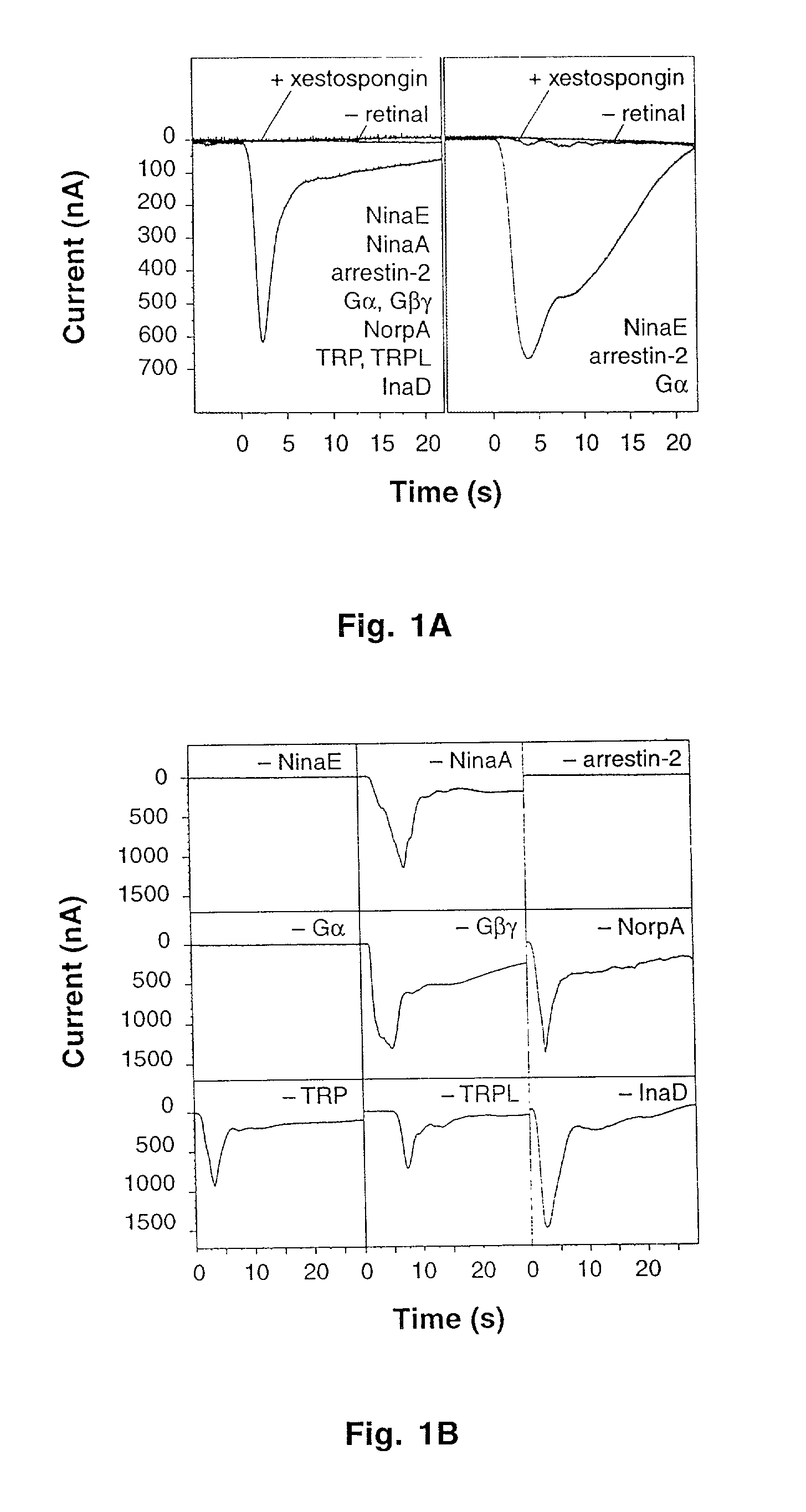
[0099]The coding regions (GenBank accession numbers in parentheses) of ninaE (K02315), ninaA (M62398), arrestin-2 (M32141), Gα (dgq; M58016), Gβ (gbe; M76593), Gγ (AJ250440), norpA (J03138), trp (M34394), trpl (M88185) and inaD (U15803) were amplified by PCR from the Drosophila head cDNA library GH (Berkeley Drosophila Genome Project) and ligated to pXES43, a derivative of pGEMHE (Liman, Tytgat et al. “Subunit stoichiometry of a mammalian K+ channel determined by construction of multimeric cDNAs,”Neuron 861–71 (1992)). Capped transcripts for injection into Xenopus oocytes were synthesized directly from these templates after linearization (MEGAscript T7, Ambion). Plasmids for expression in neurons (pChARGe-1 and pChARGe-2) were based on the pCI-neo backbone (Promega). pChARGe-1 carried ninaE and Gα under CMV- and SV40-control, respectively; pChARGE-2 contained sequences encoding EGFP (with a 20-amino acid N-terminal GAP-43 tag that confers plasma membrane ...

example 2

Xenopus Oocytes

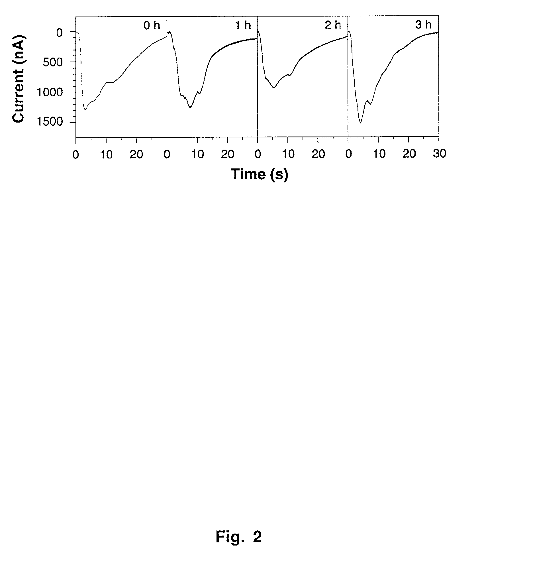
[0101]Stage VI oocytes were microinjected with the specified mRNA mixtures, which were adjusted to keep the doses of individual messages constant at 2–4 fmol / oocyte. Photocurrents were recorded at −80 mV with a two-electrode voltage-clamp amplifier (Axoclamp-2B, Axon Instruments) 2–4 days after mRNA injection. Electrodes (2–3 MΩ) were filled with 3 M KCl; the extracellular recording solution (Barth's Saline) contained, in mM, 87.5 NaCl, 2 KCl, 2.4 NaHCO3, 2 CaCl2, 1 MgCl2, 5 Tris-HCl, pH 7.2. Signals were externally amplified (CyberAmp 380, Axon Instruments) before digitization at 100 Hz (Digidata 1200, Axon Instruments). To block IP3-sensitive Ca2+ stores where indicated, 20 μM xestospongin C (Gafni et al., 1997; Calbiochem) was present throughout the experiment, beginning with the retinal load.

example 3

Rat Hippocampal Neurons

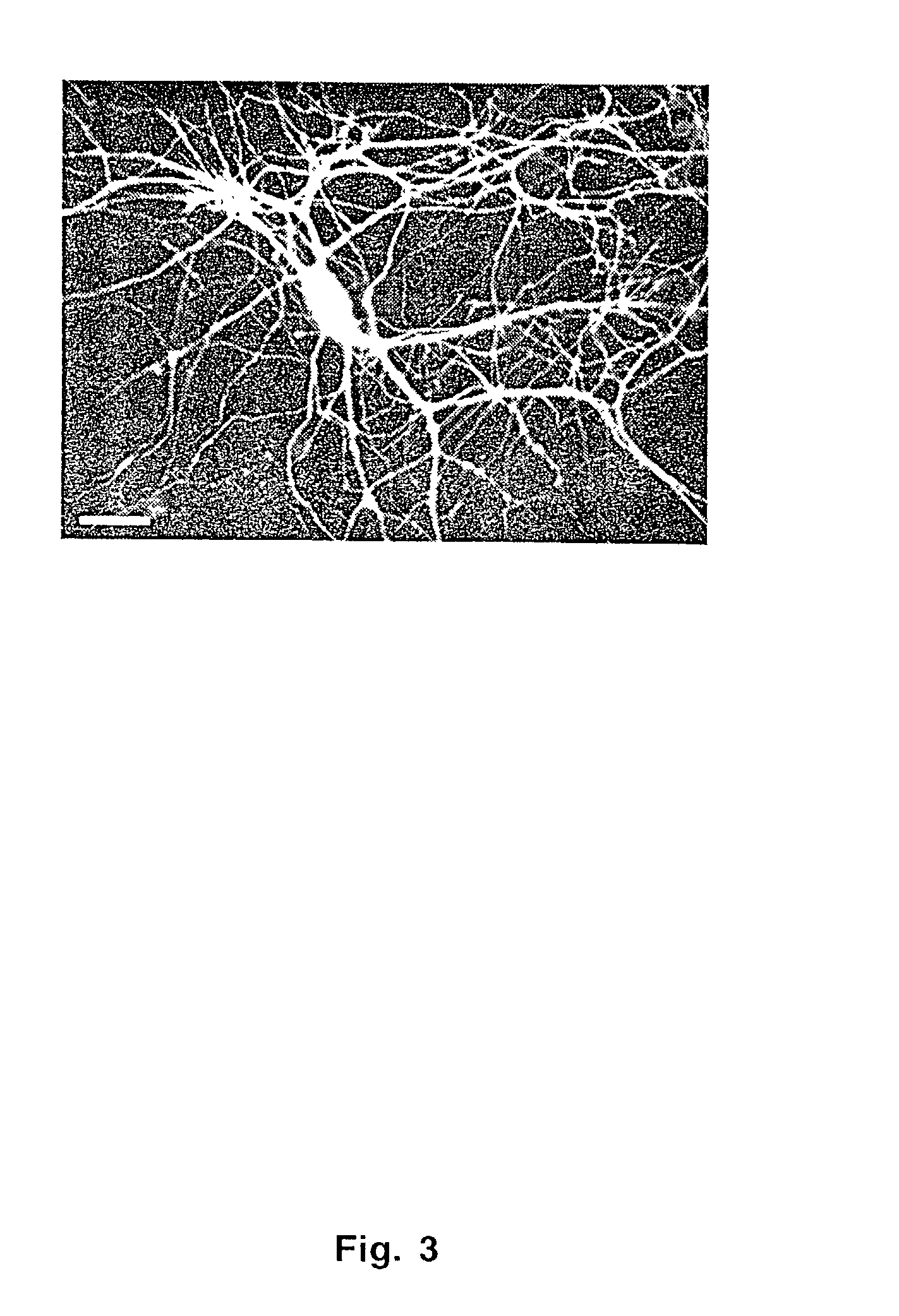
[0102]Hippocampal neurons obtained from E19 rats were grown in dissociated cultures (Yuste, Miller et al. “Synapto-pHluorins: chimeras between pH-sensitive mutants of green fluorescent protein and synaptic vesicle membrane proteins as reporters of neurotransmitter release,”Methods Enzymol 522–46 (2000)) and transfected with a calcium phosphate precipitate (pH 7.08) formed from a 1:1 mixture of CsCl-banded pChARGe-1 and pChARGe-2. Neurons were exposed to 4.2 μg / cm2 of precipitated DNA for 20 minutes. Transfections were done on day 8 after plating, recordings on days 6–10 after transfection. Neurons were identified under DIC and epifluorescence illumination (to distinguish transfected from untransfected cells; FIG. 3) and placed under whole-cell current clamp before reconstitution of NinaE with retinal. Patch pipettes (˜2.5 MΩ) contained, in mM, 120 K-gluconate, 10 KCl, 5 ATP, 0.3 GTP, and 10 K-HEPES, pH 7.2. The extracellular recording solution contained, in mM...


PUM

PropertyMeasurementUnit
Concentrationaaaaaaaaaa
Wavelengthaaaaaaaaaa

Abstract

Cells are rendered sensitive to stimulation by introducing into a non-photoreceptor cell nucleic acid sequences encoding at least an opsin gene product, an arrestin gene product, and the alpha subunit of the heterotrimeric G protein of the Gq family. The introduced sequences are expressed by the cell to yield at least the opsin gene product, the arrestin gene product, and the alpha subunit of the heterotrimeric G protein of the Gq family. Retinal or a derivative thereof capable of bonding with the opsin gene product to form a rhodopsin is provided to the cell. The cell is then irradiated with light having a wavelength capable of converting the rhodopsin to metarhodopsin. The conversion of rhodopsin to metarhodopsin triggers a cascade of intracellular responses within the cell resulting in an increased intracellular concentration of IP3 and calcium ions.

Description

CROSS-REFERENCE TO RELATED APPLICATIONS[0001]The present application claims the benefit under 35 U.S.C. 119(e) of U.S. Provisional Patent Application Ser. No. 60 / 312,707, filed Aug. 16, 2001, and U.S. Provisional Patent Application Ser. No. 60 / 345,741, filed Dec. 31, 2001; the disclosures of which are incorporated herein by reference.FEDERAL FUNDING LEGEND[0002]This invention was made using federal funds under Grant No. 08748 by the National Cancer Institute. Accordingly, the Government has certain rights in this invention.BACKGROUND OF THE INVENTION[0003]1. Field of the Invention[0004]The present invention relates generally to the fields of cellular biochemistry and neurophysiology. Generally, the present invention relates to techniques for optically stimulating genetically designated cells throughout the body. More specifically, it relates, inter alia, to techniques for direct activation of neural circuits by optically stimulating groups of genetically designated neurons.[0005]2. ...

Claims


Application Information

Patent Timeline
IPC IPC(8): C12N15/63C12N15/85C12N15/87A01N35/00A61K31/11C07K14/435C12N5/02
CPCC07K14/43581
Inventor MIESENBOCK, GEROZEMELMAN, BORIS V.
Owner SLOAN KETTERING INST FOR CANCER RES
Why Eureka
Try Eureka