
Method of screening

Inactive Publication Date: 2006-10-19
WALTER & ELIZA HALL INST OF MEDICAL RES
View PDF0 Cites 2 Cited by
  • Summary
  • Abstract
  • Description
  • Claims
  • Application Information

AI Technical Summary

Benefits of technology

[0077] Accordingly, yet another aspect of the present invention is directed to an isolated pathogen, which pathogen expresses a non-wild-type form of one or more antigens derived from said pa

Problems solved by technology

However, such assays do not discriminate between the presence of immunointeractive molecules which bind but which do not further impact on the functional activity of the pathogen to which they bind versus immunointeractive molecules which do impact on this functioning.

Method used


Image

  • Method of screening
  • Method of screening
  • Method of screening

Examples


example 1

Antibodies Against Merezoite Surface Protein (MSP)-119 are a Major Component of the Invasion Inhibitory Response in Individuals Immune to Malaria

Materials And Methods

Plasmids

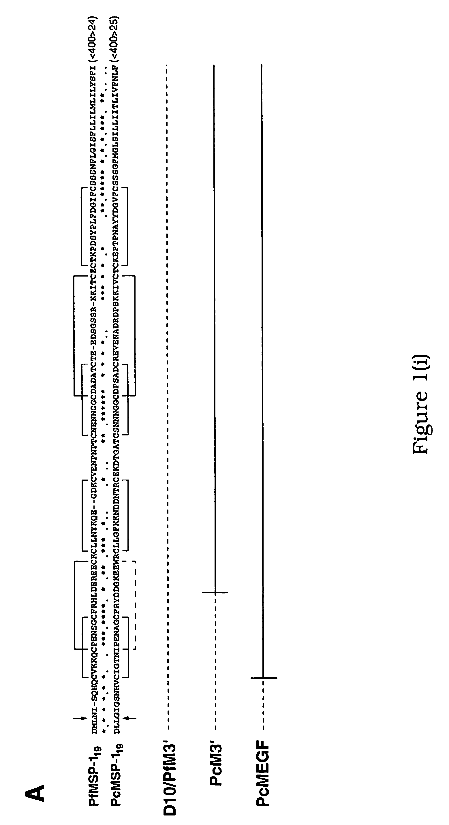
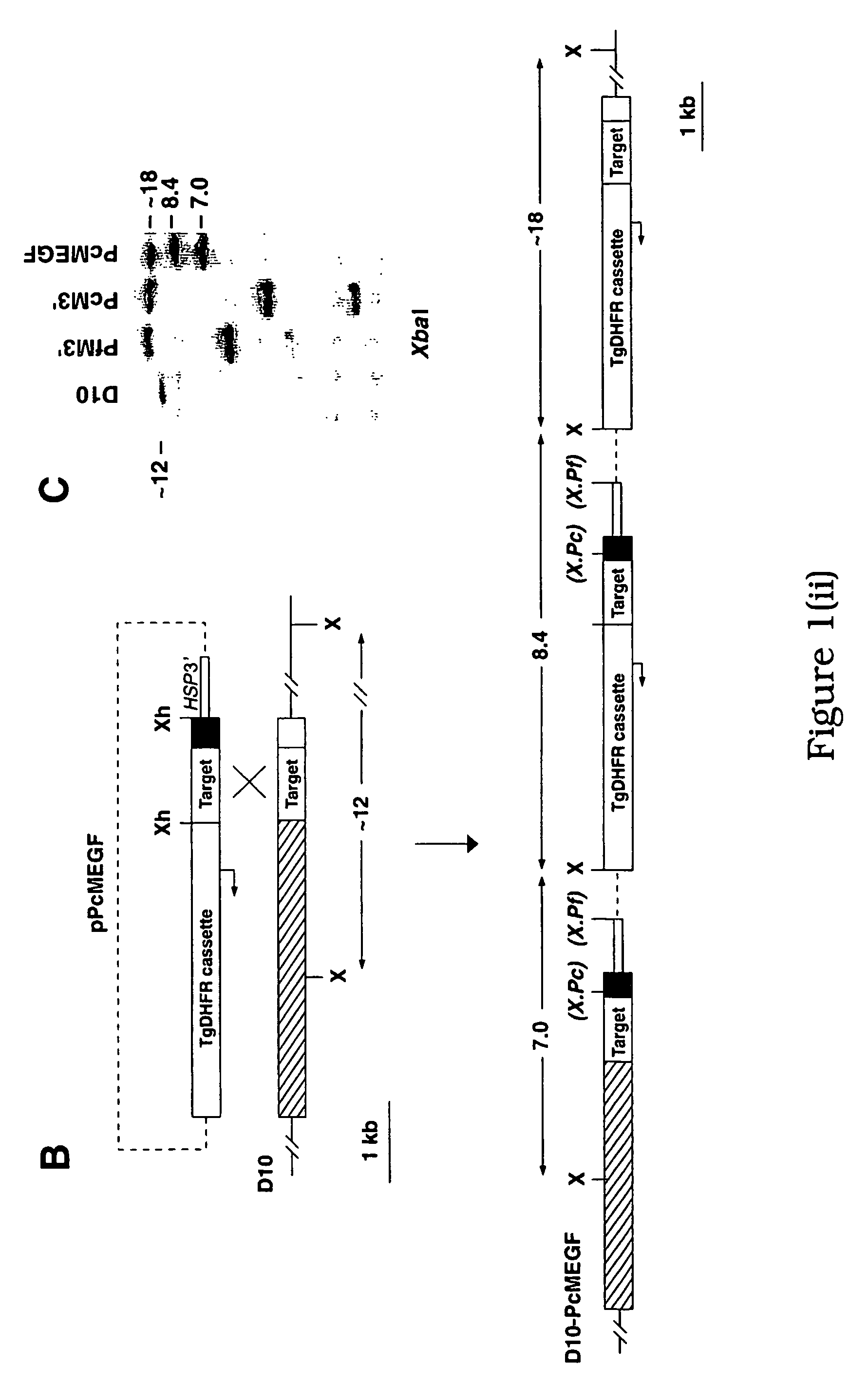
[0230] Construction of the plasmids pFfM3′ and pPcM3′ has been described previously (O'Donnell, R. A. et al. 2000 supra). The plasmid pPcMEGF was constructed by the insertion of a 1,200-bp XhoI fragment into the unique XhoI site of a plasmid pHC2 (Triglia, T., Healer, J., Caruana, S. R., Hodder, A. N., Anders, R. F., Crabb, B. S. and Cowman, A. F. (2000) Mol. Microbiol. 38:706-718). This target fragment comprises a 900-bp internal region of the P. falciparum MSP-1 gene fused in frame to the MSP-119 region of P. chabaudi. The fragment was generated by PCR amplification from P. falciparum (D10) and P. chabaudi (adami DS) genomic DNA (gDNA) using the oligonucleotide pairs Pf#1 5′-ATTTCTCGAGAATCCGAAGATAATGACG-3′ (1), PfEGF-R 5′-GAAACATCCAGCATTTTCTGGAAGTTTGTTCCTATGCATTGGTGTTGTGAAATG-3′ (2). The resulting amplico...

example 2

Generation of Recombinant Parasites

Methods

Plasmids

[0253] The pHCl plasmid vector has been described (Crabb, B. S. et al. 1997 supra). XhoI insers for cloning into this plasmid were amplified from the relevant genomic DNA using the following oligonucleotides (restriction endonuclease sites are bolded): Pf#1,5′-ATTTCTCGAGAATCCGAAGATAATGACG-3′ (5); Pf#2,5′-ATTGCTCGAGATCGATGTTTAACATATCTTGGAATTTTTCC-3′ (6); Pf#3, 5′-TTTAACTCGAGCATTTTTTAAATGAAACTG-3′ (7); Pf#4,5′-CATCTAGATGTCTGAAACATCCAG-3′ (8); Pc#1,5′-GGATGTTTCAGACATCTAGATGGTAAAG-3′ (9); Pc#2,5′-TCACTCGAGTTAAAATAAATTAAATACAATTAATGTG-3′ (10). To derive the pAMSPI and pPfM3′ fragments, Pf#1 / Pf#2 and Pf#1 / Pf#3 were used, respectively. to derive the pPcM3′ insert, amplicons from Pf#1 / Pf#4 and Pc#1 / Pc#2 were first digested with XbaI and ligated. The pPcM3′ vector is identical to pHCl except that it has a litmus 28 (NEB) backbone.

Parasite Transfection

[0254] Plasmids were transfected into P. falciparum parasites (D10 line) essentially ...

example 3

A New Rodent Model to Assess Blood-Stage Immunity to the Plasmodium Falciparum Antigen MSP-119 Reveals a Protective Role for Invasion Inhibitory Antibodies

Materials And Methods

Plasmids

[0259] To create the pPb-PfM19 replacement plasmid, 1.3 Kb of P. berghei MSP-1 targeting sequence was firstly fused in frame to the MSP-119 region of P. falciparum upstream from the first cysteine residue of EGF domain 1. This was achieved by PCR amplification of P. berghei ANKA and P. falciparum D10 genomic DNA (gDNA) using the oligonucleotide pairs PbF (5′-CGGGGTACCATCGATAAATACTTTACCTCTGAAGCTGTTCC (15)) and PbR1 (5′-TACATGCTTAGGGTCTATACCTAATAAATC (16)), and PbPfF (5′-GGTATAGACCCTAAGCATGTATGCGTAAAAAAACAATGTCCAGAA (17)) and PfR (5′-TGCTCTAGATTAAATGAAACTGTATAATATTAAC (18)), respectively, and sewing the products together via PCR using the primers PbF and PfR. The insertion of KpnI (underlined) and XbaI sites (boldface) into the oligonucleotides facilitated cloning of the resulting fragment into the ...


PUM

PropertyMeasurementUnit
Immunogenicityaaaaaaaaaa
Strain pointaaaaaaaaaa
Inhibitionaaaaaaaaaa

Abstract

A method of detecting the presence of a functionally inhibitory immunointeractive molecule in a biological sample. Preferably, the immunointeractive molecule is directed to a pathogen derived antigen and, more particularly, a parasite derived antigen and, even more particularly, a Plasmodium drived antigen. The method of the present invention facilitates detection of the presence of functionally inhibitory immunointeractive molecules, both in vitro and in vivo, and is useful for qualitatively and / or quantitatively assessing the immune status of individuals who have been previously infected with a parasite, predicting the immune status of individuals vaccinated with an antigen based vaccine, determining the relative contribution of a specific immunoreactivity of antibody to the total inhibitory antibody elicited by combination vaccines which include two or more antigens, assessing vaccines to determine the efficacy of different forms of an antigen, determining vaccine potency, assessing the protective potential of certain immunoreactivities of antibodies and determining the importance of parasite inhibitory antibodies.

Description

FIELD OF THE INVENTION [0001] The present invention relates generally to a method of detecting the presence of an immunointeractive molecule in a biological sample. More particularly, the present invention relates to a method of detecting the presence of a functionally inhibitory immunointeractive molecule in a biological sample. Preferably, said immunointeractive molecule is directed to a pathogen derived antigen and, even more particularly, a parasite derived antigen. The method of the present invention facilitates detection of the presence of functionally inhibitory immunointeractive molecules, both in vitro and in vivo, and is useful, inter alia, for qualitatively and / or quantitatively assessing the immune status of individuals who have been previously infected with a parasite, predicting the immune status of individuals vaccinated with an antigen based vaccines, determining the relative contribution of a specific immunoreactivity of antibody to the total inhibitory antibody eli...

Claims


Application Information

Patent Timeline
IPC IPC(8): A61K49/00G01N33/569C12N1/10C07K14/445C07K16/20C12N1/11C12N15/30G01N33/68
CPCC07K14/445C07K16/205G01N2333/445G01N33/6854G01N33/56905Y02A50/30
Inventor O'DONNELL, REBECCADE KONING-WARD, TANIACRABB, BRENDAN
Owner WALTER & ELIZA HALL INST OF MEDICAL RES
Learn More