

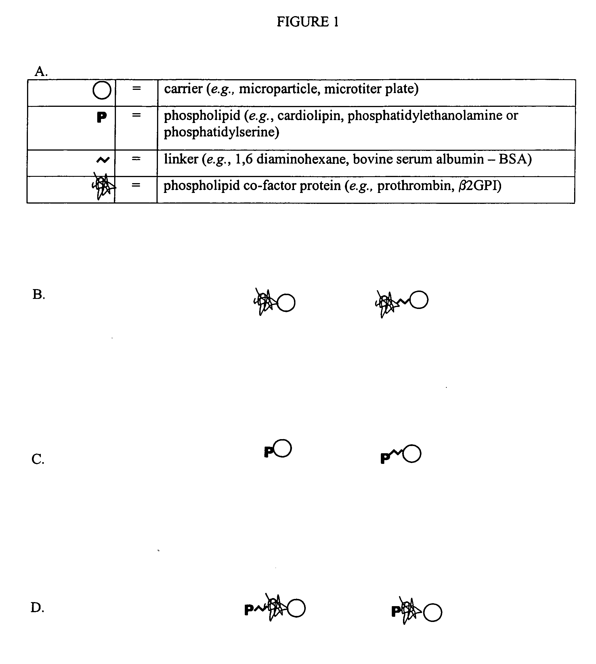
Solid phase immobilization of phospholipids and cofactor proteins via covalent attachment
a technology of cofactor proteins and solid phase immobilization, which is applied in the field of solid phase immobilization of phospholipids and cofactor proteins via covalent attachment, can solve the problems of heterogeneous passive configuration, difficult reproducible manufacture of passive configuration, and inability to detect the configuration of this format,
- Summary
- Abstract
- Description
- Claims
- Application Information
AI Technical Summary
Benefits of technology
Problems solved by technology
Method used
Image

Examples
example 1
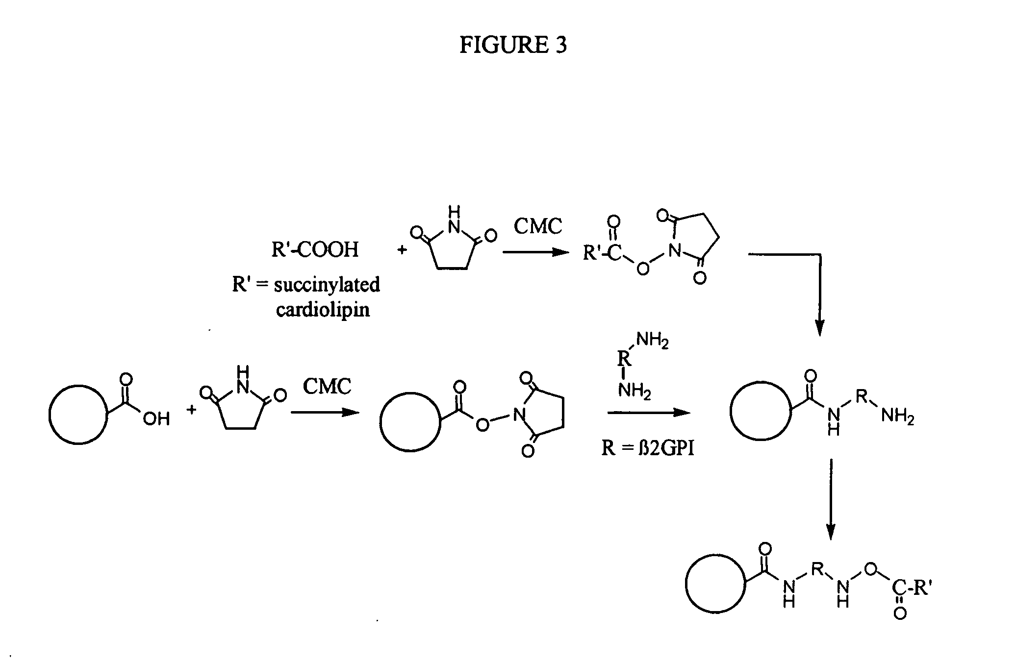
Synthesis of Cardiolipin-β2GPI Protein-Beads
[0096] Into a 1.8 mL microfuge tube we placed 10 mg of 8 μm carboxylate-modified paramagnetic microspheres. We washed once (using magnetic separation) with about 1 mL of 167 mM MES at a pH of about 6.1. We added about 300 μL of 668 mM sulfo-N-hydroxysuccinimide (NHSS) in 167 mM MES at a pH of about 6.1 and 700 μL of 144 mM N-cyclohexyl-N′-(2-morpholinoethyl)carbodiimide metho-p-toluenesulfonate (CMC) in ethanol. We incubated with mixing (e.g., rotation) for about 30 minutes at room temperature. We magnetically separated and discarded the supernatant. We washed once with about 1 mL of 50 mM 4-(2-hydroxyethyl)-1-piperazinepropanesulfonic acid (EPPS), at a pH of about 8.1. We added 634 μL deionized water, 200 μL of 250 mM EPPS of a pH of about 8.1 and 166 μL of a 1.51 mg / mL solution of β2GPI. We incubated with mixing (e.g., rotation) for about 60 minutes at room temperature. We washed the beads about 4 times with about 1 mL of 50 mM PBS / 0.1%...
example 2
Synthesis of Phosphatidylserine-β2GPI Beads
[0100] Into a 1.8 mL microfuge tube we placed about 10 mg of 8 μm carboxylate-modified paramagnetic microspheres. We washed once (using magnetic separation) with about 1 mL of 167 mM 2-(N-Morpholino)ethanesulfonic acid (MES), at a pH of about 6.1. We added about 300 μL of 668 mM sulfo-N-hydroxysuccinimide (NHSS) in 167 mM MES at a pH of about 6.1 and about 700 μL of 144 mM N-cyclohexyl-N′-(2-morpholinoethyl)carbodiimide metho-p-toluenesulfonate (CMC) in ethanol. We incubated the samples with mixing (e.g., rotation) for about 30 minutes at room temperature. We magnetically separated beads from the supernatant, which was discarded. Beads were washed once with about 1 mL of 50 mM 4-(2-hydroxyethyl)-1-piperazinepropanesulfonic acid (EPPS), at a pH of about 8.1. We added to the beads about 634 μL deionized water, about 200 μL of 250 mM EPPS pH 8.1 and about 166 μL of a 1.51 mg / mL solution of β2GPI and incubated with mixing (e.g., rotation) for ...
example 3
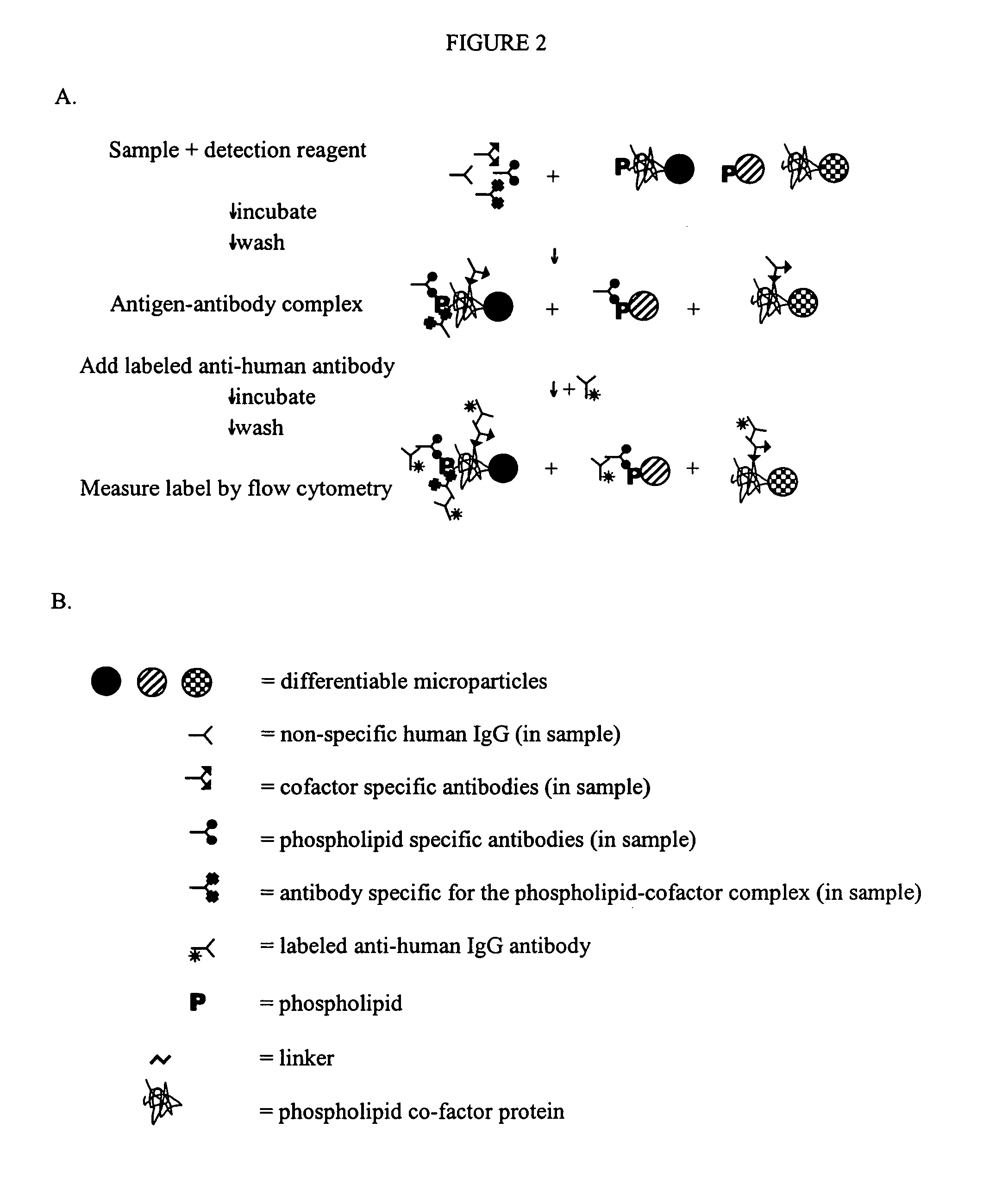
Diagnosis of APS Using Cardiolipin or Phosphatidylserine-β2GPI Beads
[0103] As seen in Table 3, blood samples were collected from ninety-seven healthy blood donors and 106 patients previously diagnosed with either primary or secondary Anti-Phospholipid Syndrome (APS). A panel of multiplexed differentiable particles coated (i.e., covalently attached) with 1) β2GPI only, 2) succinylated cardiolipin only, 3) succinylated cardiolipin and β2GPI and 4) phosphatidylserine and β2GPI was generated. For 3 and 4, both succinylated cardiolipin and phosphatidylserine were covalently bound to the β2GPI protein. Wahses were performed in buffers that included a detergent. The blood samples were assayed on the four differentiable particles by flow cytometry. No samples from the healthy donors exhibited binding to any of the four tested particles. In contrast, samples from sixty-one of the APS patients (58%) exhibited binding to at least one of the four particles. Conventional APS diagnostic tests de...
PUM
 Abstract
Description
Claims
Application Information