Ultrasonographic device and ultrasonographic method

a technology of ultrasonography and ultrasonography, which is applied in the field of ultrasonography diagnostic equipment and ultrasound diagnostic methods, can solve problems such as the difficulty of finding turbulence itself, and achieve the effect of reducing transparency and increasing the variance of blood flow

Inactive Publication Date: 2006-10-26
HITACHI MEDICAL CORP
View PDF19 Cites 18 Cited by
  • Summary
  • Abstract
  • Description
  • Claims
  • Application Information

AI Technical Summary

Benefits of technology

[0006] The display means displays information synthesized from the color display and the transparent display. The ultrasound diagnostic apparatus further comprises selection means selecting one of the color display and the transparent display, wherein the display means displays the information selected by the selection means. The ultrasound diagnostic apparatus further comprises transparency control means controlling a degree of transparency of the color Doppler image of the transparent display.
[0007] The transparency control means controls a degree of the transparency based on the blood flow information of the color Doppler image. The transparency control means controls a degree of the transparency based on a variance of a blood flow of the color Doppler image. The transparency control means sets the transparency of the color Doppler image in such a manner that the transparency is reduced with an increase in the variance of the blood flow. The transparency control means obtains the variance as a relative value to display the color Doppler image as: an opaque image when the variance is maximum; a transparent image when the variance is nul

Problems solved by technology

In conventional color Doppler image display, when a flow like turbulence is present in a blood flow, only a portion of the blood flow around the

Method used


Image

  • Ultrasonographic device and ultrasonographic method
  • Ultrasonographic device and ultrasonographic method
  • Ultrasonographic device and ultrasonographic method

Examples


Embodiment Construction

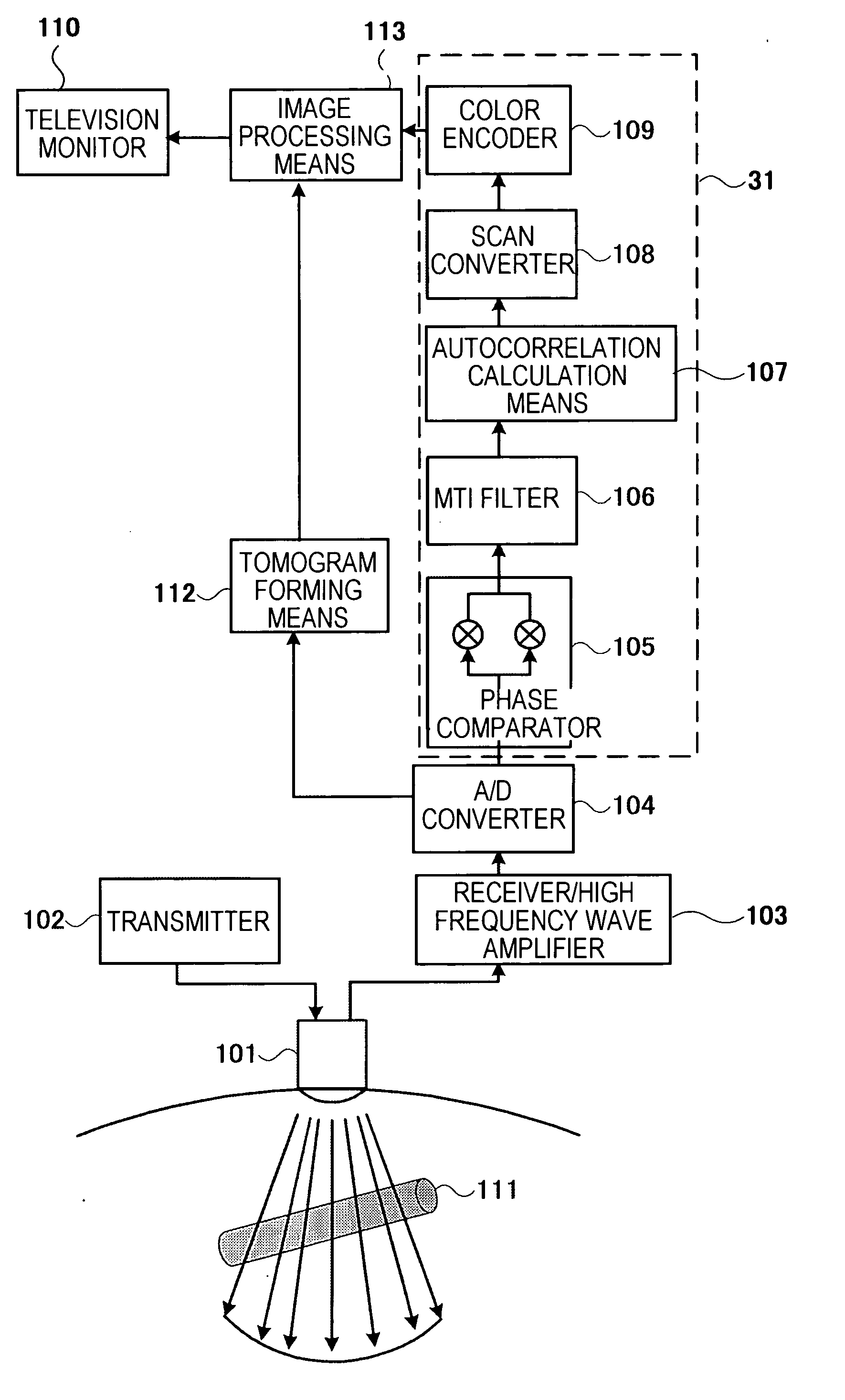
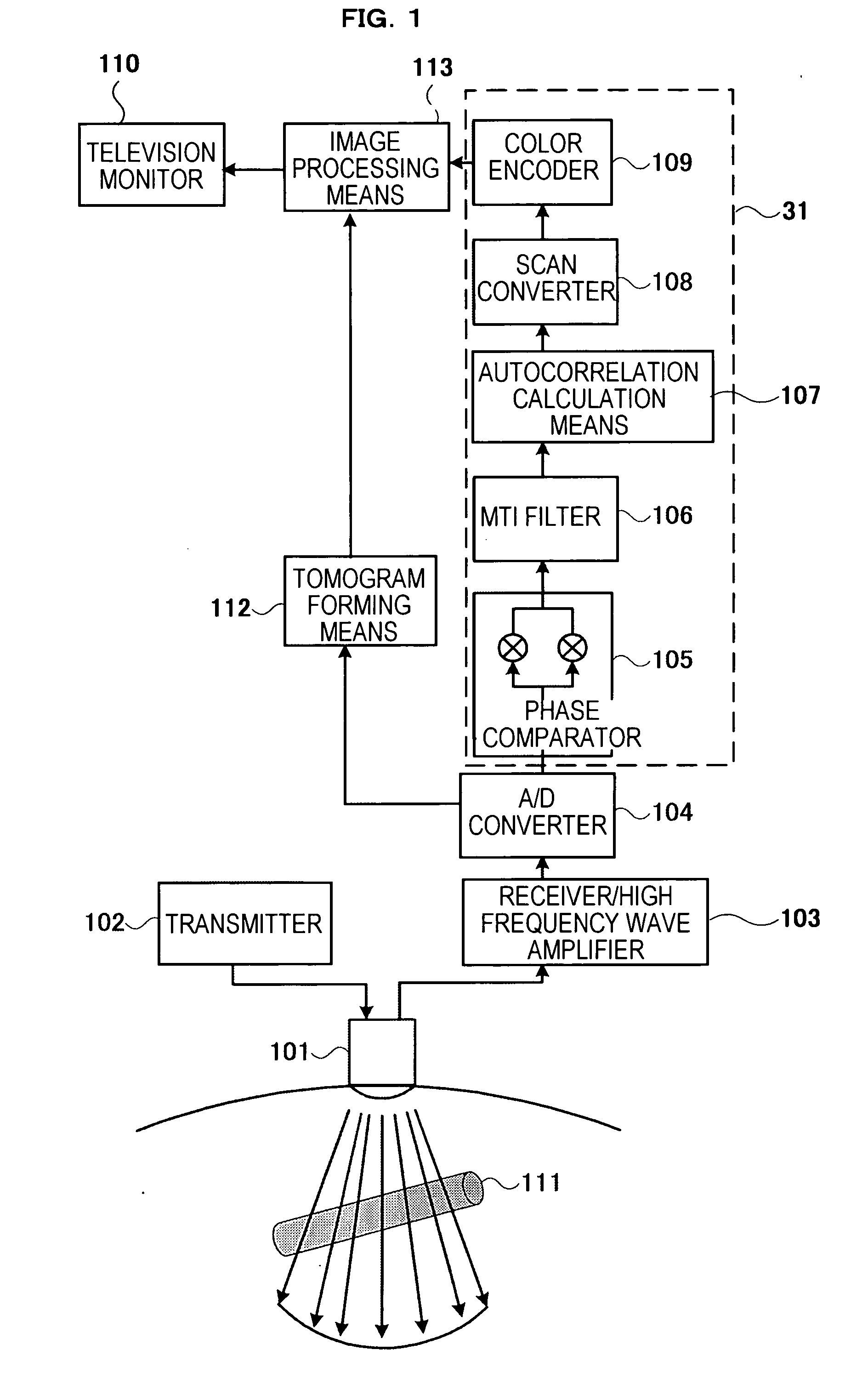
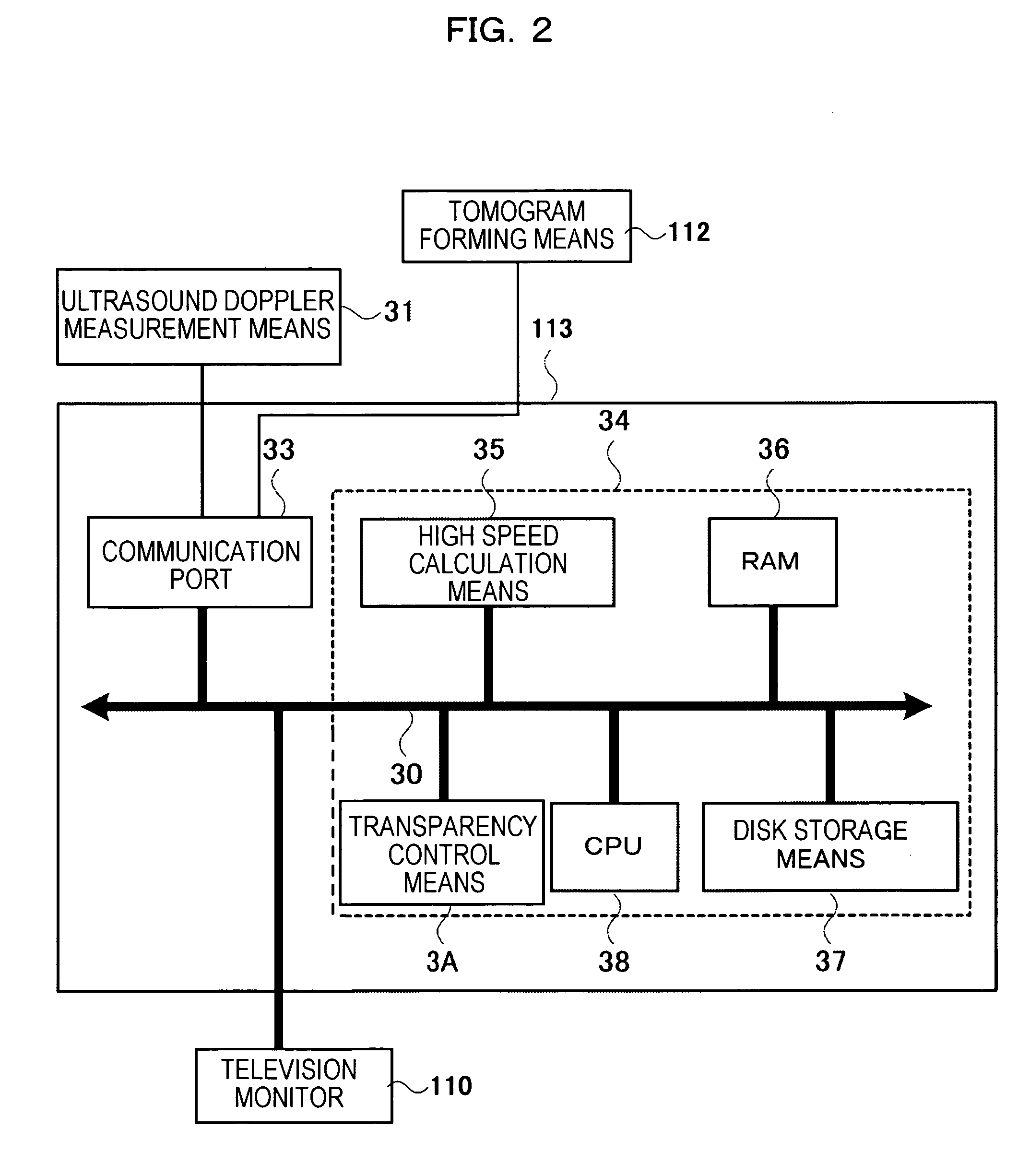
[0016] An ultrasound diagnostic apparatus of this invention having a color Doppler measurement function will be described with reference to FIG. 1. An ultrasound pulse transmitted from a transmitter 102 is sent repeatedly from an ultrasound probe 101 to a reflection object 111 at a constant interval T. Ultrasound pulses reflected by the reflection object 111 are received by a receiving circuit 103 to be converted into digital signals by an A / D converter 104, so that digital signal outputs of a cosine component and a sine component are obtained from a phase comparator 105. A low frequency component (clatter component) the cosine component signal and the sine component signal is attenuated by a high pass MTI filter 106 so as to extract a high frequency component (blood flow component) therefrom, and then an average speed, a variance, and power of the blood flow are calculated by an autocorrelation calculation means 107. The calculation results are rearranged in accordance with a telev...


PUM


Abstract

An ultrasound diagnostic apparatus including: a tomogram forming means forming a tomogram of a diagnosis portion of an examinee by transmitting/receiving an ultrasound wave to/from the examinee via an ultrasound probe; color Doppler image forming means forming a color Doppler image based on a Doppler signal obtained from the diagnosis portion; image processing means performing image processing on the tomogram and the color Doppler image; and display means displaying images obtained by the image processing means, the tomogram and the color Doppler image being color displayed on the display means, wherein the image processing means causes the color Doppler image to be displayed transparently.

Description

TECHNICAL FIELD [0001] This invention relates to an ultrasound diagnostic apparatus and an ultrasound diagnosing method enabling to display turbulence information together with blood flow information, the ultrasound diagnostic apparatus having a color Doppler measurement function and forming and then displaying a plurality of color Doppler images obtained by measuring a diagnosis portion of an examiner. BACKGROUND ART [0002] In displaying a color Doppler image, particularly in three-dimensional display, perspective information indicating a position of a blood flow whether is interior or near to a projection surface is required. Patent Literature 1 discloses a technique of displaying the perspective on a screen as a color bar by means of a gradation of luminance. Patent Literature 1: JP-A-11-299784 [0003] In conventional color Doppler image display, when a flow like turbulence is present in a blood flow, only a portion of the blood flow around the turbulence is displayed, and the tu...

Claims


Application Information

Patent Timeline
IPC IPC(8): A61B8/00A61B8/06
CPCA61B8/13A61B8/06
Inventor HAYASHI, TETSUYAKANDA, HIROSHIARAI, OSAMU
Owner HITACHI MEDICAL CORP
Why Eureka
Try Eureka