
Transurethral ultrasonic imaging system

a transurethral ultrasonic imaging and ultrasound technology, applied in the field of ultrasound scanning systems, can solve the problems of inability to completely depend on trus for accurate imaging of the entire prostate gland, and inability to produce high-quality images of the prostate gland, so as to reduce the required number of tissue biopsy samples

Inactive Publication Date: 2006-11-02
FRW 1
View PDF55 Cites 8 Cited by
  • Summary
  • Abstract
  • Description
  • Claims
  • Application Information

AI Technical Summary

Benefits of technology

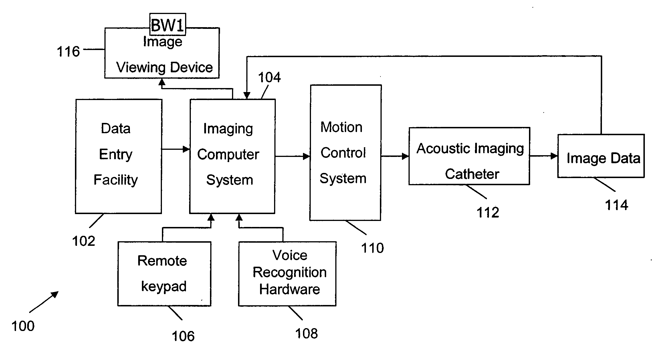
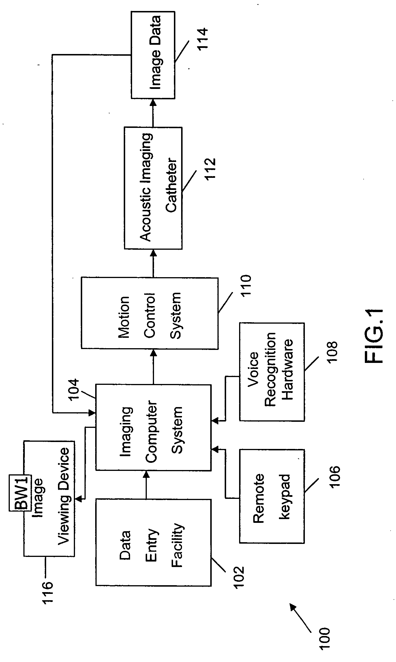
[0008] The present invention is directed to an ultrasound scanning system for the transurethral imaging of a patient's genitourinary system, and in one embodiment, the male prostate gland. The ultrasound scanning system is capable of three-dimensional imaging of the patient's prostate gland, is capable of producing multiple arrays of transverse slice images of the selected section of the patient's prostate gland, which results in complete scanning of the patient's prostate gland, and is capable of facilitating digitally positioned targeted biopsies based on the image-apparent focal tissue abnormalities, potentially reducing the required number of tissue biopsy samples. In one preferred form, the ultrasound scanning system for imaging the patient's prostate gland comprises an acoustic imaging catheter, a motion control system, and a computer system. The acoustic imaging catheter is capable of being inserted within the patient's prostatic urethra. The acoustic imaging catheter comprises an ultrasonic transducer. The ultrasonic transducer is rotated inside the acoustic imaging catheter, which enables production of the scan data representative of a section of the patient's prostate gland. The acoustic imaging catheter is moved to different positions inside the patient's prostatic urethra, in relation to a fixed anatomical landmark, to generate images of the selected sections of the patient's prostate gland. The motion control system controls the axial and rotational motion of the acoustic imaging catheter. The computer system is in communication with the acoustic imaging catheter and the motion control system. The computer system processes signals received from the acoustic imaging catheter and generates selected images including, if desired, a three-dimensional image of the patient's prostate gland, which is displayed on an associated image viewing device. The image viewing device is positioned in a location, which allows substantially simultaneous viewing of both a patient's pelvic region and the image viewing device to the physician, while ultrasonically imaging the patient's prostate gland.
[0009] It follows that the ultrasound scanning system embodying a preferred form of the present invention is capable of three-dimensional imaging of the patient's prostate gland. Further, because the acoustic imaging catheter is moved to different positions inside the patient's prostatic urethra in relation to a fixed anatomical landmark, the ultrasound scanning system embodying a preferred form of the present invention is capable of producing multiple arrays of transverse slice images of the selected sections of the patient's prostate gland. This provides a complete scanning of the patient's prostate gland. Further, because the image data of the patient's prostate gland is stored in the computer system, the ultrasound scanning system, in accordance with the present invention, provides the retrieval of the required information anytime. Further, the ultrasound scanning system embodying a preferred form of the present invention facilitates digitally positioned targeted biopsies based on the image-apparent focal tissue abnormalities, potentially reducing the required number of tissue biopsy samples.
[0010] Accordingly, it is an object of the present invention to provide an improved ultrasound scanning system which is capable of producing a three-dimensional rendering of the patient's genitourinary system.
[0011] It is a further object of the present invention to provide an improved ultrasound scanning system which is capable of producing a three-dimensional rendering of the patient's prostate gland.
[0012] It is still another object of the present invention to provide for a complete scanning of the patient's prostate gland an improved ultrasound scanning system which is capable of producing multiple arrays of transverse slice images of the selected section of the patient's prostate gland.
[0013] It is yet another object of the present invention to provide for facilitating digitally positioned targeted biopsies based on the image-apparent focal tissue abnormalities an improved ultrasound scanning system which is capable of imaging the patient's prostate gland.

Problems solved by technology

However, TRAS and TPUS fail to produce high-quality images of the prostate gland.
However, TRUS cannot be completely relied upon for accurate imaging of the entire prostate gland.
However, it will be noted by those skilled in the art that most current medical interventional procedures, for example, surgery, biopsy, and ablation still require “blind” approaches, i.e., the clinicians cannot directly see the target and / or pathway to the target during the ultrasound imaging of the prostate gland.
A visualization of the target during ultrasound scanning of the prostate gland, if available, are often limited to two-dimensional, slow and / or off-line displays.

Method used


Image

  • Transurethral ultrasonic imaging system
  • Transurethral ultrasonic imaging system
  • Transurethral ultrasonic imaging system

Examples


Embodiment Construction

[0020] For the sake of convenience, the terms used to describe various human anatomical structures and embodiments of the invention are defined below. It should be understood that these are provided merely to aid the understanding of the description, and that the definitions should in no way limit the scope of the invention, which is defined by the appended claims.

[0021] Anterior: Situated at the front or the front surface of an organ.

[0022] Apex of the prostate: The end of the prostate gland located farthest away from the urinary bladder.

[0023] Axial / Longitudinal: Along the centerline of the urethra, regardless of patient position.

[0024] Biopsy: The removal of small sample(s) of tissue for examination under a microscope.

[0025] Bladder: The hollow organ that stores and discharges urine from the body.

[0026] Bladder neck: The outlet area of the bladder. It is composed of circular muscle fibers (bladder sphincter), and helps control urine flow from the bladder into the urethra.

[...


PUM


Abstract

An ultrasound scanning system and methods of using the same. In one preferred form, an ultrasound scanning system comprises an acoustic imaging catheter comprising an ultrasonic transducer, a motion control system and an imaging computer system for imaging a patient's genitourinary system. In another preferred form, an ultrasound scanning system is used for imaging a patient's prostate gland.

Description

FIELD OF THE INVENTION [0001] The field of the present invention is imaging systems and their methods of use, and more particularly, ultrasound scanning systems and their use in imaging a patient's genitourinary system. BACKGROUND OF THE INVENTION [0002] Ultrasound is an imaging technique, which uses high-frequency sound waves to produce images of the organs and structures of the body. The ultrasound technique involves sending sound waves into the body. These sound waves reflect off the internal organs and are recorded by special instruments that create images of the anatomic parts of the internal organs. The ultrasound technique uses no ionizing radiation, and provides real-time imaging. Ultrasound is used to detect and monitor the growth of the fetus, examine many of the body's internal organs, for example, heart, liver, gallbladder, spleen, pancreas, kidneys, and bladder. [0003] Recently, ultrasound has been used to detect possible disorders within a man's prostate gland. For exa...

Claims


Application Information

Patent Timeline
IPC IPC(8): A61B8/14
CPCA61B8/12
Inventor ROSE, HAROLD B.WOSKOW, ROBERT M.GRUNDFEST, WARREN G.MACKAY, SPENCER L.RUBENSTEIN, DAVID J.ELIST, JAMES J.CRAWFORD, ALAN D.
Owner FRW 1
Learn More