System and methods for rapid and automated screening of cells

a cell and automated technology, applied in the field of automated cell screening in drug discovery, can solve the problems of small number of best-qualified hits, instruments and robotics used for hts cannot accommodate tissues, and the number of tests that could be done has become very large and will continue to grow

Active Publication Date: 2006-11-16
GLOBAL LIFE SCI SOLUTIONS USA LLC
View PDF4 Cites 56 Cited by
  • Summary
  • Abstract
  • Description
  • Claims
  • Application Information

AI Technical Summary

Problems solved by technology

Therefore, the number of tests that could be done has become very large and will continue to grow.
Most commonly, the instruments and robotics used for HTS do not accommodate tissues.
With these factors characterized, a small number of the best-qualified hits (“leads”) can be moved into very costly and time-consuming pre-clinical and clinical trials.
Unfortunately, post-primary testing is more complex and much slower than primary testing.
It is not enough to simply detect molecular interactions between compounds and isolated target molecules.
A potential problem with cell assays is the relatively low level of throughput that most evidence.
However, research systems lack the automation and throughput which would make them useful for industrial drug discovery or clinical diagnosis.
For example, Bacus (U.S. Pat. No. 5,018,209) discloses one such operator-assisted diagnostic system, which is useful with small numbers of samples, but which would not be useful in a high throughput environment.
Ideally, a simple staining process yields unambiguous detection of cells or cellular components, wherein each stained object marks a feature of interest, and other image components are unstained.
In practice, this goal is rarely achieved.
Simple intensity or color thresholding is rarely adequate for industrial applications in that only some of the segmented pixels are valid and the segmented image needs operator editing.
(1986) report that simple intensity thresholding resulted in lower precision for cell detection than was attained by flow cytometry.
There are many problems, including cell and background intensities that vary from location to location in a single image or set of images.
Automation and throughput can only be achieved within a system integrating specialized software and hardware.
The difficulty of creating such an automated and high throughput system is not to be underestimated, and is demonstrated by the very small number of such systems which have been disclosed or reduced to practice (e.g. Proffit et al., 1996; Ramm et al., 2001, 2002; U.S. Pat. No. 5,989,835; U.S. Pat. No. 6,103,479).

Method used


Image

  • System and methods for rapid and automated screening of cells
  • System and methods for rapid and automated screening of cells
  • System and methods for rapid and automated screening of cells

Examples


Embodiment Construction

[0087] The denotations and abbreviations used in this description are defined in Table 1.

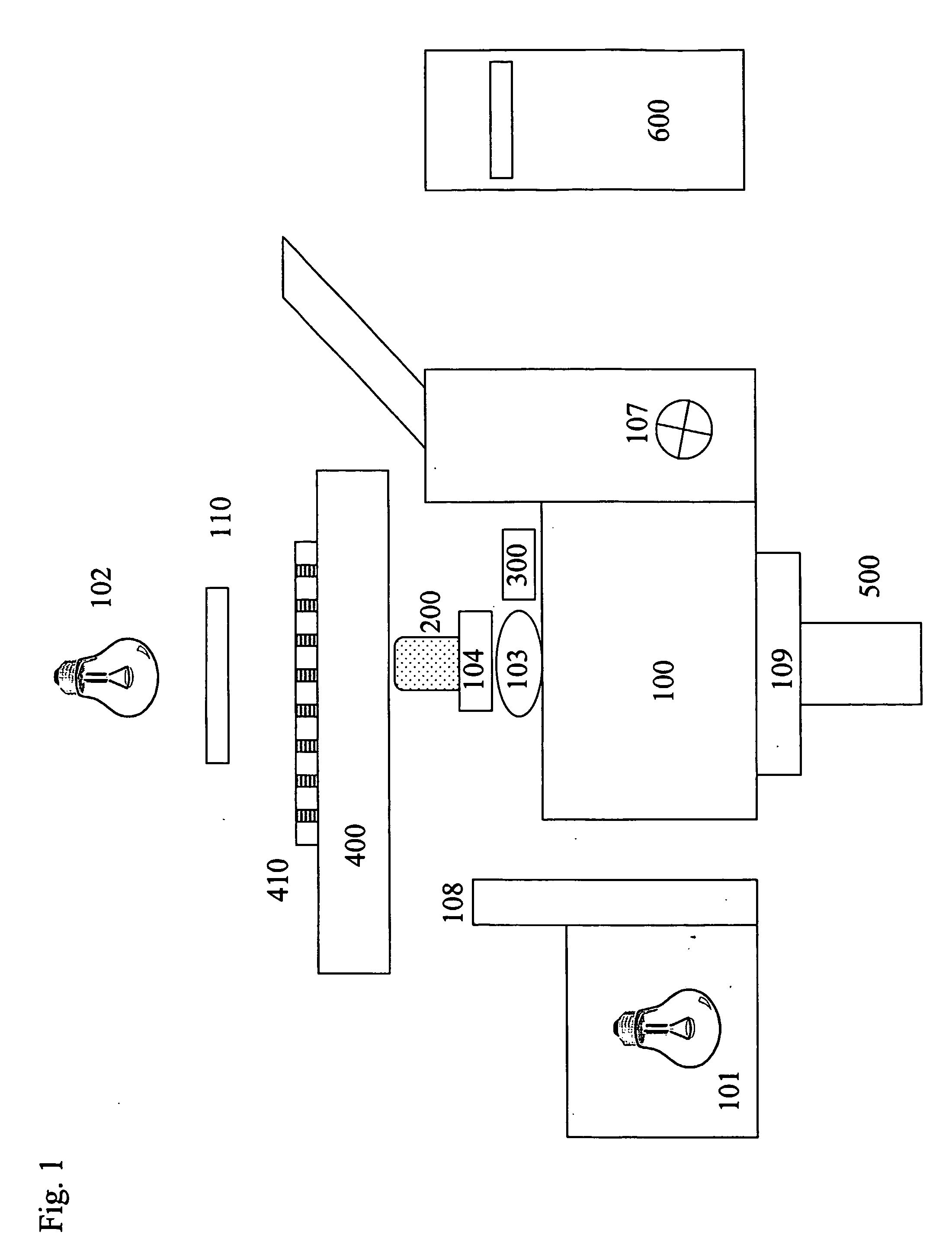
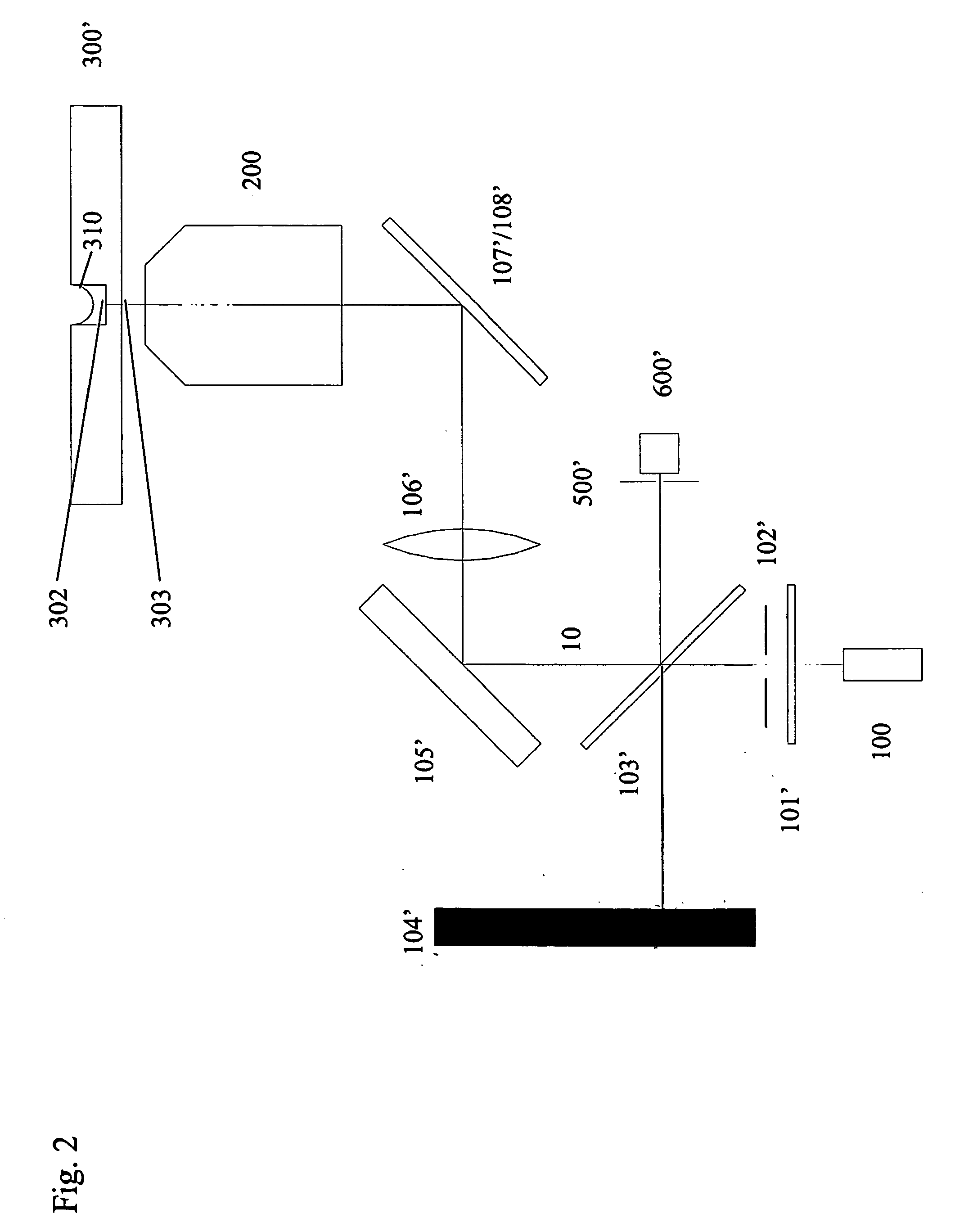
[0088] Turning now to the details of the drawings, FIG. 1 is a schematic block diagram illustrating the optical, mechanical and electrical components of the system of the present invention. Inverted microscope stand 100 is equipped with fluorescence epi-illuminator 101 and tungsten halogen transilluminator 102. Mounted on objective turret 103 is fast motor drive 104, preferably of the piezoelectric kind. Motor 104 moves objective 200 in the Z-dimension (vertically) so as to reach the best focus position. The best focus position is defined by confocal autofocus device 300 as monitored by digital computer 600. Microscope Z-focus drive 107 may also be used to move objective 200 in the Z-dimension, when software autofocus is selected. Filter changer 108 is positioned so as to present filters in the illumination path of illuminator 101, thereby selecting narrow band excitation illumination. Optional...


PUM

PropertyMeasurementUnit
distanceaaaaaaaaaa
diameteraaaaaaaaaa
diameteraaaaaaaaaa

Abstract

A system for performing automated cell screening in drug discovery, including an automated microscope, a fast autofocus device and a digital imaging system. Processes are implemented in software through which relevant cellular material is segmented and quantified with minimal user interaction. Improvements in the following areas: known methods for image processing are implemented in such a way that automated segmentation is achieved; sets of known measurements (pixel counting, etc.) are implemented as methods which demonstrate aspects of biology in a reliable fashion; components for automated positioning, focusing, imaging and processing of a multiplicity of samples are integrated as systems within which the segmentation and measurement methods may be mounted; and components and methods are adapted into systems which yield more highly automated and more rapid cell screening.

Description

CROSS REFERENCE TO RELATED APPLICATIONS [0001] This application is a filing under 35 U.S.C. § 371 and claims priority to international patent application number PCT / IB03 / 01821 filed May 9, 2003, published on Nov. 20, 2003 as WO 03 / 095986 and also claims priority to U.S. provisional patent application No. 60 / 380,822 filed on May 14, 2002; the disclosures of which are incorporated herein by reference in their entireties.FIELD OF THE INVENTION [0002] The present invention relates generally to automated cell screening in drug discovery and, more particularly, concerns a system for performing such screening, including an automated microscope, a fast autofocus device, and a digital imaging system; as well as processes implemented in software through which relevant cellular material is segmented and quantified with minimal user interaction. BACKGROUND OF THE INVENTION [0003] New drug candidates are discovered by testing compounds against targets, a process termed screening. Traditionally, ...

Claims


Application Information

Patent Timeline
Patent Type & Authority Applications(United States)
IPC IPC(8): G06K9/00G01N15/00G01N15/14G01N21/17G01N21/64G01N33/48G06T1/00
CPCG01N15/1475G01N21/6428G01N21/6458G01N33/5026G01N2015/1497G06T2207/30024G06K9/00127G06T7/0012G06T2207/10056G06T2207/20141G01N2333/4603G06T7/187G06V20/69
Inventor RAMM, PETERALEXANDROV, YURIYCYBUCH, JURICHDONDERS, PAULZARATE, CARLOSSOLTYS, BOHDAN J.
Owner GLOBAL LIFE SCI SOLUTIONS USA LLC
Why Eureka
Try Eureka