Method and apparatus for locating the fossa ovalis, creating a virtual fossa ovalis and performing transseptal puncture

a technology of fossa ovalis and transseptal puncture, which is applied in the field of methods and apparatus for locating the fossa ovalis, creating the virtual fossa ovalis and performing transseptal puncture, can solve the problems of cardiac tamponade and death, the use of puncture technique is in decline, and the complications are serious and life-threatening, so as to reduce the impedance, the effect of reducing the slew rate and reducing the pacing threshold

Active Publication Date: 2006-12-07
ST JUDE MEDICAL ATRIAL FIBRILLATION DIV
View PDF14 Cites 80 Cited by
  • Summary
  • Abstract
  • Description
  • Claims
  • Application Information

AI Technical Summary

Benefits of technology

[0016] By way of example, the His bundle may be located by positioning an electrode-containing catheter across the tricuspid valve annulus and recording a His bundle electrogram while observing the location of this His catheter on the display screen. If desired, the user may provide input to the system which identifies that the displayed data points representing the three-dimensional locations of the electrodes on the His catheter as defining the location of the His bundle. The plane of the interatrial septum may then be located on the basis of the position of the His bundle catheter, particularly by rotating the displayed data to an LAO view until the tip of the His catheter is “end on” (the tip is displayed on the screen as directly facing the user). Since a His catheter typically has a number of electrodes positioned along its length, the location of these additional electrodes will define interatrial septum. Alternatively, or in addition thereto, the His catheter, as it is advanced towards the His bundle may be used to identify the location of the posteroseptal tricuspid annulus, and its location marked in the navigation system. By connecting points from the His catheter with the location of the posteroseptal tricuspid annulus, the septal plane may be further identified. The coronary sinus ostium may similarly be located by positioning an electrode-containing catheter in the coronary sinus and observing the location of the coronary sinus catheter on the display screen. As is well known to users of these navigational systems, an electrode-containing catheter can be readily observed as it enters the coronary sinus ostium, thereby allowing the user to readily identify the location of the ostium. This location may be identified by the user so that the three-dimensional location of the ostium may be stored in the system.

Problems solved by technology

This development, along with the occurrence of complications from the transseptal puncture technique led to a decline in the use of the puncture technique.
The danger of the transseptal puncture technique lies in the potential development of complications which can be serious and life-threatening.
These include perforation of adjacent structures such as the aorta, the coronary sinus or the free wall of the atrium resulting in cardiac tamponade and death.
Concern over the potentially grave complications has given the procedure an aura of “danger and intrigue.” The complications almost always are due to unintentional puncture of the wrong structure.
It is believed that some of the procedure-related complications may be due to insufficient anatomical landmarks and individual variations in the position of the heart with respect to the chest wall.
However, fluoroscopy has significant limitations.
Due to the limitations of fluoroscopy and the potential for life threatening complications during transseptal puncture, single plane fluoroscopy alone is not enough.
Biplane fluoroscopy is considered to be expensive and many institutions cannot afford to install such a system in their laboratories.
However, echocardiography also has limitations.
If TEE is used to guide the puncture, a different operator has to operate the TEE system and therefore errors can occur, especially in the interpretation of the data.
In fact, cardiac tamponade and other serious complications can still occur during transseptal puncture, despite the use of ultrasound guidance.
The additional time and expense of using ultrasound catheters is considerable and routine use of these is impractical.
In summary, the above-described techniques have significant limitations & shortcomings.

Method used


Image

  • Method and apparatus for locating the fossa ovalis, creating a virtual fossa ovalis and performing transseptal puncture
  • Method and apparatus for locating the fossa ovalis, creating a virtual fossa ovalis and performing transseptal puncture
  • Method and apparatus for locating the fossa ovalis, creating a virtual fossa ovalis and performing transseptal puncture

Examples


Embodiment Construction

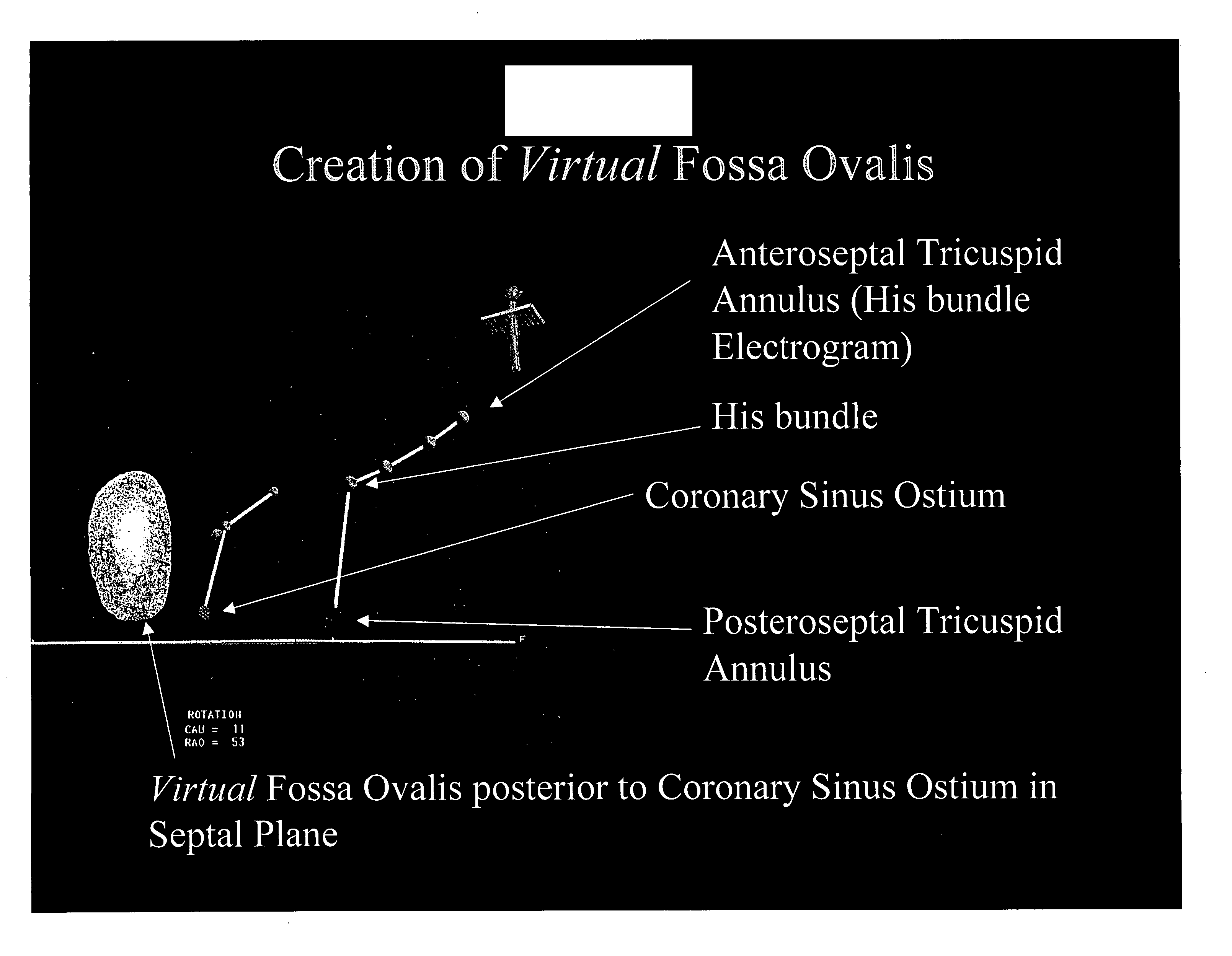
[0037] In the methods and apparatus described in the '844 Application, the fossa ovalis may be identified by changes in electrogram morphology, pacing threshold and / or impedance values. In particular, the fossa ovalis may be located by measuring the electrophysiological (“EP”) activity of the fossa ovalis and surrounding heart tissue. By observing differences in the EP activity of tissue at various locations, the operator may determine the location of the fossa ovalis. The lower muscle content and higher fibrous tissue content of the fossa ovalis with respect to the rest of the interatrial septum, as well as the relative “thinning” of the fossa, results in changes in EP activity which may be readily observed via an intracardiac electrogram. For example, the fossa ovalis will record broader, fractionated electrograms of lower amplitude and lower slew rates. Based upon these surprising findings, one or more electrodes for acquiring EP data may be incorporated into a catheter / dilator u...


PUM


Abstract

A method of locating the fossa ovalis in a patient by locating the His bundle, plane of the interatrial septum, and coronary sinus ostium in a patient, and thereafter locating the fossa ovalis on the basis of one or more predetermined distances between the fossa ovalis and the His bundle and the coronary sinus ostium. An apparatus for locating the fossa ovalis and performing a transseptal puncture is also provided.

Description

CROSS-REFERENCES TO RELATED APPLICATIONS [0001] This application claims the benefit of U.S. Provisional Application No. 60 / 658,111, filed Mar. 3, 2005, which is hereby incorporated by reference.FIELD OF THE INVENTION [0002] The present invention relates generally to methods and apparatus for locating a patient's fossa ovalis in the atrium of a human heart, creating a virtual fossa ovalis using an electroanatomical mapping system and using the information to perform transseptal punctures. In particular, these methods and apparatus may be based, at least in part, on anatomical measurements. BACKGROUND OF THE INVENTION [0003] Transseptal puncture refers to needle puncture of the interatrial septum through the fossa ovalis and is the standard technique for percutaneous introduction of catheters into the left atrium. The technique was described simultaneously by Ross and Cope in 1959. Brockenbrough and colleagues modified the design of the needle and guiding catheter apparatus in the 196...

Claims


Application Information

Patent Timeline
Patent Type & Authority Applications(United States)
IPC IPC(8): A61B5/05
CPCA61B5/042A61B5/06A61B5/1076A61B19/50A61B19/5244A61M25/0041A61B2017/00053A61B2017/00243A61B2019/505A61B2019/5251A61B2017/00044A61B34/20A61B34/10A61B2034/105A61B2034/2051A61B5/063A61B5/283
Inventor KRISHNAN, SUBRAMANIAM C.
Owner ST JUDE MEDICAL ATRIAL FIBRILLATION DIV
Why Eureka
Try Eureka