System and method for coronary artery segmentation of cardiac CT volumes

a segmentation system and cardiac ct volume technology, applied in image enhancement, image analysis, medical science, etc., can solve the problems of limited processing time and memory cost, and achieve the effects of robust and accurate segmentation, limited time and memory cost, and easy implementation

Inactive Publication Date: 2007-02-08
SIEMENS MEDICAL SOLUTIONS USA INC
View PDF1 Cites 83 Cited by
  • Summary
  • Abstract
  • Description
  • Claims
  • Application Information

AI Technical Summary

Benefits of technology

[0010] Exemplary embodiments of the invention as described herein generally include methods and systems for a locally adaptive method for the segmentation of coronary arteries from cardiac CTA volumes. Given a user-provided seed-point at the arterial root, an algorithm according to an embodiment of the invention automatically retrieves the entire arterial tree by combining successive local segmentations. Vessel segments are extracted by an automatic threshold selection with a connected-filter-like approach, followed by post-processing steps which are performed within a fast level-set framework. A skeletonization process extracts an approximate vessel centerline, providing new seed points to re-launch the algorithm. A method according to an embodiment of the invention can be easily implemented and can produce robust and accurate segmentation for a very limited time and memory cost, and requires one parameter, a “local volume jump threshold”, which can be set intuitively with regard to the size of target structures. Furthermore, a method according to an embodiment of the invention induces a limited cost in processing time and memory, typically less than 20 sec and 10 MB on average, on a 1.5 GHz Pentium IV with 1 GB RDRAM. By combining a method according to an embodiment of the invention with other aorta segmentation methods, a one-click segmentation of the entire aorta and coronary tree can be achieved. Provided one seed point, a dedicated algorithm can robustly and quickly extract the aorta and detect the aortic valve and the coronary branchings. A segmentation method according to an embodiment of the invention can be launched automatically to retrieve both coronaries. The entire process can take as little as 10 to 40 seconds on average.

Problems solved by technology

Furthermore, a method according to an embodiment of the invention induces a limited cost in processing time and memory, typically less than 20 sec and 10 MB on average, on a 1.5 GHz Pentium IV with 1 GB RDRAM.

Method used


Image

  • System and method for coronary artery segmentation of cardiac CT volumes
  • System and method for coronary artery segmentation of cardiac CT volumes
  • System and method for coronary artery segmentation of cardiac CT volumes

Examples


Embodiment Construction

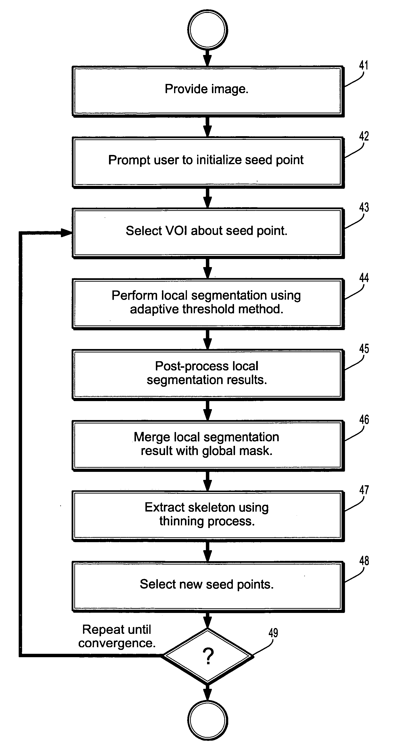
[0038] Exemplary embodiments of the invention as described herein generally include systems and methods for local and adaptive tracking algorithm. To account for the variability of the characteristics (size, intensity distribution) along the vessel, local segmentations are combined in small sub-volumes of interest, performed by an adaptive thresholding process. These local masks are post-processed using mathematical morphology and smoothing techniques and the approximate vessel centerline is extracted in order to determine new seed points to re-launch the process.

[0039] As used herein, the term “image” refers to multi-dimensional data composed of discrete image elements (e.g., pixels for 2-D images and voxels for 3-D images). The image may be, for example, a medical image of a subject collected by computer tomography, magnetic resonance imaging, ultrasound, or any other medical imaging system known to one of skill in the art. The image may also be provided from non-medical contexts...


PUM


Abstract

A method for segmenting coronary vessels in digitized cardiac images includes providing a digitized cardiac image, providing a seed point in the image, selecting a volume-of-interest about the seed point, performing a local segmentation in the volume-of-interest, including initializing a connected component with the seed point and a threshold intensity value to the intensity of the seed point, adding a point to the connected component if the point is adjacent to the connected component and if the intensity of the point is greater than or equal to the threshold value, lowering the threshold intensity value, and computing an attribute value of the connected component, wherein if a discontinuity in the attribute value is detected, the local segmentation is terminated, wherein a local segmentation mask of a vessel is obtained.

Description

CROSS REFERENCE TO RELATED UNITED STATES APPLICATIONS [0001] This application claims priority from “Coronary Artery Segmentation on Cardiac CT Volumes”, U.S. Provisional Application No. 60 / 703,187 of Lesage, et al., filed Jul. 28, 2005, the contents of which are incorporated herein by reference.TECHNICAL FIELD [0002] This invention is directed to segmentation of digitized medical images. DISCUSSION OF THE RELATED ART [0003] Blood vessel segmentation is important for clinical tasks such as diagnosis of vascular diseases, surgery planning and blood flow simulation. Although this field has been widely explored in the past, no solution has proved to be fully satisfactory for the segmentation of CT coronary arteries. [0004] Research in the cardiovascular imaging field is motivated by the number of people suffering from related diseases. Congestive heart failure, resulting from blocked coronary arteries, is of particular concern. Nearly 5 million American people already have congestive he...

Claims


Application Information

Patent Timeline
Patent Type & Authority Applications(United States)
IPC IPC(8): G06K9/00G06K9/34
CPCG06T7/0081G06T7/0091G06T7/60G06T2207/30101G06T2207/20012G06T2207/20044G06T2207/20101G06T2207/10072G06T7/11G06T7/155Y10S128/92Y10S128/922
Inventor LESAGE, DAVIDRASCH, MATTHIAS
Owner SIEMENS MEDICAL SOLUTIONS USA INC
Why Eureka
Try Eureka