
Automatic detection method of spectral Doppler blood flow velocity

a technology of automatic detection and blood flow velocity, applied in ultrasonic/sonic/infrasonic diagnostics, instruments, applications, etc., can solve the problems of low estimation accuracy, poor repeatability, and laborious operation of the operator, so as to reduce the error of envelope detection, perform the envelope detection accurately and robustly, and the envelope detection is more robust.

Active Publication Date: 2007-02-22
SHENZHEN MINDRAY BIO MEDICAL ELECTRONICS CO LTD
View PDF10 Cites 23 Cited by
  • Summary
  • Abstract
  • Description
  • Claims
  • Application Information

AI Technical Summary

Benefits of technology

The present invention provides a method and system for accurately detecting the spectral Doppler blood flow velocity, which is important for calculating blood flow parameters continuously and accurately. The method involves analyzing the spectrum of the Doppler signals, determining a threshold, and then using the threshold to determine the frequency shift parameters or blood flow velocity corresponding to the current power spectral line. The system includes means for obtaining and analyzing the Doppler signals, as well as means for updating the threshold to perform accurate envelope detection. The technical effects of the invention include reducing the effect of signal-to-noise ratio and bandwidth, and facilitating accurate calculation of blood flow parameters.

Problems solved by technology

The disadvantages of such manual detection are that the tracing of peak velocities by the operator is tedious and time consuming, the repeatability is poor, and the estimation accuracy is low; furthermore, the operator must stop the acquisition of Doppler signals in order to trace peak velocities during detection, thus it is unable to estimate in real time.
In clinical applications, this method is severely affected by SNR and bandwidth.
The mean threshold according to this method may vary with each of the spectral lines due to the randomness of noise, which results in variation of the estimated threshold level therewith.
However, the actual mean noise level is relatively stable, thus the method for automatically tracking envelope may cause unpredicted results.
The main disadvantage of the above-mentioned techniques is that: the mixed method is based on the hypothesis that the maximum frequency shift in the frequency spectrum diagram shall be less than the cut-off frequency.
In addition, since the result of maximum frequency shift estimation is greatly affected by the actual estimated noise and the integral curve, when the noise threshold is set to a small value, the maximum frequency shift estimation will be relatively small, and when the noise threshold is set to a big value, the maximum frequency shift estimation will be relatively big; thus this method is greatly affected by the actual SNR and accurate estimation of noise.
This method does not estimate the noise level directly, but it is severely affected by the integral curve; when the SNR is relatively low, the integral curve fails to guarantee a relatively rapid variation when the frequency is low, and a slow variation when the frequency is high, the error of the maximum frequency estimated then will be relatively high.
Similar to the geometric method, when the signal energy is relatively high and SNR is relatively low, the integral curve fails to guarantee that it varies relatively rapid when the frequency is low, and relatively slow when the frequency is high, the error of the maximum frequency estimated then will be relatively high.
In the adaptive threshold method, since the noise threshold is obtained by averaging the SNRs, and the SNRs are relatively severely affected by the signal intensity, the maximum frequency shift estimation will be affected by an incorrect estimation of the noise level.
Further more, since the threshold is in relation to the result of the previous cardiac cycle envelope detection, when the Doppler spectrum diagram is relatively stable, the adjustment of the adaptive threshold can not guarantee that the threshold will converge to a stable value.
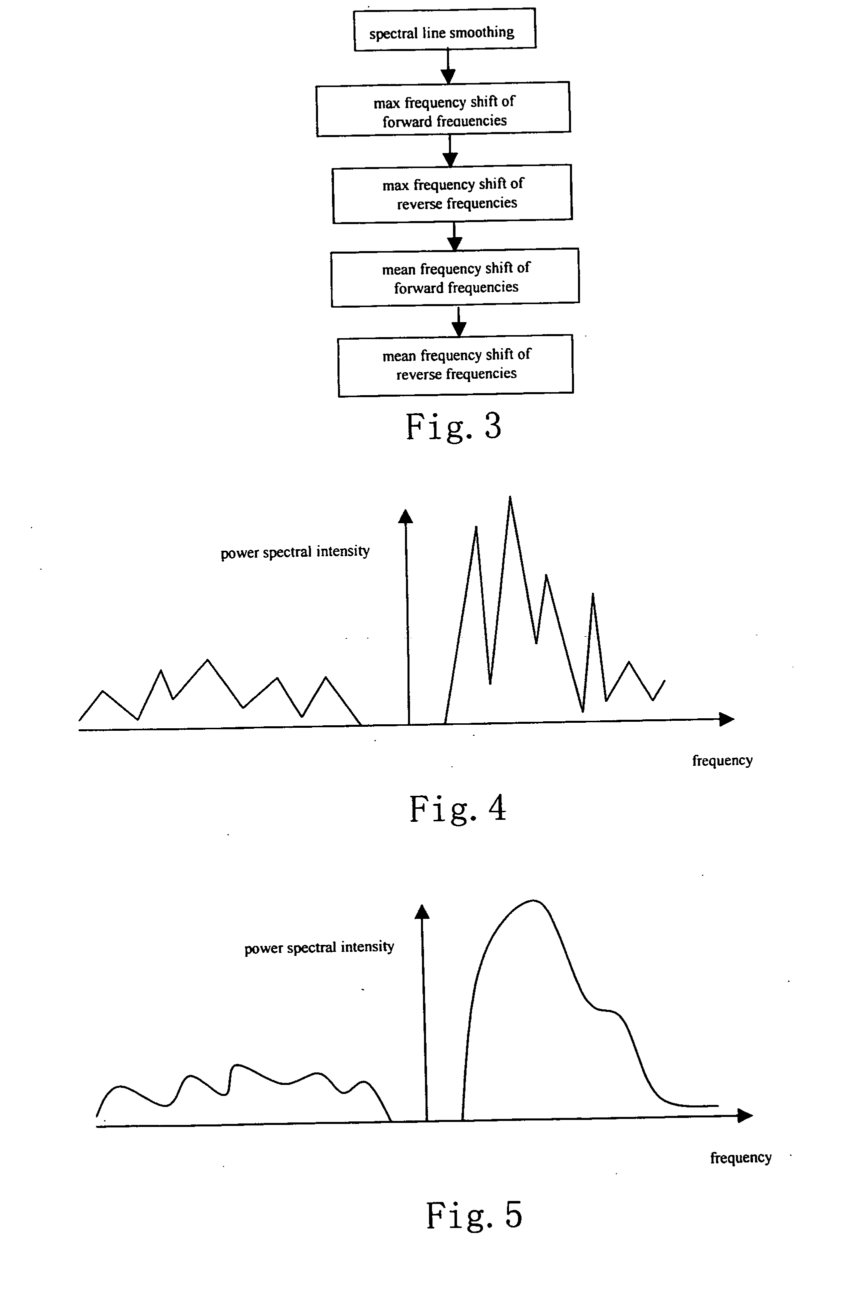
A simple threshold determination method is used in the determination of the envelope, the robustness of the envelope detection will also be impaired by the relatively great undulation of the power spectrum calculated by FFT.

Method used


Image

  • Automatic detection method of spectral Doppler blood flow velocity
  • Automatic detection method of spectral Doppler blood flow velocity
  • Automatic detection method of spectral Doppler blood flow velocity

Examples


Embodiment Construction

[0028] The present invention is further described with reference to the preferred embodiments shown in the accompanying figures.

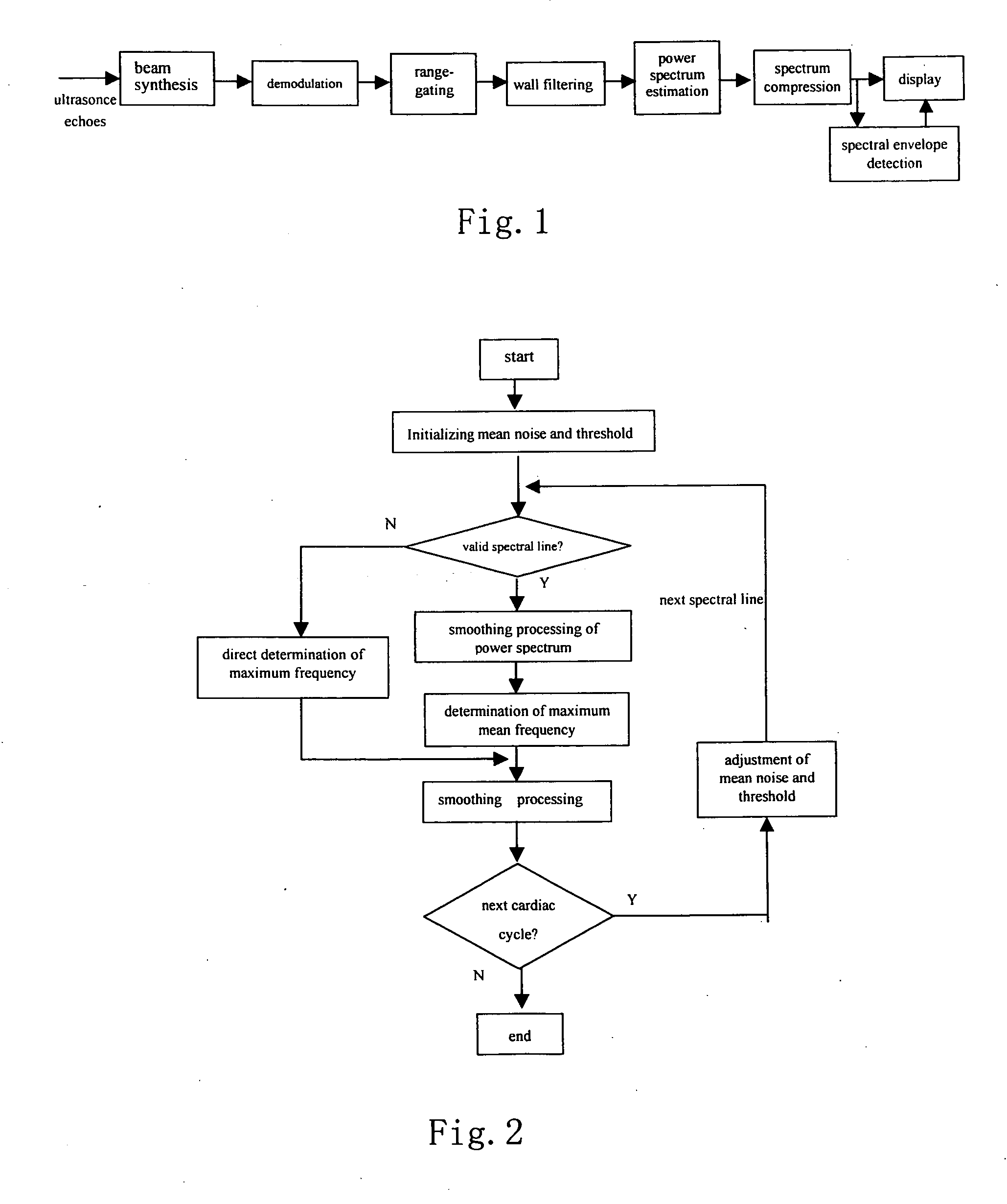
[0029] According to an embodiment of the present invention, an automatic detection method of spectral Doppler blood flow velocity is used for measuring blood flow velocity in an ultrasonic system, comprises the steps of: [0030] A. obtaining Doppler signals of the blood flow by demodulating, filtering and analog-to digital converting RF ultrasonic echoes; [0031] B. analyzing the spectrum of said Doppler signals to obtain each of the power spectral lines of the Doppler signal varying with time; [0032] C. determining a threshold; and [0033] D. determining the frequency shift parameters or the blood flow velocity corresponding to the current power spectral line based on said threshold and the current power spectral line, until the measurement procedure is ended or all of the power spectral lines have been processed;

wherein, said threshold is correlated to th...


PUM


Abstract

A method and system for detecting spectral Doppler blood flow velocity are disclosed. The method comprises the steps of: obtaining Doppler signals of the flow by demodulating, filtering, and analog-to-digital converting RF ultrasonic echoes; analyzing the spectrum of the Doppler signals to obtain each of the power spectral lines of the Doppler signal varying with time; determining a threshold; and determining the frequency shift parameters or the blood flow velocity corresponding to the current power spectral line based on the threshold and the current power spectral line.

Description

FIELD OF THE INVENTION [0001] The present invention relates to ultrasound technique and, particularly to ultrasound technique of measurement of fluid velocity by the use of spectral Doppler effect and, more particularly to a method for obtaining blood flow velocity by ultrasonic diagnostic instruments and envelope detection. BACKGROUND OF THE INVENTION [0002] In ultrasonic imaging systems, Doppler effect has been widely used in blood flow detection. For example, in an ultrasonic system for examining heart, artery and vein by the use of ultrasonic Doppler technique, it is necessary to extract related parameters from the Doppler spectrum diagram to estimate the dynamic states of blood flow of the heart and vessels. Those parameters include peak velocity, diastolic minimal velocity, resistance index, systolic / diastolic index, and etc. The determination of many of those parameters depends on the peak velocity and mean velocity of blood flow. While the detection of the peak velocity and ...

Claims


Application Information

Patent Timeline
Patent Type & Authority Applications(United States)
IPC IPC(8): A61B8/00
CPCA61B8/06A61B8/08A61B8/488G01F1/663G01S7/52026G01S15/8979
Inventor LI, YONG
Owner SHENZHEN MINDRAY BIO MEDICAL ELECTRONICS CO LTD
Learn More