
Modeling of systemic inflammatory response to infection

a systemic inflammatory response and model technology, applied in the field of model of the systemic inflammatory response to infection, can solve the problems of insufficient treatment options, limited search for new effective treatments for sepsis, and death of hospitalized patients

Inactive Publication Date: 2007-04-12
JANSSEN PHARMA NV
View PDF18 Cites 36 Cited by
  • Summary
  • Abstract
  • Description
  • Claims
  • Application Information

AI Technical Summary

Problems solved by technology

Septic shock is among the leading causes of death of hospitalized patients and is a condition for which insufficient treatment options are available.
The search for new effective treatments for sepsis has been limited.
During bacterial infections, bacteria and its products can cause septic shock that can result in death.
Sepsis, including all stages through septic shock, results from the inability of the immune system to properly control a bacterial infection.
In some instances, however, bacteria gain access to the circulation, resulting in mis-regulated production of inflammatory cytokines, sepsis syndrome, septic shock, and eventually death.
Staging sepsis to identify points at which the clinician can intervene with preventive measures has been and continues to be a very challenging task.
Broad disease definitions have limited the ability of clinicians to identify appropriate therapies for patients who have sepsis and who are at high risk for developing sepsis.
In addition, these definitions do not permit the clinician to differentiate between an at-risk patient who may derive a net benefit from a new therapy and a patient who will either not benefit, given his / her underlying disease co-morbidities, or who may be placed at higher risk from the therapy's inherent safety profile.
Additionally, the variability of disease progression and sequelae have made staging sepsis very difficult.
The difficulty in staging sepsis, combined with the contrasting results obtained with treatments tested, have made it very difficult to identify candidate drugs for treating sepsis and sepsis syndrome.
Such efforts have been largely unsuccessful—an alarming result for a disease syndrome with a current mortality rate of 30 to 50%.
Furthermore, even with respect to that drug, Xigris® (Lilly), there is not a straightforward way to determine when the drug should be administered to a sepsis patient.
Obstacles for developing sepsis therapies include incomplete understanding of the syndrome, inadequacies in staging the syndrome, and lack of adequate animal models.
Currently, animal models for sepsis syndrome do not mimic the human disease and have been considered an important cause behind the failure of proposed therapies.
Murine models have been used extensively with limited success to evaluate the efficacy of therapeutics in development for septic shock.
Moreover, the SICS scoring system and other scoring systems have not provided effective modeling to predict outcome or to detect when and if a given patient has become septic.

Method used


Image

  • Modeling of systemic inflammatory response to infection
  • Modeling of systemic inflammatory response to infection
  • Modeling of systemic inflammatory response to infection

Examples


example 1

Infectious Immunocompromised Mouse Model

[0122] Initially, C3H / HeJ mice were compared with C3H / HeN normal mice in a pouch model for their ability to survive infection. Mice of strain C3H / HeJ are defective in the TLR4 receptor and do not undergo LPS-induced shock. The mice were anesthetized with isofluorane, shaved in the area caudal to the ears, and a pouch was created by subcutaneous injection of 2-3 ml of air followed by the subcutaneous injection of 0.2 ml of a 0.5% solution of croton oil in olive oil. Either four days (d4) or five days (d5) later, animals were checked for the presence of a pouch. The number of animals observed to have pouches at these times are shown in Table 1 below, under the columns “d4” and “d5.” Animals without pouches were discarded. E. coli bort was injected in the pouches as reported in the first column of Table 1.

[0123] All animals of the HeJ strain were euthanized due to terminal health conditions, starting at 18.5 h and lasting until 48 h post-inject...

example 2

Identification of a Biomarker Panel in an Immunocompromised Mouse Model at 22 Hours Post-Infection

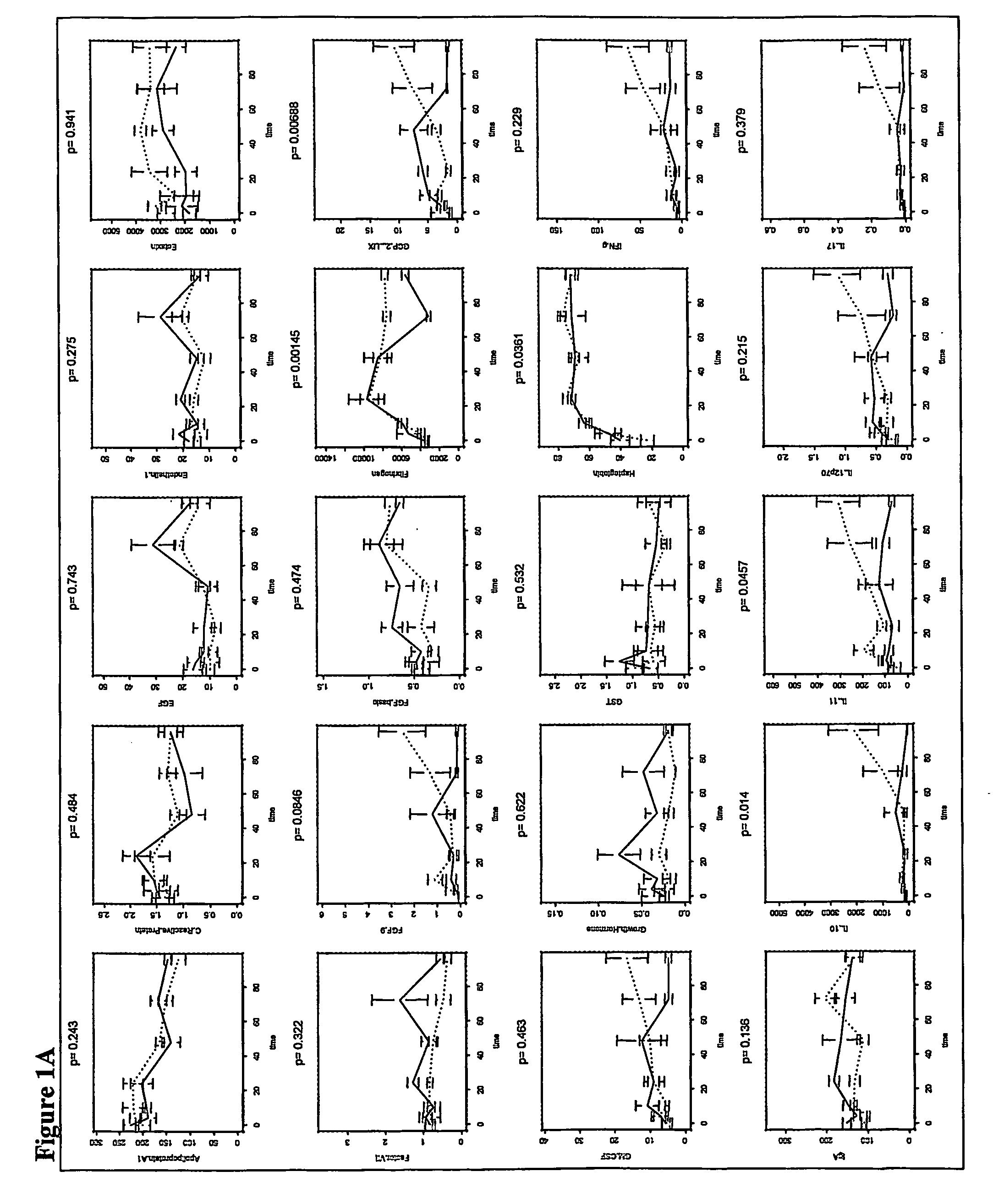
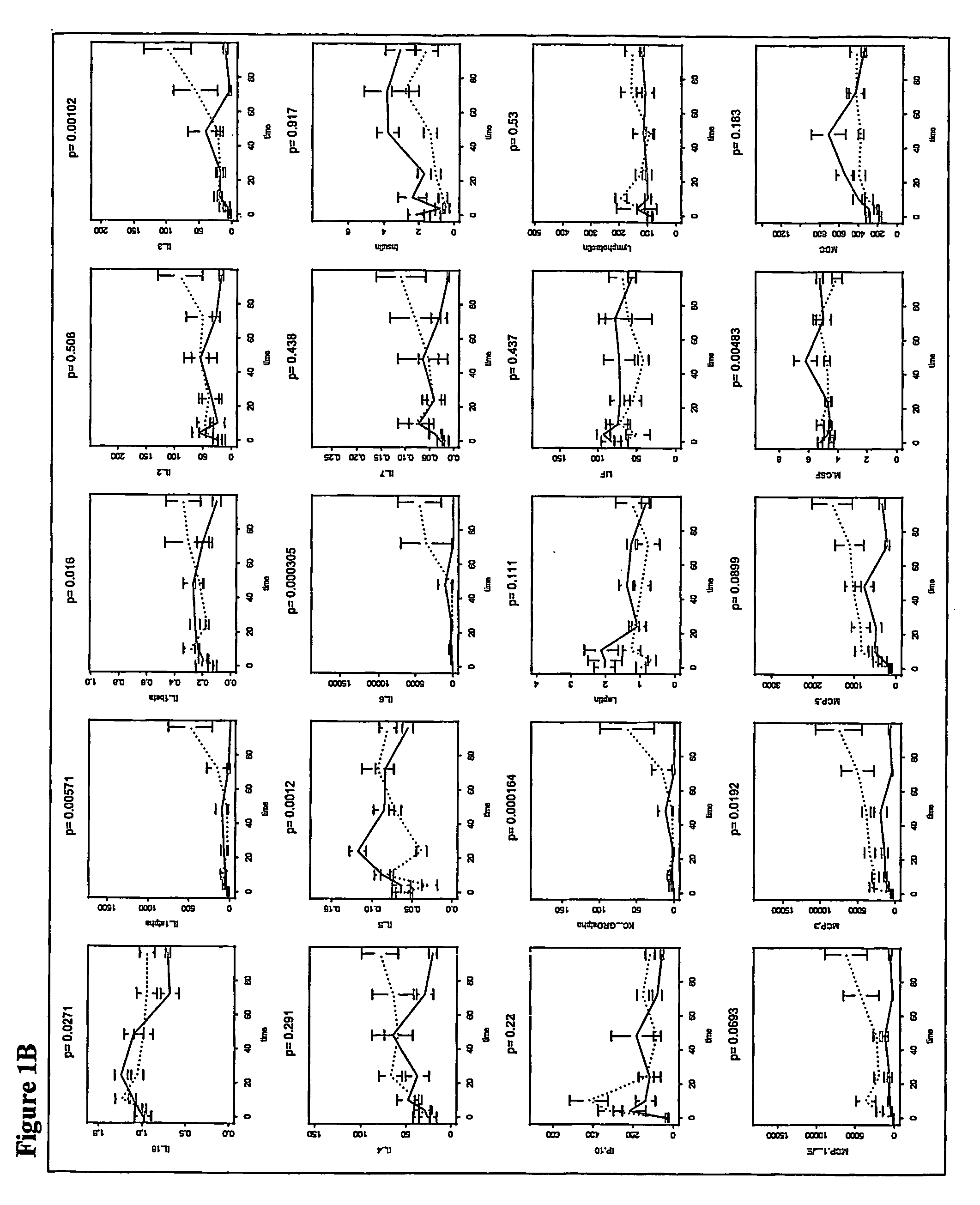
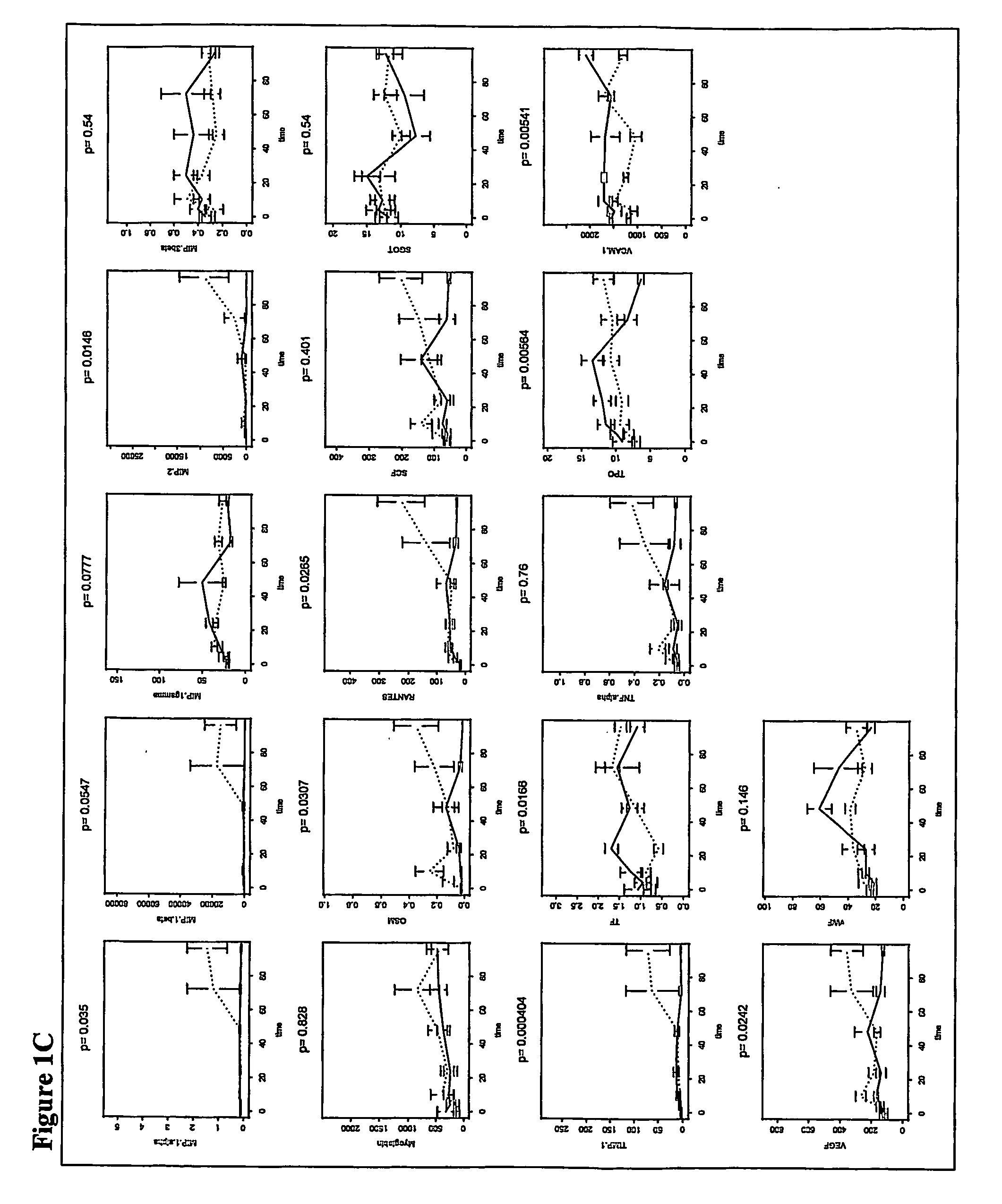
[0133] In an experiment using mice immunocompromised as described above, 22 mice were tested. Of these animals, 8 were doomed and 8 survived. As described in the survival study in Example 1, blood samples were taken from mice at 22 hours after infection. These samples were analyzed and used to derive a model to predict the outcome, i.e., survived or doomed, for animals that were both irradiated and infected with bacteria.

[0134] The 59 analytes measured in the samples were Apolipoprotein A1, β2 Microglobulin, C Reactive Protein, D-dimer, EGF, Endothelin-1, Eotaxin, Factor VII, FGF-9, FGF-Basic, Fibrinogen, GCP-2, LIX, GM-CSF, Growth Hormone, GST, Haptoglobin, IFN-α, IgA, IL-10, IL-11, IL-12p70, IL-17, IL-18, IL-1α, IL-1β, IL-2, IL-3, IL-4, IL-5, IL-6, IL-7, Insulin, IP-10, KC-GRO, Leptin, LIF, Lymphotactin, MCP-1-JE, MCP-3, MCP-5, M-CSF, MDC, MIP-1α, MIP-1β, MIP-1α, MIP-2, MIP-3β, Myog...

example 3

Use of Biomarker Panel Identified in Immunocompromised Mouse Model to Predict Disease Outcome-I

[0140] The discrimination function derived as described in Example 2 was applied to a set of mice. The discrimination model correctly predicted 100% doomed and 100% survived animals.


PUM

PropertyMeasurementUnit
body temperatureaaaaaaaaaa
temperatureaaaaaaaaaa

Abstract

Models for the systemic inflammatory response to infection, which involve the use of immunocompromised animals, and methods of using the models are described. These models can be used in identifying analytes or biomarker panels that can be used in staging or monitoring sepsis. The models can also be used for predicting an animal's disease outcome or in providing a prognosis for sepsis patients. Further, the invention relates to methods for evaluating potential treatments for sepsis.

Description

CROSS-REFERENCE TO RELATED APPLICATION [0001] This application claims priority to U.S. Provisional Application No. 60 / 523,296, the disclosure of which is incorporated by reference herein.FIELD OF THE INVENTION [0002] This invention relates to models for the systemic inflammatory response to infection comprising immunocompromised mice. The invention also relates to methods of using the models to identify biomarkers correlated with the systemic inflammatory response to infection, to identify biomarker panels useful in staging the disease, and to predict disease outcome. Further, the invention relates to methods for evaluating potential treatments for sepsis. BACKGROUND OF THE INVENTION [0003] Septic shock is among the leading causes of death of hospitalized patients and is a condition for which insufficient treatment options are available. The search for new effective treatments for sepsis has been limited. The incidence of sepsis is expected to increase sharply in the near future due...

Claims


Application Information

Patent Timeline
Patent Type & Authority Applications(United States)
IPC IPC(8): G06F19/00A61BC12Q1/68G01N33/53G01N33/68G16B20/00G16B40/10
CPCG01N33/6842G01N33/6893G01N2800/26G06F19/18G06F19/24G16B20/00G16B40/00G16B40/10
Inventor VITIELLO, MARIA ANTONIAZHANG, YIAMARATUNGA, DHAMMIKA JAYANATHSHI, TAOWARD, CHRISTINE K.
Owner JANSSEN PHARMA NV
Learn More