
A system for intelligent analysis of corneal nerve fibers using in vivo confocal microscopy images

A technology of confocal microscopy and nerve fibers, which can be used in image analysis, nervous system evaluation, image enhancement, etc., and can solve problems such as identifying systemic multi-system neuropathy

Active Publication Date: 2021-05-18
THE PEOPLES HOSPITAL OF GUANGXI ZHUANG AUTONOMOUS REGION
View PDF4 Cites 0 Cited by
  • Summary
  • Abstract
  • Description
  • Claims
  • Application Information

AI Technical Summary

Problems solved by technology

At present, there is no related technology that uses corneal nerves to identify systemic multisystem neuropathy.

Method used


Image

  • A system for intelligent analysis of corneal nerve fibers using in vivo confocal microscopy images
  • A system for intelligent analysis of corneal nerve fibers using in vivo confocal microscopy images
  • A system for intelligent analysis of corneal nerve fibers using in vivo confocal microscopy images

Examples


Embodiment Construction

[0059] The preferred embodiments of the present invention will be described in detail below in conjunction with the accompanying drawings, so that the advantages and features of the present invention can be more easily understood by those skilled in the art, so as to define the protection scope of the present invention more clearly.

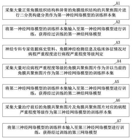
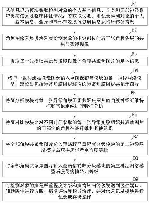
[0060] Such as figure 1 As shown, the embodiment of the present invention is a system for intelligently analyzing corneal nerve fibers using in vivo confocal microscope images, including an information recording module, a corneal image acquisition module, an information extraction module, an image preliminary screening module, a feature analysis module, a feature comparison module, Disease grading module and result sending module.

[0061] Information recording module: used to record or store the basic personal information of the test object, systemic and local nervous system disease information, treatment status and clinical signs. Basic person...


PUM


Abstract

The invention discloses a system for intelligently analyzing corneal nerve fibers by using confocal microscope images in vivo, including: an information recording module; a corneal image acquisition module for collecting several confocal microscope images of each layer of the cornea; an information extraction module; Sieve module, locate confocal images of abnormal corneal tissue from each corneal confocal image through the first neural network model; feature analysis module; feature comparison module; disease grading module, including disease course severity grading module and disease outcome grading module The course severity grading module is that all corneal confocal pictures pass through the second neural network model to obtain the course severity level; Return to grade; result sending module. The invention analyzes the characteristics of corneal nerve fibers, the degree of damage and the changing process thereof, and evaluates the state of local or systemic neuropathy.

Description

technical field [0001] The present invention relates to the technical field of artificial intelligence, and more specifically, relates to a system for intelligently analyzing corneal nerve fibers using in vivo confocal microscope images. Background technique [0002] Peripheral neuropathy is defined as a syndrome of sensory loss, muscle weakness and atrophy, decreased tendon reflexes, and vasomotor symptoms, alone or in any combination. In the early stage of the disease, sensory disturbance is the main symptom. Auxiliary examination methods for accurately judging the scope and degree of nerve damage in the early stage of the disease are necessary to help clinicians take correct treatment methods. In addition to clinical history and signs, the current clinical methods for evaluating the extent and degree of peripheral nerve injury mainly use invasive nerve electrophysiological examination, muscle biopsy and expensive MRI examination. [0003] The corneal nerve is composed of...

Claims


Application Information

Patent Timeline
Patent Type & Authority Patents(China)
IPC IPC(8): G06T7/00G16H30/20G16H50/20G06N3/04A61B3/13A61B5/00
CPCG06T7/0012G16H30/20G16H50/20A61B3/13A61B5/40A61B5/7267G06T2207/30041G06N3/045
Inventor 徐帆吕健陈琦崔凌陈青曾思明李敏何文静唐芬蒋莉唐宁宁陈丽妃周舟黄慧
Owner THE PEOPLES HOSPITAL OF GUANGXI ZHUANG AUTONOMOUS REGION
Learn More