
Reduction of Streak Artifacts In Low Dose CT Imaging through Multi Image Compounding

Inactive Publication Date: 2007-06-21
SIEMENS MEDICAL SOLUTIONS USA INC
View PDF32 Cites 12 Cited by
  • Summary
  • Abstract
  • Description
  • Claims
  • Application Information

AI Technical Summary

Benefits of technology

[0011] In accordance with an embodiment of the present invention, an FBP method, referred to below as Compounded FBP (CFBP), has been developed to improve the reconstruction quality for low-dose CT scans where signal-to-noise (SNR) ratio is low and the number of projections may be restricted. The inventive method exploits complementary information from projection subsets to discriminate between angle dependent streaking noise and angle invariant true edges. This improves the image quality of the filtered back projection. The CFBP method produces image reconstruction of a quality that is typically similar to the quality of images produced by IR methods, but without the computational disadvantages of IR.

Problems solved by technology

However, it is becoming more challenging for known conventional reconstruction methods to meet the stringent constraints of current imaging applications.
However, to lower the X-ray dosage (i.e., in low-dose CT imaging), the signal-to-noise ratio and the number of projections are reduced, which often results in undesirable “streaking artifacts”.
There are typically two main sources of streaking artifacts First, the filter may amplify the imaging noise in the sinogram, which often produces streaking lines during back projection.
Second, strong sharp edges, caused by, e.g., metal or bones, are back projected along the projection direction, causing streaking artifacts, especially when the number of projections is limited.
Streaking noise is signal dependent and spatial variant, often making it extremely difficult to model this artifact accurately and distinguish the noise from true edges in the image.
In the field of medical radiology, this typically results in difficult and / or inconsistent clinical interpretation(s).
Although conceptually this approach is typically simpler than FBP, for medical applications, it has traditionally lacked the speed of implementation and accuracy.
This is due to the slow convergence of the algorithm and high computational demands.

Method used


Image

  • Reduction of Streak Artifacts In Low Dose CT Imaging through Multi Image Compounding
  • Reduction of Streak Artifacts In Low Dose CT Imaging through Multi Image Compounding
  • Reduction of Streak Artifacts In Low Dose CT Imaging through Multi Image Compounding

Examples


Embodiment Construction

[0028] In one aspect of the present invention, a new FBP method results in improved reconstruction quality. The invention may be used beneficially for low dose CT scans where signal-to-noise (SNR) ratio is low and the number of projections may be restricted. As noted, this improved method is referred to herein as Compounded FBP (CFBP). The inventive method exploits complementary information from projection subsets to discriminate between angle dependent streaking noise and angle invariant true edges. To aid in understanding the invention, we will discuss below: 1. Filtered Back Projection (FBP); 2. Compounded Filtered Back Projection (CFBP); 3. CFBP Background; 4. FBP Derived from MAP Framework; and 5. CFBP Based on Cross Group Regularization.

1. Filtered Back Projection

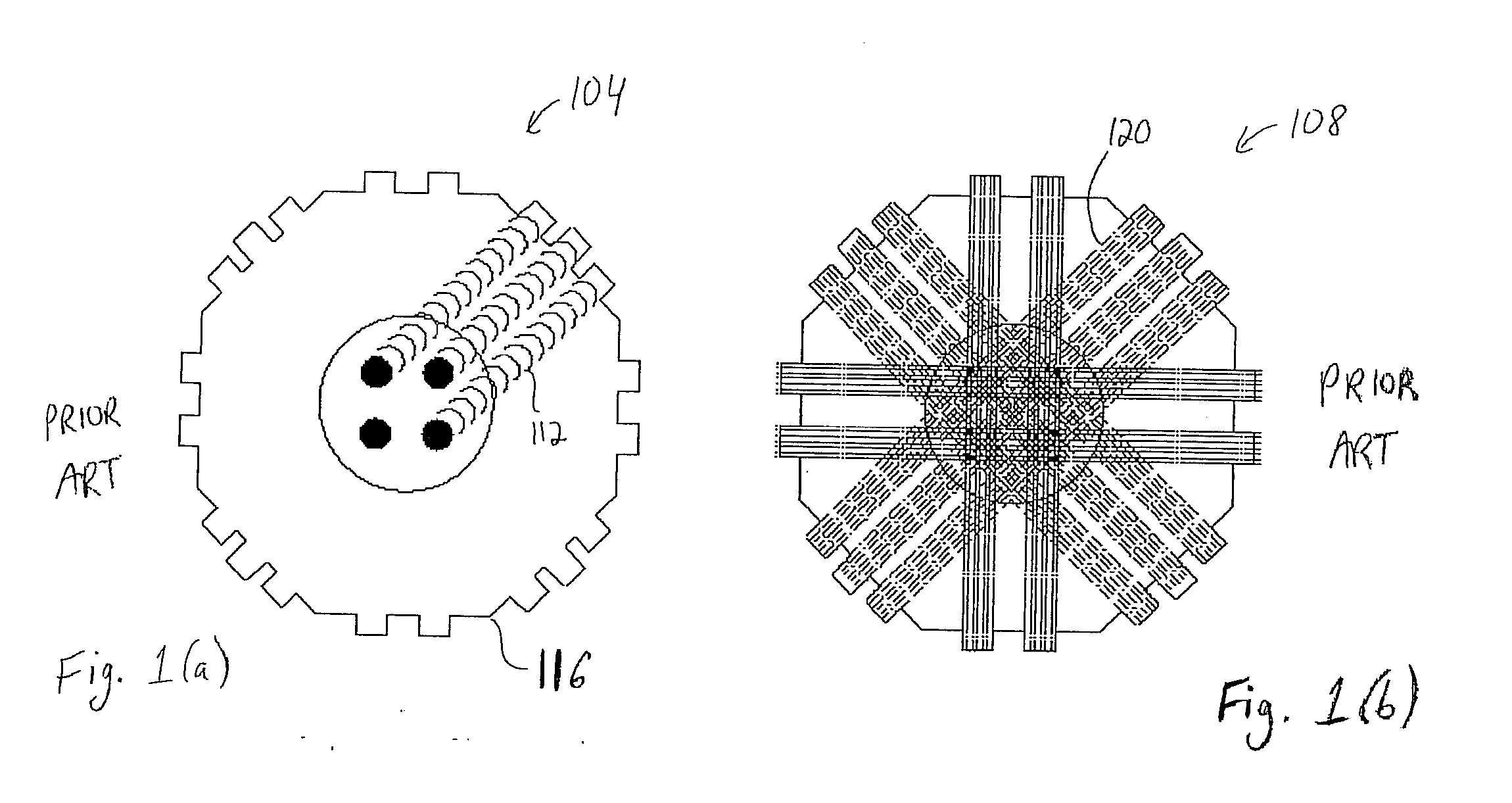
[0029] As discussed above, FBP involves the construction of an image by analyzing projection images obtained from many different viewing angles. The technique is shown schematically in FIG. 1. FIG. 1(a) shows a pri...


PUM


Abstract

Disclosed is a method and system for constructing, from a computerized tomography (CT) scan, an image relating to a physical structure. Projection data associated with the image is obtained and divided into a plurality of subsets. Filtered back projection (FBP) is then applied to each subset in the plurality of subsets. The image is constructed based on the application of the FBP to each subset in the plurality of subsets.

Description

[0001] This application claims the benefit of U.S. Provisional Application No. 60 / 725,945, filed Oct. 12, 2005, which is incorporated herein by reference.BACKGROUND OF THE INVENTION [0002] Computer Axial Tomography (CAT), sometimes known generally as Computerized Tomography (CT), is used in many applications, especially medical radiology, to obtain two or three dimensional views of the interior of three dimensional bodies (CT or CAT Scans). The technique involves subjecting a three dimensional body to radiation that enters the body from many different angles. The amount of radiation that is scattered or reflected by the body is then detected as a function of the angle of scattering. The scattered data is then analyzed to construct an image of the interior of the body. A two dimensional “slice” of the interior can be “reconstructed”, for example on a screen, and viewed. The slice can be reconstructed for any desired angle of intersection with the body. [0003] While computerized tomog...

Claims


Application Information

Patent Timeline
IPC IPC(8): H05G1/60A61B6/00G01N23/00G21K1/12
CPCG06T11/005G06T2211/421A61B6/5258
Inventor CHEN, YUNQIANGFANG, TONG
Owner SIEMENS MEDICAL SOLUTIONS USA INC
Learn More