
Biological artificial cornea and method of making

a biochemical and artificial cornea technology, applied in the field of medical prosthesis, can solve the problems of failure of transplantation, failure of additional research on the preparation of artificial corneas from animal corneas by low-temperature freezing and simple sterilization treatment, and failure of direct animal corneal transplantation. to achieve the effect of minimizing antigens

Inactive Publication Date: 2007-06-21
SUMMIT GD BIOTECH
View PDF35 Cites 40 Cited by
  • Summary
  • Abstract
  • Description
  • Claims
  • Application Information

AI Technical Summary

Benefits of technology

[0006] It is an object of the present invention to provide safe and reliable biological artificial corneas having high biocompatibility, stability, which can be degraded and absorbed, and which are capable of inducing cornea regeneration.
[0008] In order to accomplish the objects of the present invention, the present invention provides an artificial cornea for implantation into a human body which is made by a method that includes the steps of providing a natural animal cornea that has a substrate, crosslinking and fixing the substrate, minimizing the antigens from the substrate, and coupling an active layer to the substrate.

Problems solved by technology

However, transplantation of a cornea not only has difficulties such as securing the source of donation, but immunological rejection often leads to failures in the transplantation.
However, such direct animal corneal transplantations were unsuccessful because of immunological rejection.
Additional research on preparations of artificial corneas from animal corneas by low-temperature freezing and simple sterilization treatment were also unsuccessful because the elimination of antigens was not complete and the patients' bodies could not accept the transplants due to poor tissue compatibility.

Method used


Image

  • Biological artificial cornea and method of making
  • Biological artificial cornea and method of making
  • Biological artificial cornea and method of making

Examples


example 1

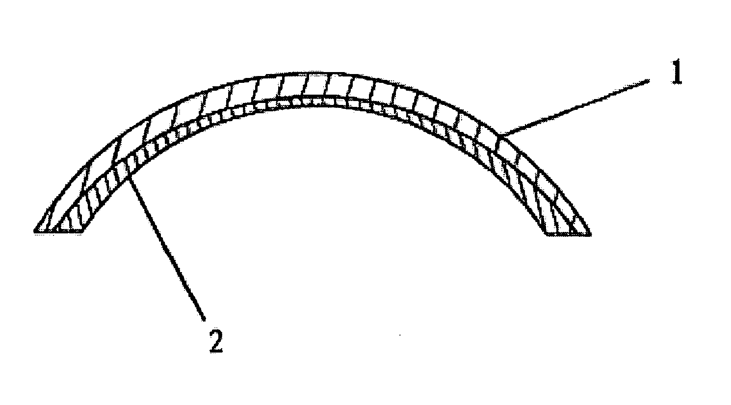
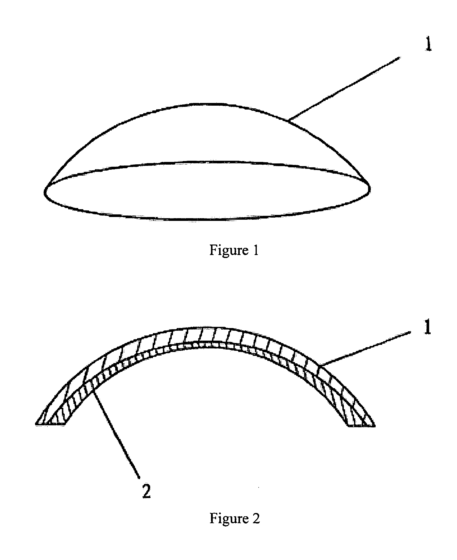
[0034] As shown in FIGS. 1 and 2, the biological artificial cornea comprises a substrate 1 prepared from an animal cornea by crosslinking and fixing with a non-aldehyde fixative and minimizing antigens. An active surface layer 2 is formed by coupling the inner (eyeball-facing) surface of substrate 1 with an active component consisting of a polypeptide or glycosaminoglycan capable of adhering to growth factors. One example of the polypeptide is the polypeptide obtained from the condensation of 16 lysines (K16), glycine (G), arginine (R), asparagic acid (D), serine (S), proline (P) and cysteine (C), and said glycosaminoglycan is hyaluronic acid, chondroitin sulfate, dermatan sulfate, heparin, acetylheparin sulfate or keratan sulfate. This biological artificial cornea can be made from the following steps: [0035] 1. Selection of materials: Fresh eyeballs are collected from healthy pigs and frozen in special preservation bottles before being transported. [0036] 2. Pretreatment: The anima...


PUM

PropertyMeasurementUnit
water-solubleaaaaaaaaaa
hydrogen bonding poweraaaaaaaaaa
temperatureaaaaaaaaaa

Abstract

An artificial cornea for implantation into a human body is made by a method that includes the steps of providing a natural animal cornea that has a substrate, crosslinking and fixing the substrate, minimizing the antigens from the substrate, and coupling an active layer to the substrate.

Description

BACKGROUND OF THE INVENTION [0001] 1. Field of the Invention [0002] The present invention relates to a medical prosthesis for human implantation, and in particular, to a device for reconstructing a damaged cornea. [0003] 2. Description of the Prior Art [0004] Loss of sight caused by corneal damage or pathological changes is one of the most common ophthalmologic diseases, and the current treatment method relies on transplantation of a cornea donated from a cadaver. However, transplantation of a cornea not only has difficulties such as securing the source of donation, but immunological rejection often leads to failures in the transplantation. Accordingly, scientists have attempted to use animal corneas to treat corneal diseases in humans, including studies performed on the direct transplantation of animal corneas. However, such direct animal corneal transplantations were unsuccessful because of immunological rejection. Additional research on preparations of artificial corneas from ani...

Claims


Application Information

Patent Timeline
Patent Type & Authority Applications(United States)
IPC IPC(8): A61F2/14
CPCA01N1/00A61F2/14A61F2/142A61L27/3604A61L27/3687A61L2430/16C12N5/0697A61F2240/001A61F2210/0004
Inventor XU, GUO-FENG
Owner SUMMIT GD BIOTECH
Learn More