Ultrasonograph and ultrasonography

a technology of ultrasonography and ultrasonography, applied in the field of ultrasonography, can solve the problems of affecting the diagnosis of patients, and affecting the diagnosis of vascular endothelial function, and achieves the effect of high sensitivity and high accuracy in diagnosing vascular endothelial function

Inactive Publication Date: 2007-09-20
PANASONIC CORP
View PDF3 Cites 34 Cited by
  • Summary
  • Abstract
  • Description
  • Claims
  • Application Information

AI Technical Summary

Benefits of technology

[0035] According to the present invention, the changes of elastic modulus of vascular wall caused by vascular endothelial reaction after the stopping of avascularization is measured with high accuracy by using ultrasonic waves. As a result, it is possible to provide an ultrasonic diagnostic apparatus, which can makes it possible to diagnose vascular endothelial function with high sensitivity.

Problems solved by technology

In recent years, patients suffering from diseases of circulatory organs such as myocardial infarction, cerebral infarction, etc. have been increasing, and it is now an important problem to prevent and treat such diseases.
More concretely, when atheroma is formed on arterial wall or new cells of artery are not formed due to various reasons such as hypertension, artery loses elasticity and is hardened and becomes fragile.
However, to perform such diagnosis, it is necessary to insert vascular catheter into blood vessel, and this means that heavier burden is applied on the patient.
For this reason, the examination using vascular catheter has been limited on the use to specify the affected site on a patient, who has been definitely determined to have such atherosclerotic lesion.
For instance, this procedure has not been performed for health examination.
However, these values do not directly indicate the severity of atherosclerosis.
However, once atherosclerosis develops and advances, it is difficult to completely recover the affected artery even though it is possible to suppress further development of atherosclerosis.
However, in the ultrasonic diagnostic apparatus used in the past, as represented by the ultrasonic diagnostic apparatus used for observing the conditions and the shape of a fetus or for auscultation of heart sounds of fetus, information on shape or information on movement is not given with very high resolution.
For this reason, it has not been possible to identify elastic property of artery, which expands and contracts in association with heart beat by using conventional type ultrasonic diagnostic apparatus.
For example, as the apparatus disclosed in the Patent Document 1 as given below, the accuracy of the measurement on displacement of the object to be measured has not been very satisfactory.
On the other hand, risk factors such as hypertension, hyperlipide, smoking, diabetes, etc. may decrease the functions of vascular endothelial cells.
That is, there is a problem in measurement accuracy in the method described in the Non-Patent Document 1.
That is, problems may also arise in the measurement sensitivity in the method described in the Non-Patent Document 1.

Method used


Image

  • Ultrasonograph and ultrasonography
  • Ultrasonograph and ultrasonography
  • Ultrasonograph and ultrasonography

Examples


Embodiment Construction

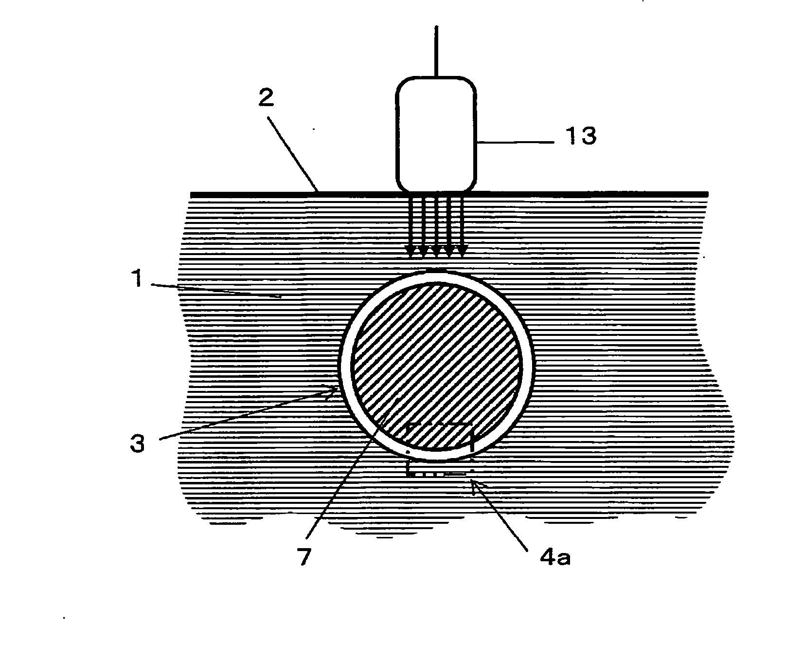
[0043] The ultrasonic diagnostic apparatus according to the present invention determines moving speed of each region of an object to be measured and amount of expansion and contraction and elastic modulus in micro-size region. The object to be measured itself does not move. In particular, the ultrasonic diagnostic apparatus of the present invention is suitable for measurement of elastic modulus in each region of living body and has high spatial resolution. For this reason, it can be preferably used for measuring expansion and contraction and elastic modulus of vascular wall. Description will be given below on a case to measure expansion and contraction and elastic modulus of vascular wall.

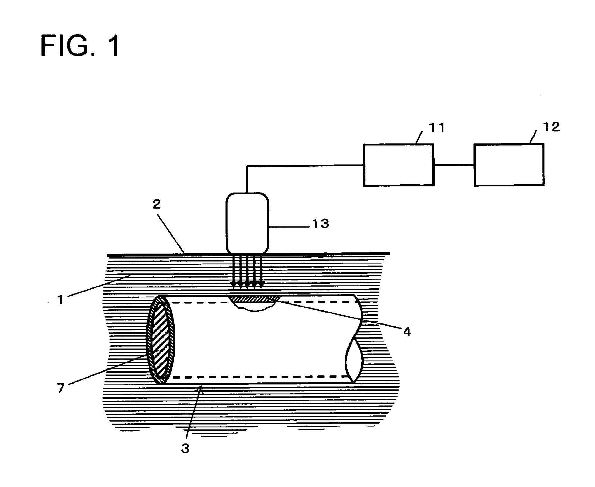
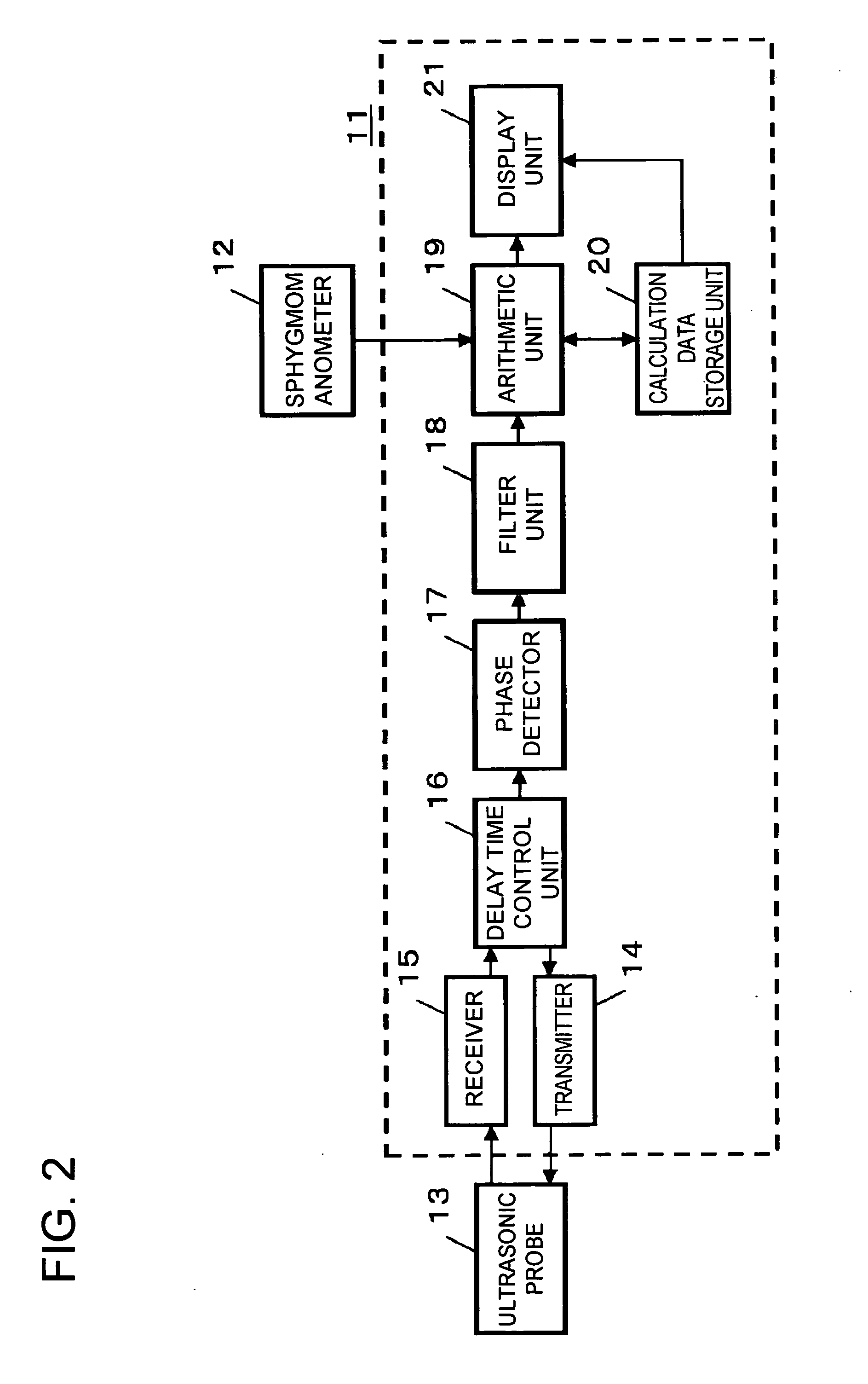
[0044] Brief description will be given below on an embodiment of the ultrasonic diagnostic apparatus according to the present invention. FIG. 1 is a schematical block diagram showing an arrangement of diagnosis of conditions and behaviors of tissues in vascular wall using the ultrasonic diagnostic...


PUM


Abstract

A new technique is disclosed for providing an ultrasonic diagnostic apparatus and an ultrasonic diagnostic method, by which it is possible to diagnose vascular endothelial function with high sensitivity through measurement of changes of elastic modulus of vascular wall in the region of tunica intima and tunica media with high precision by using ultrasonic waves in the diagnosis of vascular endothelial reaction after the stopping of avascularization. According to this technique, the apparatus comprises an arithmetic unit 19 for obtaining elastic modulus of a vascular wall 4, and the apparatus is provided with at least one of a calculation data storage unit 20 for storing changes over time of elastic modulus of vascular wall when artery is avascularized and the avascularization is then stopped, or a display unit 21 for displaying changes over time of elastic modulus of the vascular wall.

Description

TECHNICAL FIELD [0001] The present invention relates to an ultrasonic diagnostic apparatus for diagnosing conditions and behaviors of tissues in blood vessel wall using ultrasonic waves. The invention also relates to an ultrasonic diagnostic method. BACKGROUND ART [0002] In recent years, patients suffering from diseases of circulatory organs such as myocardial infarction, cerebral infarction, etc. have been increasing, and it is now an important problem to prevent and treat such diseases. [0003] The development of myocardial infarction or cerebral infarction is deeply related with atherosclerosis. More concretely, when atheroma is formed on arterial wall or new cells of artery are not formed due to various reasons such as hypertension, artery loses elasticity and is hardened and becomes fragile. When blood vessel is blocked at a portion where atheroma is formed or when vascular tissues enclosing atheroma are ruptured and atheroma flows into blood vessel. Artery may be blocked at oth...

Claims


Application Information

Patent Timeline
Patent Type & Authority Applications(United States)
IPC IPC(8): A61B8/00A61B8/08
CPCA61B8/485A61B8/0858
Inventor KANAI, HIROSHIHASEGAWA, HIDEYUKI
Owner PANASONIC CORP
Why Eureka
Try Eureka