
Fluorogenic Nucleic Acid Probes Including Lna for Methods to Detect and/or Quantify Nucleic Acid Analytes

a nucleic acid analyte and fluorogenic technology, applied in the field of molecular biology, can solve the problems of increased detector fluorescence, false positives, and prone to contamination, and achieve the effect of quenching the detector fluorescence, increasing the fluorescence of the detector dye, and not being quenched

Inactive Publication Date: 2007-11-22
SIGMA ALDRICH CO LLC
View PDF17 Cites 1 Cited by
  • Summary
  • Abstract
  • Description
  • Claims
  • Application Information

AI Technical Summary

Benefits of technology

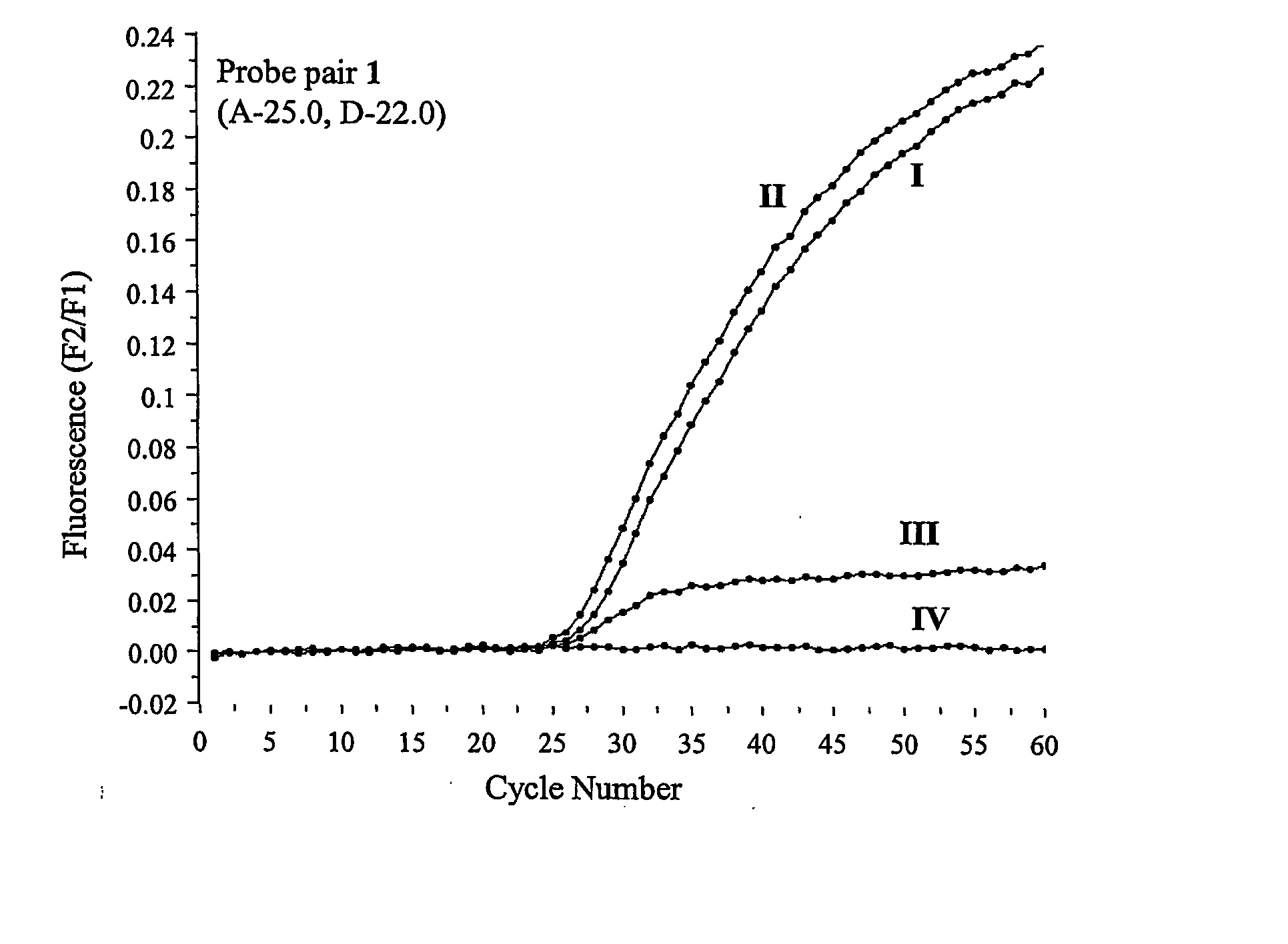
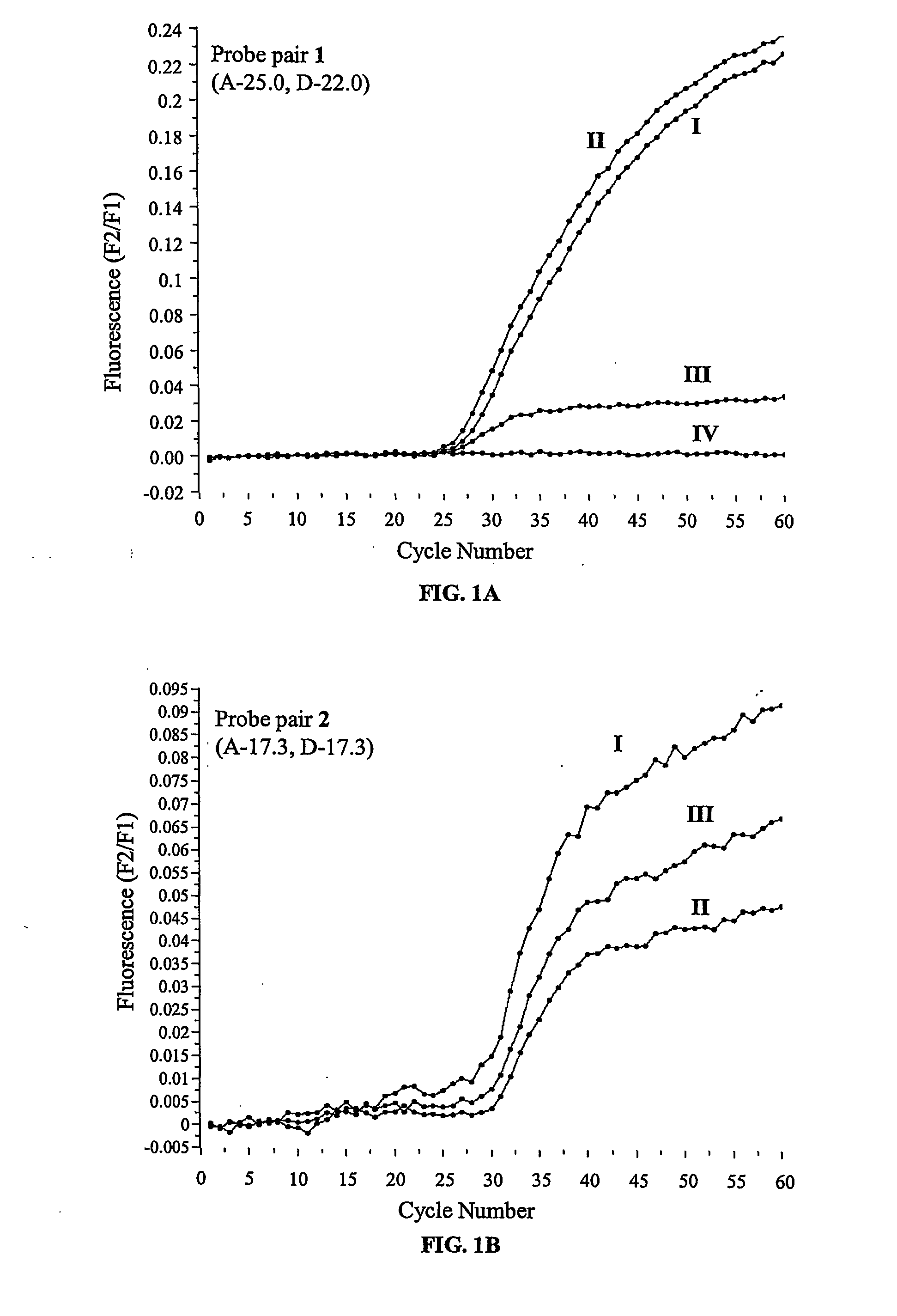
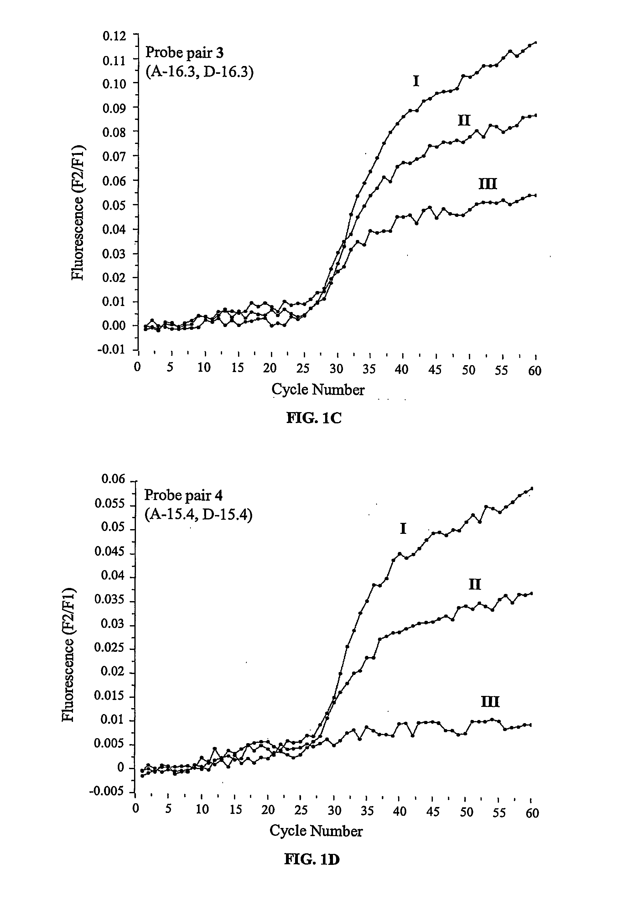
[0032]FIGS. 3B to 3D depict the observed relative fluorescence intensities in real-time PCR experiments as a function of the PCR cycle number using nucleic acid probe pairs 7, 8 and 9, respectively, and DNA templates related to the human CF SNP G542X. Nucleic acid probe pairs 7, 8 and 9 are comprised of 3 or 4 monomeric LNA moieties in the probe carrying the donor dye only. The corresponding experiments are described in Example 5. The plots for the wild type template, the heterozygous template, the mutant type template and the control without template are represented by lines I, II, III and IV, respectively.
[0033]FIGS. 3E and 3F depict the observed relative fluorescence intensities in real-time PCR experiments with the nucleic acid probe pairs 10 and 11, respectively, and DNA templates related to the human CF SNP G542X as a function of the PCR cycle number. The corresponding experiments are described in Example 5. Both probe pairs are identical in length and sequence, but the Red 640 derivatized probe of probe pair 10 is comprised of 5 monomeric LNA moieties, whereas the probes of probe pair 11 are comprised of deoxynucleotides only. The plots for the wild type template; the heterozygous template, the mutant type template and the control without template are represented by the lines I, II, III and IV, respectively.
[0034]FIGS. 4A to 4F display the time derivatives of the melting curves for the probe pairs 1 and 7 to 11, respectively, measured according to Example 5 subsequent to the PCR experiments described in Example 5 with nucleic acid templates related to the human CF SNP G542X. The plots of the derivative melting curves for the wild type derived amplicon, the heterozygous type derived amplicon, the mutant type derived amplicon and the control are represented by the lines I, II, III and IV, respectively.

Problems solved by technology

Such procedures, as well as, other methods that similarly rely on end-point analysis are generally labor intensive, require several separate and distinct handling processes and skilled personnel, are relatively slow to produce a result, and are prone to contamination and false positives due to the open system.
As a result the two blocks of DNA nucleotide sequence of the probe are separated, which in turn results in an increased fluorescence of the detector dye, which is no longer quenched by the acceptor.
The detector and quencher dyes are in close proximity to one another in this conformation, which results in quenching of the detector fluorescence.
The described fluorescence based methods are all limited in that they lack specificity and discrimination capability e.g. towards certain types of mutations.
Thus, they can not cope with the growing demand for methods allowing the rapid screening of complete genomes for mutations, particularly single base mutations, in a high-throughput format.
However, Jakobsen et al. do not disclose how to use their invention and completely fail in reducing it to practice.
Furthermore, the design of the probes described by Jakobsen et al. is very limited in that at least the second mono-nucleotidic position from the 3′- and / or the 5′-end has to be a LNA moiety.

Method used


Image

  • Fluorogenic Nucleic Acid Probes Including Lna for Methods to Detect and/or Quantify Nucleic Acid Analytes
  • Fluorogenic Nucleic Acid Probes Including Lna for Methods to Detect and/or Quantify Nucleic Acid Analytes
  • Fluorogenic Nucleic Acid Probes Including Lna for Methods to Detect and/or Quantify Nucleic Acid Analytes

Examples


example 1

General Procedure for the Preparation of the Labelled Oligo Probes

[0087] Oligonucleotide primers and probes were synthesized on a UFPS-24 synthesizer (Proligo LLC, Boulder, Colo., USA) using standard phosphoramidite chemistry, as known in the art. The 3′-fluorescein-labelled probes were synthesized using a fluorescein labelled CPG (Roche Diagnostics, Indianapolis, Ind., USA). The resulting labelled probes were purified by reverse phase high pressure liquid chromatography (HPLC) (Waters Symmetry Column, 5 μm, 3.9×150 mm, C18). The purities of these probes were determined by analytical reversed phase HPLC with monitoring at wavelengths of 260 nm and 495 nm.

[0088] The LC Red 640 labelled probes were synthesized using a phosphate-on CPG (Proligo LLC, Boulder, Colo., USA). Amino functionalization of the 5′-terminus was achieved by adding an amino modifying amidite with a C6 linker (Glen Research, Sterling, Va., USA) in the last synthetic cycle. After the deprotection and desalting step...

example 2

General Procedure for Performing Real-Time PCR Experiments with Hybridization Probe Pairs

[0089] The experiments were performed on a LightCycler™ thermal cycler (Roche Diagnostics, Indianapolis, Ind., USA). The PCR reactions were set up in a total volume of 25 μL with each tube containing standard PCR buffer (10×, 2.5 μL), MgCl2 (4 mM), the deoxynucleotide triphosphates dATP, dGTP, dCTP and dTTP (200 μM each), the forward and reverse primers 5′-agg aag atg tgc ctt tca-3′ (SEQ ID NO:1) and 5′-aaa tgc ttg cta gac caa t-3′ (SEQ ID NO:2) (500 nM each), template DNA (10 ng), FastStart™ Taq DNA-Polymerase (1 unit, Roche Diagnostics, Indianapolis, Ind., USA), BSA (0.5 mg / mL), the probe derivatized with fluorescein (200 nM) and the probe derivatized with LC Red 640 (400 nM). The reactions were initiated at 95° C. for 7 minutes, followed by 60 cycles of denaturation at 95° C. for 10 seconds, annealing at 72° C. for 10 seconds and elongation at 72° C. for 15 seconds. The fluorescence intensit...

example 3

General Procedure for Measuring Melting Curves of Hybridization Probe Pairs

[0090] Following real time PCR experiments as performed according to Example 2 the samples, still located in the LightCycler™ thermal cycler, were subjected to the following temperature profile: 95° C. for 30 seconds, 40° C. for 30 seconds and heating from 40 to 75° C. at a rate of 0.1° C. per second. The fluorescence intensities were recorded as a function of temperature in relation to the background fluorescence of a sample that was processed as specified above except that no template was added.


PUM

PropertyMeasurementUnit
wavelengthsaaaaaaaaaa
wavelengthaaaaaaaaaa
Tmaaaaaaaaaa

Abstract

The present invention relates to novel methods for detecting or quantifying nucleic acid analytes through their interactions with a nucleic acid probe or a pair of nucleic acid probes, wherein the probe or the pair of probes is comprised of at least one monomeric LNA moiety and two or more dyes, wherein at least one of said dyes is fluorescent. Preferably the probe or the pair of probes is comprised of a combination of two dyes, wherein either both are fluorescent dyes that coactively function as the donor dye and the acceptor dye of a FRET system, or wherein one of said dyes is a fluorescent dye and the other is a corresponding non-fluorescent quencher dye. Included in the present invention are novel nucleic acid probes for use in the detection and quantification of analytes according to the methods of this invention. The novel nucleic acid probes of the invention are comprised of an n-meric nucleic acid comprising any number of 1 to n monomeric locked nucleic acid (LNA) moieties that may be located in any position(s) of the nucleic acid sequence. The nucleic acid probes are further characterized in that they are derivatized with one or more dyes, wherein said dyes are independently selected from fluorescent dyes or non-fluorescent quencher dyes. The methods provided by the invention are based on the change of fluorescence resulting from the hybridization of the inventive nucleic acid probes or pairs of nucleic acid probes with nucleic acid analytes.

Description

FIELD OF INVENTION [0001] The present invention relates to the field of molecular biology. More specifically, the present invention relates to the field of assays that utilize nucleic acid probes to detect and / or quantify nucleic acid analytes. The subject invention will be useful in any application where it is desired to detect or quantify a nucleic acid analyte. BACKGROUND OF THE INVENTION [0002] Advances in DNA technology and sequencing, specifically the sequencing of whole genomes including the human genome, have resulted in a significantly increased need to detect and / or quantify specific nucleic acid sequences. Applications include the fields of in vitro diagnostics, including clinical diagnostics, research in the fields of molecular biology, high throughput drug screening, veterinary diagnostics, agricultural-genetics testing, environmental testing, food testing, industrial process monitoring and insurance testing. In vitro diagnostics and clinical diagnostics is related to t...

Claims


Application Information

Patent Timeline
Patent Type & Authority Applications(United States)
IPC IPC(8): C12Q1/68C07H21/00
CPCC12Q1/6876C12Q2600/156
Inventor ARAR, KHALIL
Owner SIGMA ALDRICH CO LLC
Learn More