Eureka AIR delivers breakthrough ideas for toughest innovation challenges, trusted by R&D personnel around the world.

Mammary stem cell marker

a stem cell and marker technology, applied in the field of mammary stem cell markers, can solve the problem of not having a single cell surface biomarker that allows substantial enrichment of somatic mammary stem cells, and achieve the effect of increasing expression

Active Publication Date: 2007-12-06
VAN ANDEL RES INST +1
View PDF2 Cites 13 Cited by
  • Summary
  • Abstract
  • Description
  • Claims
  • Application Information

AI Technical Summary

Benefits of technology

[0018]FIG. 6 shows in vivo stem cell activity of FACS sorted LRP5 high (top 5.13%) and negative mammary epithelial cells. Mammary epithelial cells were isolated from 10-week, virgin Balb / c mice and stained for LRP5. LRP5 high (top 5.13%), negative, and total population mammary epithelial cells were FACS sorted (see description for FIG. 5 above). The sorted cells were then transplanted into cleared fat pads of 3-week Balb / c recipient mice. Following 8 weeks, mammary glands were harvested, carmine stained, and scored for primary outgrowths.
[0019]FIG. 7 shows immunohistochemical staining of adult mammary tissue of a wild-type B6 mouse using an LRP5-specific polyclonal antibody. The arrow indicates a representative cell with positive staining (brown). The tissue was counterstained with hematoxylin. Hence, cells that lack Lrp5 expression are blue. Lrp5 is found to be expressed on the cellular surface of a small fraction of mammary ductal cells. Some LRP5 staining can also be observed in the stroma surrounding the mammary ducts.
[0020]FIG. 8 shows normalized Lrp5 expression levels (mRNA) in 275 breast cancer patients grouped into the following two classes: class 1 (196 patients)-breast cancer patients who are cancer-free at the 5 year time point from first diagnosis; and class 2 (79 patients)—breast cancer patients who still have cancer (either the original cancer or recurrence) or have died at the 5 year time point from first diagnosis. The line near the middle of the box for each class is the median normalized expression value of Lrp5. Each box captures 25th percentile to 75th percentile of the patients in terms of normalized Lrp5 expression level. The top and bottom bars indicate the 100 percentile and 0 percentile, respectively. The dot above and below the box indicate outliers. The scale is a log2 scale which means that from 0 to 1 there is a 2-fold increase in expression, 0 to 2 4-fold increase, 0 to 3 8-fold increase, and 0 to 4 16-fold increase. T-test was performed to analyze the difference in Lrp5 expression between the two classes of patients and we obtained a P value of 8.8×10⁻⁵.
[0021]FIG. 9 shows normalized Lrp5 expression levels (mRNA) in 295 breast cancer patients grouped into the following two classes: class 1 (194 patients)—breast cancer patients who are metastasis-free from first diagnosis for 5 years; and class 2 (101 patients)—breast cancer patients with metastasized cancer (within 5 years of first diagnosis). The line near the middle of the box for each class is the median normalized expression value of Lrp5. Each box captures 25th percentile to 75th percentile of the patients in terms of normalized Lrp5 expression level. The top and bottom bars indicate the 100 percentile and 0 percentile, respectively. The dot above and below the box indicate outliers. The scale is a log2 scale which means that from 0 to 1 there is a 2-fold increase in expression; 0 to 2, a 4-fold increase; 0 to 3, an 8-fold increase; and 0 to 4, a 16-fold increase. A t-test was performed to analyze the difference in Lrp5 expression between the two classes of patients and we obtained a P value of 3.5×10⁻⁶.

Problems solved by technology

Currently, there is no single cell surface biomarker available that allows substantial enrichment of somatic mammary stem cells.

Method used


Image

  • Mammary stem cell marker
  • Mammary stem cell marker
  • Mammary stem cell marker

Examples


example 1

The Wnt Signaling Receptor Lrp5 is Required for Mammary Ductal Stem Cell Activity and Wnt1-Induced Tumorigenesis

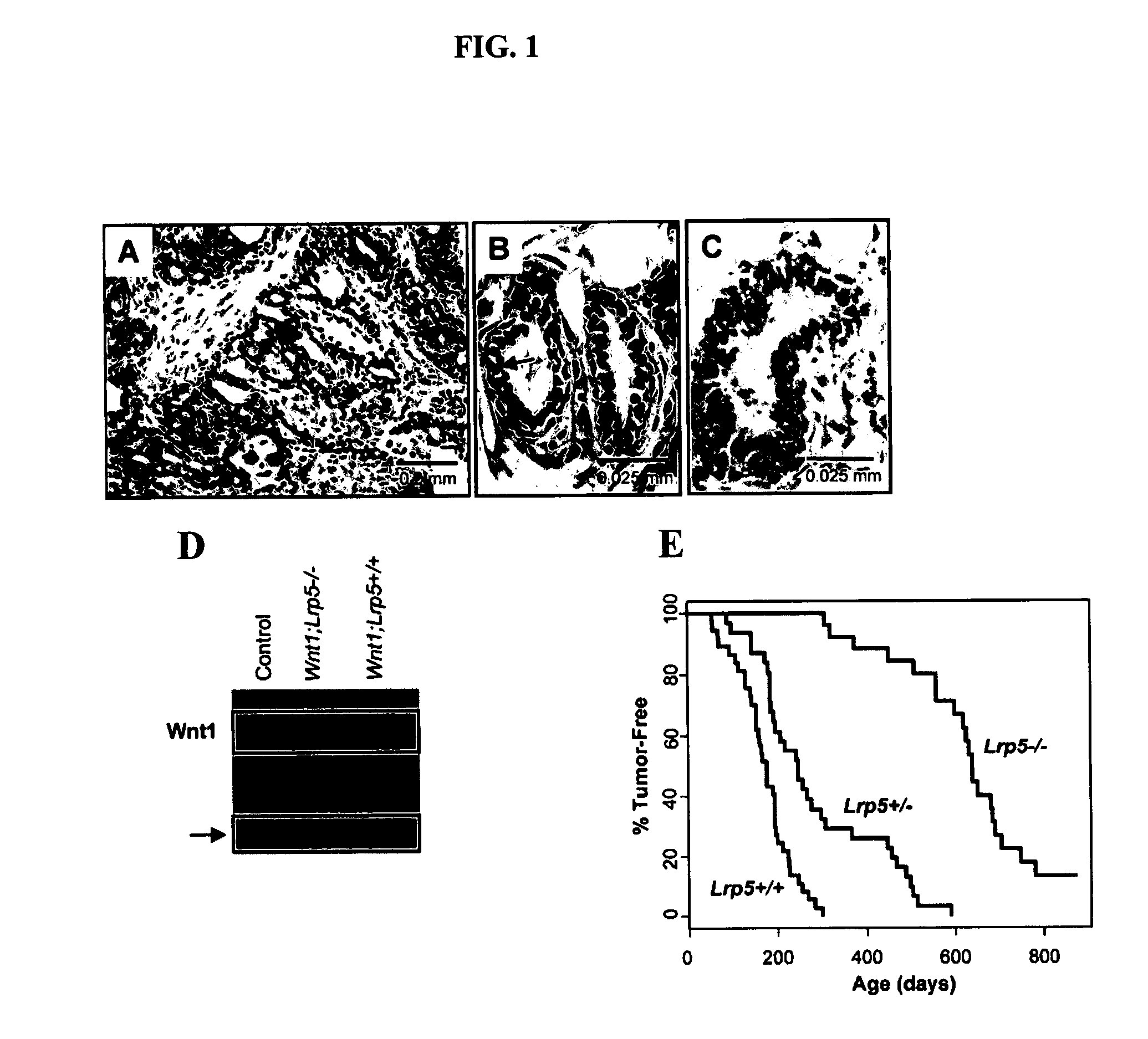
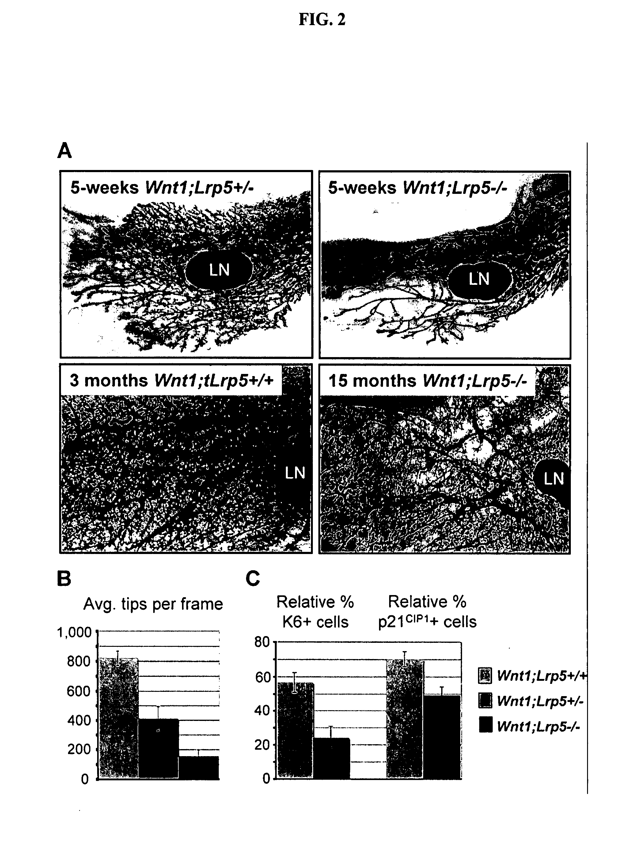
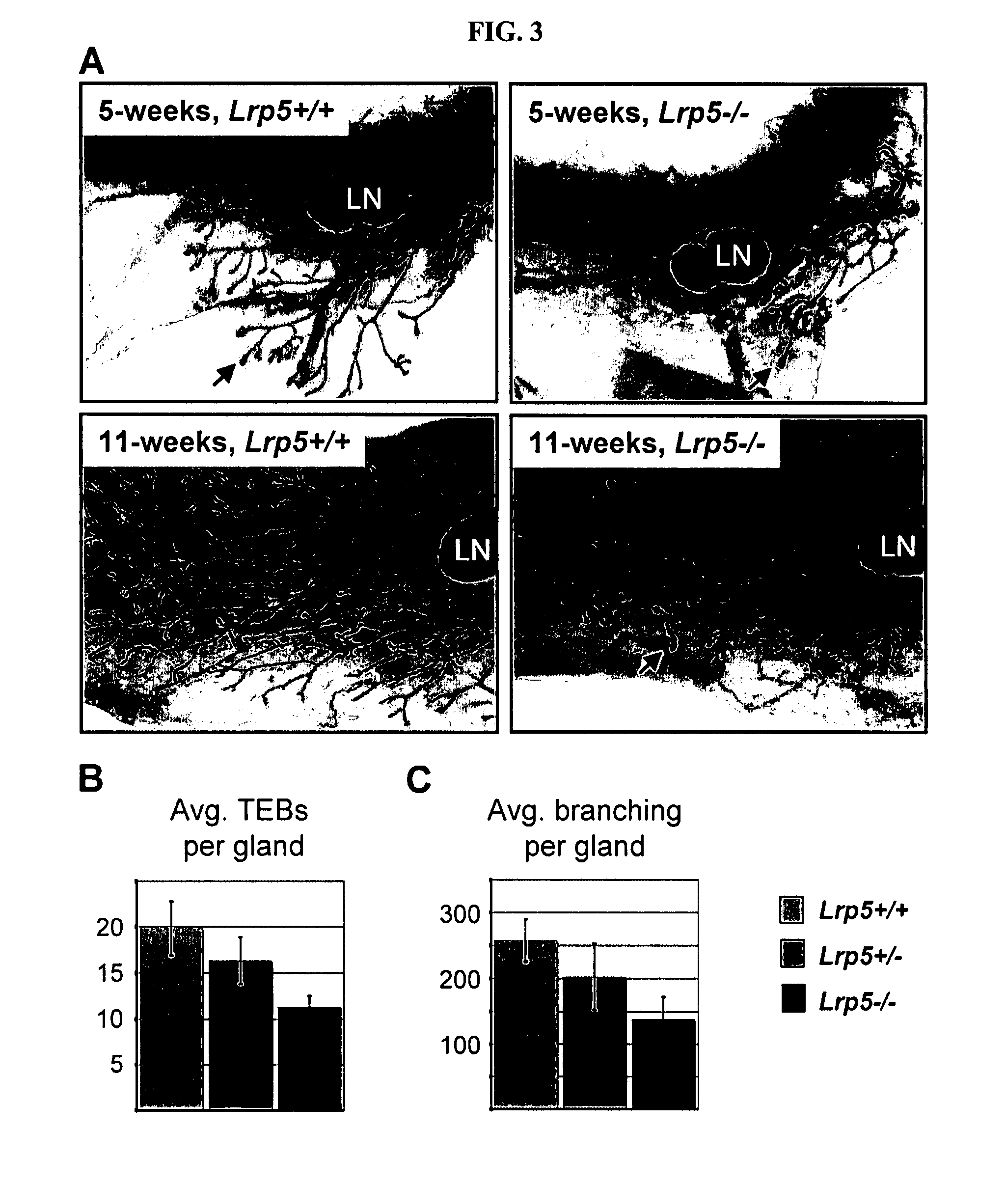
[0078] Canonical Wnt signaling has emerged as a critical regulatory pathway for stem cells. The association between ectopic activation of Wnt signaling and many different types of human cancer suggests that Wnt ligands can initiate tumor formation through altered regulation of stem cell populations. This example shows that mice deficient for the Wnt co-receptor LRP5 are resistant to Wnt1-induced mammary tumors, which have been shown to be derived from the mammary stem / progenitor cell population. These mice exhibit a profound delay in tumorigenesis that is associated with reduced Wnt1-induced accumulation of mammary progenitor cells. In addition to the tumor resistance phenotype, loss of LRP5 delays normal mammary development. The ductal trees of 5-week-old Lrp5− / − females have fewer terminal end buds, which are structures critical for juvenile ductal extension presumed to...

example 2

LRP5 is a Biomarker for Mammary Stem Cells

Materials and Methods

[0146] Mammary glands were obtained from 14 week old, virgin Balb / c mice. The glands were harvested and minced with fine scissors on ice. Mammary organoids were dissociated by enzymatic digestion with hyaluronidase and collagenase for six hours at 37° C. (reagents and protocol from Stem Cell Technologies, Vancouver, Canada). The mammary organoids were then further dissociated into single cells by brief trypsin and dispase exposures (reagents and protocol from Stem Cell Technologies). Single mammary epithelial cell preparations were then stained with the following rat antibodies (BD Biosciences, San Jose, Calif.): anti-CD45-APC (30-F11) and anti-CD31-APC (MEC 13.3) for 30 min at 4° C. In addition, the cells were also incubated for 30 min at 4° C. with rabbit anti-mouse LRP5 (Babij P. et al. J. Bone Miner. Res. 2003, 18:960-974) followed by incubation with Goat anti-rabbit IgG-Pacific Blue (Molecular Probes, Eugene, Ore...

example 3

LRP5 Expression in the Mammary Glands of Wild-Type and Lrp5-Null Mice as Well as MMTV-Wnt1 Transgenic Mice

[0148] Methods for immunohistochemistry for Lrp5 expression: Wild-type and Lrp5-null (Lrp5− / −, negative control) female mammary glands of congenic B6 mice and mammary glands of MMTV-Wnt1 transgenic mice (Tsukamoto A S et al. Cell 1988, 55:619-625) were isolated and fixed in 4% paraformaldehyde in PBS at 4° C. overnight. The mammary glands were then embedded in paraffin and cut at a thickness of 5 μm. The sections were deparaffinized and rehydrated. Immunohistochemistry for LRP5 was performed using rabbit polyclonal anti-mouse antibody G171V at a dilution of 1:5000. The localization of the primary antibody was identified by biotinylated anti-rabbit IgG, amplified with ABC reagent and visualized by 3,3′-diaminobenzidine (DAB) (Vector laboratories). The sections were counterstained with hematoxyline.

[0149] Results: FIG. 7 shows a section of mammary gland immunohistochemistry of a...


PUM

PropertyMeasurementUnit
timeaaaaaaaaaa
concentrationaaaaaaaaaa
median timeaaaaaaaaaa

Abstract

It is disclosed here that low density lipoprotein receptor-related protein 5 (LRP5) is a cell surface marker for somatic mammary stem cells and mammary tumor stem cells. The disclosure here provides new tools for enriching somatic mammary stem cells and mammary tumor stem cells. Methods of screening for agents that modulate LRP5 activity, of treating mammary tumor or breast cancer, of monitoring somatic mammary stem cells and mammary tumor stem cells in vivo are also provided, and of assessing prognosis of human breast cancer.

Description

CROSS-REFERENCE TO RELATED APPLICATION [0001] This application claims the benefit of U.S. Patent Application No. 60 / 809,281, filed on May 30, 2006, which is herein incorporated by reference in its entirety.STATEMENT REGARDING FEDERALLY SPONSORED RESEARCH OR DEVELOPMENT [0002] This invention was made with United States government support awarded by the following agency: NIH RO1 CA113869-01. The United States has certain rights in this invention.BACKGROUND OF THE INVENTION [0003] The mammary gland is a compound tubulo-alveolar gland that is composed of a series of branched ducts that, during lactation, drain sac-like alveoli (lobules). In the rodents, the mammary epithelium is embedded within a mammary fat pad, whereas in humans, it is embedded within a fibrous and fatty connective tissue. The mammary epithelium is composed of two lineages of epithelial cells: the luminal cells (which make milk during lactation) and basal positioned myoepithelial cells. Like other epithelia, the mamma...

Claims


Application Information

Patent Timeline
Patent Type & Authority Applications(United States)
IPC IPC(8): A61K39/395A61K48/00C12Q1/68G01N33/574
CPCA01K2267/0331C07K16/3015G01N33/92G01N33/57415G01N33/5041A61P43/00
Inventor WILLIAMS, BARTALEXANDER, CAROLINE M.LINDVALL, CHARLOTTAMCCONNELL, NISHA
Owner VAN ANDEL RES INST
Why Eureka
Eureka Blog
Learn More