Method For Transforming Head Surface Coordinates To Brain Surface Coordinates And Transcranial Brain Function Measuring Method Using The Transformation Data

a technology of brain surface coordinates and transformation data, applied in the direction of nmr measurement, instruments, applications, etc., can solve the problems of complex brain imaging procedure, particular equipment is required, appearance of spatial distortion, etc., and achieve the effect of more correct brain function image analysis, high processing speed and high scalability

Active Publication Date: 2007-12-06
SHIMADZU CORP +1
View PDF4 Cites 118 Cited by
  • Summary
  • Abstract
  • Description
  • Claims
  • Application Information

AI Technical Summary

Benefits of technology

[0040] When head surface coordinates are transformed to brain surface coordinates according to the present invention, it becomes possible to project arbitrary points on the head surface onto the brain surface, so that it becomes possible that data obtained by a transcranial brain function measuring / stimulating method are projected on the brain surface with high precision in a software program. As described above, the present invention can solve the problems of distortion accompanied with a conventional sphere fitting method and the applied methods therefor.
[0041] The present invention provides extremely high scalability for processing the data obtained by the transcranial brain function measuring / stimulating method. When the data obtained by the transcranial brain function measuring / stimulating method is projected on the brain surface by using the method according to the present invention, it becomes possible that the data is transformed into a format which can be normalized and used commonly in the brain function study. Through the normalization, an integration of data among a plurality of subjects may also be simply conducted. In addition, it becomes possible to integrate the data into and compare with those obtained by the other brain function measuring methods. Thus, the present invention provides a novel technology which becomes a foundation with respect to a propagation of the transcranial brain function measuring / stimulating method such as NIRS and TMS, so that it makes a remarkable contribution to developments in brain function studies and their clinical applications.
[0042] In the present invention, when coordination of standard points on a head surface with those three-dimensional brain surface projection points is previously conducted, it becomes possible to project data obtained by the transcranial brain function measuring / stimulating method onto the brain surface without taking three-dimensional head region images. Besides, it becomes possible to clearly obtain spatial error information as to the projection.
[0043] Transformation of standard points on a head surface to a brain surface three-dimensional coordinates may be expressed as a probability distribution based on measurement data of a plurality of subjects. Furthermore, when a center of the probability distribution is used, it is also possible to determine one position of the corresponding brain surface three-dimensional coordinates with respect to one standard point on the brain surface.
[0044] With respect to arbitrary points on a head surface, when a relative positional relationship with near three points of standard points on the head surface is determined, it is possible to project the head surface onto the brain surface underlying the head surface by referring to the positions of the standard points projected on the brain surface. These procedures can also be executed on a software program.
[0045] Thus, it becomes possible to project points on the head surface onto the brain surface according to the software program based on the present invention in either a case where a three-dimensional head region image is obtained, or a case where such image is not obtained. A method by which arbitrary positions on a head surface can be projected on a brain surface without taking any three-dimensional head region image has never been known. Accordingly, the present invention is a method for providing quite novel advantages. However, a software program for expressing a two-dimensional map of data measured by means of NIRS of a brain surface has been known, the method of this software is not based on a manner for correct coordination of respective pixels in the two-dimensional map with voxels on a three-dimensional coordinate. Hence, the present invention is extremely effective for realizing a more correct brain function image analysis.

Problems solved by technology

However, such brain imaging procedure is complicated, and particular equipment is required.
However, since nonspherical head surface and brain surface are applied to spheres in the sphere fitting method, there is a problem of an appearance of a spatial distortion.

Method used


Image

  • Method For Transforming Head Surface Coordinates To Brain Surface Coordinates And Transcranial Brain Function Measuring Method Using The Transformation Data
  • Method For Transforming Head Surface Coordinates To Brain Surface Coordinates And Transcranial Brain Function Measuring Method Using The Transformation Data
  • Method For Transforming Head Surface Coordinates To Brain Surface Coordinates And Transcranial Brain Function Measuring Method Using The Transformation Data

Examples


example 1

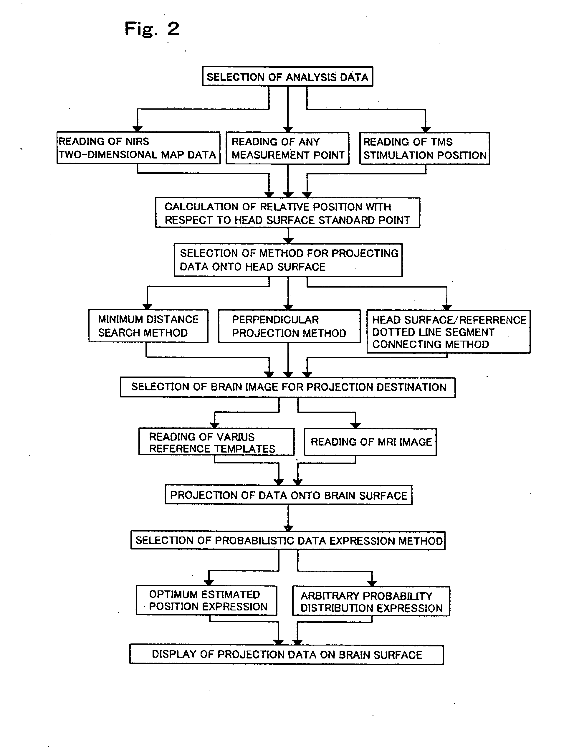
[0079] Projection points on a brain surface are determined with respect to nineteen standard points of the international 10-20 system on a head surface. More specifically, markers are disposed on the 10-20 system standard points on a head surface of a subject, and a MRI image is taken to obtain a three-dimensional image of the head. Each of projection points on the brain surface corresponding to each of the standard points on the head surface is determined with respect to the three-dimensional image of the head in accordance with the minimum distance search method.

[0080] Furthermore, the measurement as mentioned above is conducted with respect to seventeen subjects, and the data extending over all the subjects puts together to express a probability distribution by means of MINI standard brain coordinates. Namely, head images of the respective subjects are normalized into a MINI standard brain; and a probability distribution of positions in the 10-20 system standard points of brain ...

example 2

[0083] In accordance with the method of the present invention, brain activity data obtained by an actual NIRS measurement is projected on a brain surface by the use of the measuring system of FIG. 1 with taking no MRI image.

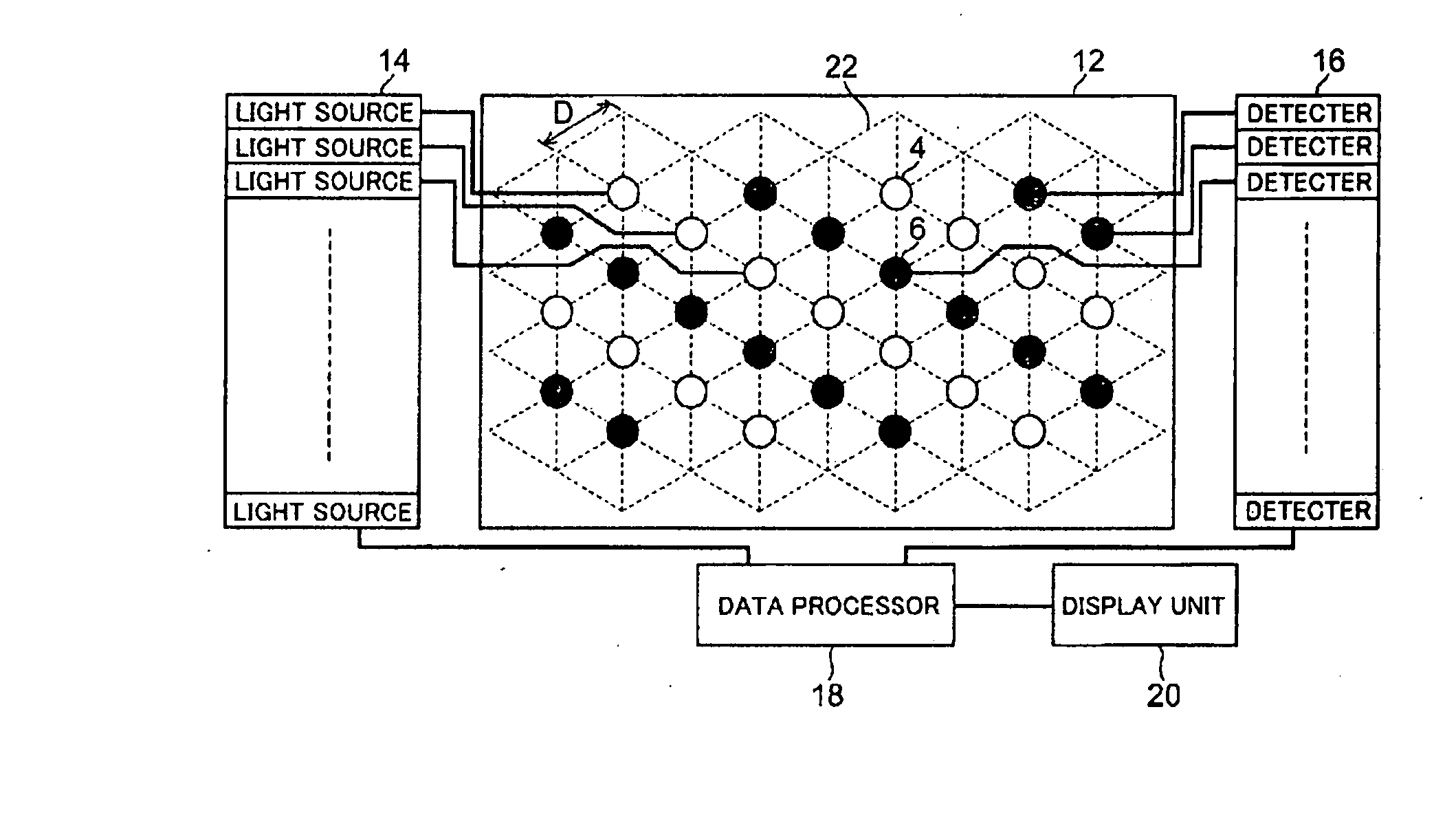
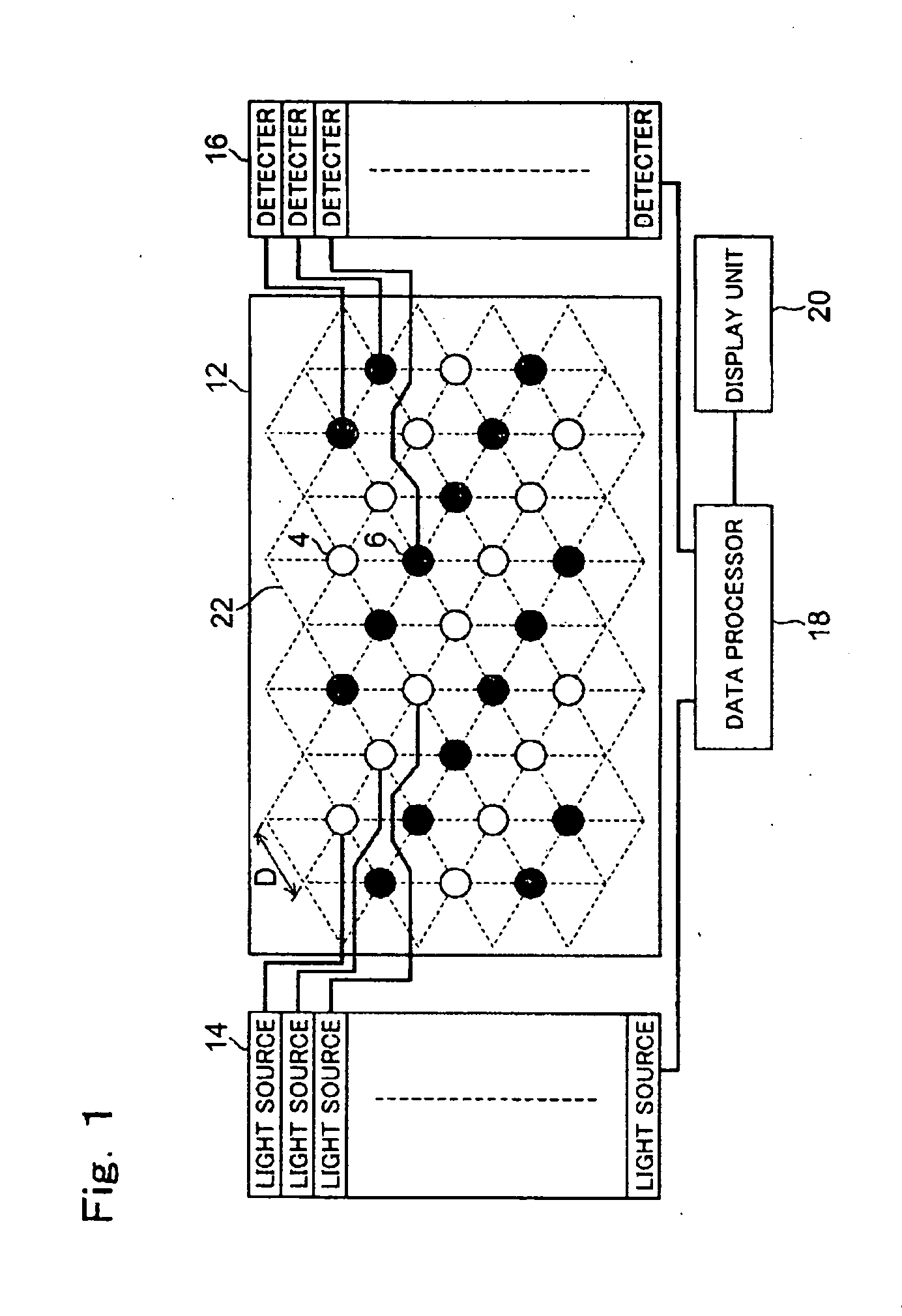
[0084] As the brain activity data, changes of an oxygenated hemoglobin concentration in a prefrontal cortex as to a case where each of ten subjects pares an apple are applied. Mutual positions of fixing points of measurement probes of the NIRS with respect to nineteen standard points of international 10-20 system have been previously measured; and then, positions of the NIRS measurement probes are obtained on the head surfaces.

[0085] Thereafter, coordinate information is integrated as to the ten subjects, whereby average position coordinates of the NIRS measurement probes are obtained on the head surface. These average positions are projected on a brain surface image of a typical subject by referring to the mutual position information with respect to the ninete...

example 3

[0088] In accordance with the method of the present invention, a comparison of simultaneous measurement data of a NIRS and a functional nuclear magnetic resonance apparatus (fMRI) is implemented. Activities in a motor area are measured simultaneously by means of the NIRS and the fMRI in the case where a subject pares imitatively an apple wherein measurement probes of the NIRS and position markers for indicating fixed positions of the probes have been fixed to the subject. A NIRS measurement region is projected on a brain surface image of a subject by applying the minimum distance search method according to the present invention. In the measurement by means of the fMRI, a region wherein significant decrease is observed in a reduced hemoglobin concentration is checked. In the NIRS measurement, changes in the oxygenated hemoglobin concentration are measured. Brain activity is measured based on a decrease in the reduced hemoglobin concentration in the fMRI measurement, while based on an...


PUM


Abstract

Data collected by a transcranial brain function measuring / stimulating method is accurately projected and displayed onto a brain surface. If there is no three-dimensional head image, data is projected and displayed onto the brain surface of a standard brain. The head surface coordinates are transformed to the brain surface coordinates of the brain surface underlying the head surface by, e.g., a minimum distance search method. The coordinates of a projected point on the brain surface of the head surface and the probability distribution are determined for a standard brain normalized with data on subjects.

Description

TECHNICAL FIELD [0001] The present invention relates to a method for estimating brain surface coordinates relating to measurement / stimulation in near-infrared spectroscopy (NIRS), a transcranial magnetic stimulator (TMS), and the other transcranial brain function measuring / stimulating methods; its software program; and a transcranial brain function measuring apparatus for displaying measuring / stimulating results using the brain surface coordinates estimated by the methods; and more particularly to a method for projecting measurement data developed on a head surface as arbitrary points on the head surface or its set onto a brain surface underlying the head surface to express them; the software program therefor; and an apparatus therefor. BACKGROUND ART [0002] In order to find positions on brain surface being objects of measurement / stimulation in a transcranial brain function measuring / stimulating method, it is required to obtain a structural image of brain in each case by using a bra...

Claims


Application Information

Patent Timeline
Patent Type & Authority Applications(United States)
IPC IPC(8): A61B6/00G01R33/54A61B5/00A61B5/055A61B10/00G06T1/00
CPCA61B5/0059A61B5/7445A61B5/14553A61B5/061A61B5/0042A61B5/4064
Inventor DAN, IPPEITASAKAMOTO, KUNIKOOKAMOTO, MASAKOISOBE, SEIICHIROKOHYAMA, KAORUSHIMIZU, KOJITAKEO, KAZUBIRO
Owner SHIMADZU CORP
Why Eureka
Try Eureka