Implantable wireless sensor for in vivo pressure measurement and continuous output determination

a wireless sensor and in vivo technology, applied in the field of wireless, unpowered, micro-machined pressure sensors, can solve the problems of limiting its use to acute settings, the technique used to fabricate the sensors does not lend itself to the necessary miniaturization, and the fabrication methods used to manufacture them do not provide sufficient miniaturization. to achieve the effect of easy, safe and accurate measurement of pressur

Inactive Publication Date: 2007-12-06
CARDIOMEMS
View PDF7 Cites 64 Cited by
  • Summary
  • Abstract
  • Description
  • Claims
  • Application Information

AI Technical Summary

Benefits of technology

[0011] Stated generally, the present invention comprises a simple apparatus and method of monitoring the pressure within the heart or the vasculature by implanting a pressure sensor in such locations utilizing catheter-based endovascular or surgical techniques and using extracorporeal electronics to measure the pressure easily, safely, and accurately.

Problems solved by technology

The drawback of this type of sensor is that there must be a wired connection between the sensor and the extracorporeal device, thus limiting its use to acute settings.
The primary limitation to these type of sensors is that the fabrication methods used to manufacture them do not provide sufficient miniaturization to allow them to be introduced and implanted into the heart using non-surgical, catheter-based techniques while maintaining the ability to communicate wirelessly with external electronics.
The primary limitation of many of these inventions is that the techniques used to fabricate the sensors do not lend themselves to the miniaturization necessary for it to be configured as an implantable medical device while maintaining the capability of communicating wirelessly with external electronics.
These sensors suffer from many of the limitations already mentioned, with the additional concerns that they require either the addition of a power source to operate the device or the need for a physical connection to a device capable of translating the sensor output into a meaningful display of a physiologic parameter.
The device described in the Chubbuck patent is large, thus requiring surgical implantation and thereby limiting its applicability to areas that are easily accessible to surgery (e.g., the skull).

Method used


Image

  • Implantable wireless sensor for in vivo pressure measurement and continuous output determination
  • Implantable wireless sensor for in vivo pressure measurement and continuous output determination
  • Implantable wireless sensor for in vivo pressure measurement and continuous output determination

Examples


example 1

[0110]FIG. 16 illustrates a surface micromachined, capacitor coupled sensor 600. The capacitor structure 602 comprises at least two plates 604, 606, at least one 604 of which is built directly atop a first wafer 608. This plate 604 will be referred to as the bottom plate. The region of the wafer 608 where the bottom plate 604 is built will be referred to as the deflective region 610. If necessary, the thickness of the wafer 608 in the region of the deflective region 610 can be reduced in thickness to enhance its deformability.

[0111] The other plate 606 is suspended above the bottom plate 604. The top plate 606 is mechanically anchored to the deflective region by pillar-like supporting elements 612 located at the periphery of the bottom plate 604. Bottom and top plates 604, 606 are electrically insulated and physically separated from one another by an air gap 614. The top electrode 606 mechanical design, material and dimensions are carefully chosen so that the suspended part of the ...

example 2

[0124] A variation on the two-wafer design is shown in FIGS. 24-28. A sensor 700 comprises a thick upper wafer 702 and a thinner lower wafer 704. The thin lower wafer 704 comprises the pressure-sensitive deflective region portion 706 of the sensor 700. A notch 708 is optionally formed in the upper wafer 702 to accommodate an anchor, such as a corkscrew, hook, barb, or other suitable stabilization means. The notch can be created on the back side of the wafer directly if the cap is sufficiently thick to accommodate the notch and a separation distance between the bottom of the notch and the coil body without causing any parasitic, deleterious electromagnetic or mechanical effects on the sensor function. Alternatively, the notch can be created by using wet or dry methods in a separate wafer or plurality of wafers and then bonded to the back side of the sensor. The notch can have a variety of regular or irregular geometries and can have rough or smooth sidewalls—any configuration achieva...

example 3

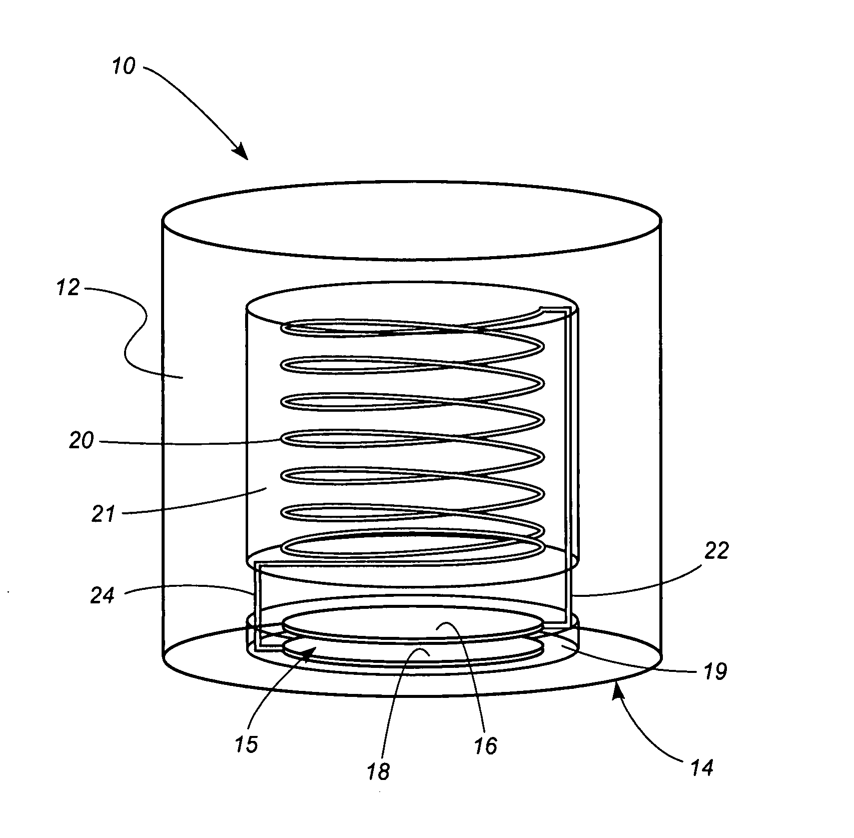
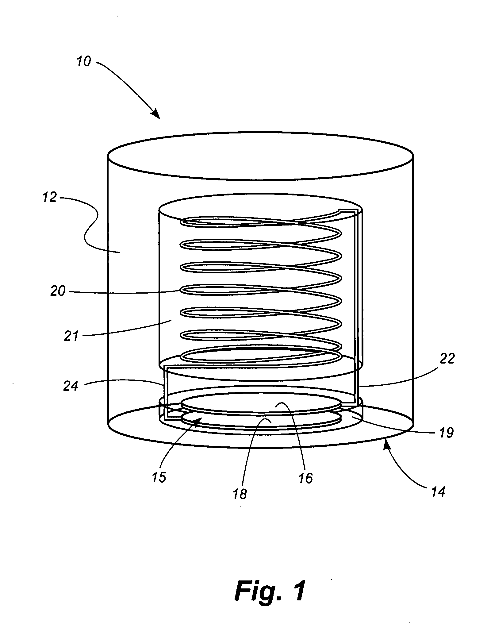
[0131]FIGS. 29-32 depict an embodiment of a sensor 800 manufactured from four stacked wafers, 802, 804, 806, and 808. The bottom wafer 802 comprises the pressure-sensitive deflective region 810 and a pair of capacitor plates 812, 814 formed on its upper surface. The second wafer 804 comprises a capacitor plate 816 formed on its lower surface and a pair of through-holes 818 for electrical connections. The third wafer 806 comprises a cylindrical cavity 820 for accommodating an inductance coil 822. Leads 824 of the inductance coil 822 extend through the holes 818 in the second wafer 804 and connect to the capacitor plates 812, 814. The fourth wafer 808 fits atop the third wafer to provide a sealed structure.

[0132]FIG. 30 illustrates a first step in the process for manufacturing the sensor 800. A recess 830 is formed in the upper surface of the bottom wafer. Then, as shown in FIG. 32, the plates 812, 814 are formed in the base of the recess 830. Referring to FIG. 32, the plate 816 is f...


PUM


Abstract

A method and apparatus for determining cardiac parameters within the body of a patient includes a wireless sensor positioned in the patient's pulmonary artery. An external RF telemetry device communicates wirelessly with the sensor and interrogates the sensor to determine changes in pressure in the pulmonary artery over time. The peak pressure difference is determined. Then, assuming zero blood flow velocity at the time of valve opening and at the time of valve closing, a velocity-time function is determined. The velocity-time function is used to determine a velocity-time integral. The velocity-time integral is then used to determine cardiac stroke volume. The cardiac stroke volume is multiplied times the heartbeat rate to determine cardiac output. The cardiac output can be monitored over time to determine continuous cardiac output.

Description

TECHNICAL FIELD [0001] This invention relates to implanted sensors for wirelessly sensing pressure, temperature and other physical properties within the human body. More particularly, the invention concerns a wireless, un-powered, micromachined pressure sensor that can be delivered using catheter-based endovascular or surgical techniques to a location within an organ or vessel. BACKGROUND OF THE INVENTION [0002] The measurement of blood pressure within the human heart and its vasculature provides critical information regarding the organ's function. Many methods and techniques have been developed to give physicians the ability to monitor heart function to properly diagnose and treat various diseases and medical conditions. For example, a sensor placed within the chambers of the heart can be used to record variations in blood pressure based on physical changes to a mechanical element within the sensor. This information is then transferred through a wire from the sensor to an extracorp...

Claims


Application Information

Patent Timeline
Patent Type & Authority Applications(United States)
IPC IPC(8): A61B5/02
CPCA61B5/0215A61B5/0031
Inventor STERN, DAVID R.
Owner CARDIOMEMS
Why Eureka
Try Eureka