Method of monitoring colorectal cancer

a colorectal cancer and colorectal cancer technology, applied in the field of colorectal cancer monitoring, can solve the problems of cancers that are not detected, patients are at an increased risk of developing additional polyps as well as colorectal cancer, and the source of aberrant methylated dna is removed, so as to achieve a high degree of sensitivity and specificity, and the probability of complete cure is virtually certain.

Inactive Publication Date: 2007-12-27
UNIV OF MARYLAND
View PDF0 Cites 6 Cited by
  • Summary
  • Abstract
  • Description
  • Claims
  • Application Information

AI Technical Summary

Benefits of technology

[0011] Methods of the invention are useful for detecting early-stage lesions in heterogeneous samples such as stool. Methods of the invention result in a high degree of sensitivity and specificity for the detection of early-stage disease. Methods of the invention are especially useful in detecting, for example, adenomas in the colon. Adenomas are non-metastatic lesions that frequently have the potential for metastasis. If all adenomas in a patient are detected and removed, the probability of complete cure is virtually certain.
[0012] Various other features and advantages of the present invention should become readily apparent with reference to the following detailed description, examples, claims and appended drawings. In several places throughout the specification, guidance is provided through lists of examples. In each instance, the recited list serves only as a representative group and should not be interpreted as an exclusive list.

Problems solved by technology

However, it is not known if methylation patterns in the promoter regions of these genes return to normal after polypectomie, which should result in the removal of the source for the aberrantly methylated DNA.
Although such lesions are removed by polypectomie, patients are at an increased risk for developing additional polyps as well as colorectal cancer in the future.
Surveillance is usually limited to repeat colonoscopies 5-10 years later, which results in some cancers that are not detected at an early curable stage.
Due to the presence of large amounts of bacterial DNA and inhibitors in stool, extraction of human DNA sufficiently clean for downstream applications is difficult.
Capture probe based approaches have been used successfully, but their use is limited as only a few target DNA fragments can be isolated at a time (Petko et al., 2005, supra).
Although successful use of a commercial kit has been described in one report (Belshaw et al., 2004, supra), we and others have not been able to repeat this consistently.
Although this test has some utility as a generic screening test for an at average risk population it is not an efficient means for detecting recurrence of preneoplastic / neoplastic lesions.

Method used


Image

  • Method of monitoring colorectal cancer
  • Method of monitoring colorectal cancer

Examples


example 1

[0026] We are currently performing a study in patients undergoing a screening colonoscopy at the UMMS GI clinic in which we enroll subjects after they have been referred for a colonoscopy, collect demographic and dietary data as well as a stool sample BEFORE the colonoscopy, obtain biopsy samples of normal colon tissue during the colonoscopy (as well as polyp tissue after pathology has been performed) and in a subset collect additional stool samples AFTER the colonoscopy / polypectomie. A total of 500 subjects are planned, so far we have completed data and sample collection in appr. 50 subjects.

[0027] Extraction of Human DNA from Feces

[0028] The largest proportion of DNA extracted from feces is of bacterial origin. In order to obtain sufficient material for methylation analysis of bisulfite converted or unconverted DNA by PCR we have optimized our extraction protocol. Instead of using bead beating for efficient cell lysis we are using a more gentle procedure that omits extensive enz...

example 2

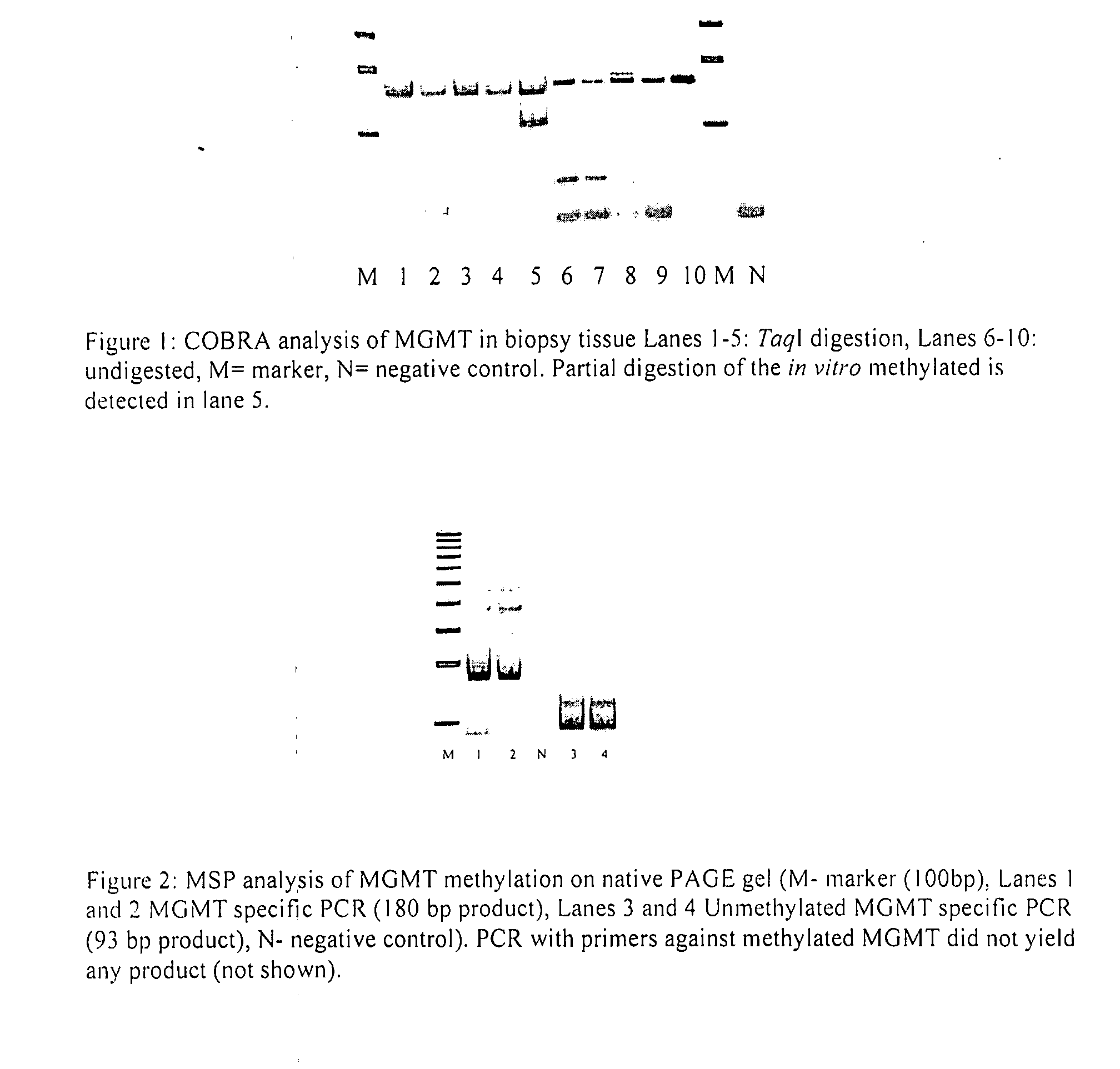
[0033] To establish methods to reliably detect aberrant methylation pattern in stool samples Analysis of aberrant methylation pattern in stool DNA requires a robust protocol for DNA extraction followed by bisulfite conversion, (quantitative) amplification and restriction analysis.

[0034] DNA Extraction, Bisulfite Conversion and Methylation-Specific PCR Analysis from Paraffin-Embedded Proximal Colon Tissue

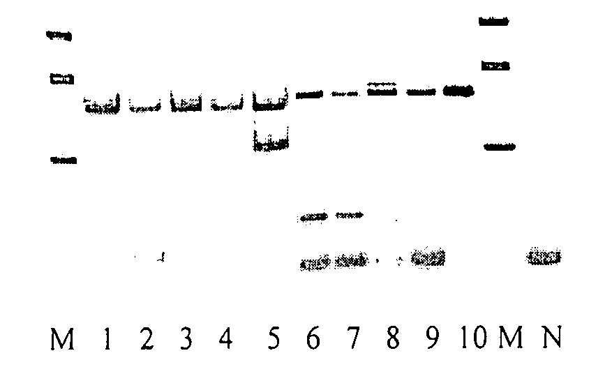
[0035] We have established a protocol to extract DNA from paraffin-embedded normal as well as polyp tissue. After removal of excessive paraffin by xylene and release of DNA from the blocks we follow the DNeasy extraction protocol for animal tissues (Qiagen). The extraction yields >0.5 μg of DNA >600 bp in length. This amount is sufficient for 4-8 bisulfite conversions with the EZ DNA kit (ZYMO Research).

[0036] Each bisulfite conversion yields sufficient template DNA for up to 10 methylation specific PCR reactions, allowing us to test a large panel of prospective markers without th...

example 3

[0044] To Determine if Aberrant Methylation Pattern Detected in Polyp Tissue can be Detected in Pre- but not Post-Colonoscopy Stool Samples Collected from the Same Subject

[0045] It is assumed that the aberrant methylation patterns detected in stool are due to shedding of cells from a preneoplastic lesion into the lumen. It is not known if methylation pattern in the promoter regions of these genes return to normal after polypectomie, which should remove the source for the aberrantly methylated DNA. If the patterns do disappear than a positive screening test for reappearance of the aberrant pattern over time might indicate the formation of new preneoplastic lesions.

[0046] We will collect a stool sample BEFORE the colonoscopy, then obtain biopsy samples of normal colon tissue during the colonoscopy (as well as polyp tissue after pathology has been performed) and in addition collect stool sample AFTER the colonoscopy / polypectomie. For the studies proposed here we will choose subjects ...


PUM

PropertyMeasurementUnit
timeaaaaaaaaaa
Methylation frequencyaaaaaaaaaa
temperatureaaaaaaaaaa

Abstract

A method and kit is described for individualized stool surveillance for occurrence/recurrence of preneoplastic or neoplastic lesions based on the analysis of genetic mutations and methylation pattern detected in biopsy tissue removed during polypectomie as compared to normal colon mucosa.

Description

[0001] This application claims the benefit of priority from U.S. Provisional Application Ser. No. 60 / 815,126 filed on Jun. 20, 2007INTRODUCTION [0002] Epigenetic changes are now known to contribute to early steps in carcinogenesis and especially changes in methylation pattern of CpG islands in promoter regions of relevant genes have been well studied in colorectal carcinogenesis (Das and Singal, 2004, J. clin. Oncol. 22, 4632-42). Methylation pattern was found to significantly correlate in DNA extracted from colorectal mucosa and from fecal samples in the same individual (Belshaw et al., 2004, Cancer Epidemiol. Biomarkers Prev. 13, 1495-501). Methylation frequency of MGMT and CDKN4 and MLH1 differed in individuals with adenomas as compared to normal controls (Petko et al., 2005, Clin. Cancer Res. 11, 1203-9), characteristic methylation pattern were also detected in other cancers including esophagal adenocarcinoma (Eads et al., 2001, Cancer Res. 61, 3410-8). [0003] It is assumed that...

Claims


Application Information

Patent Timeline
Patent Type & Authority Applications(United States)
IPC IPC(8): C12Q1/68
CPCC12Q1/6886C12Q2600/154C12Q2600/118
Inventor MAI, VOLKER
Owner UNIV OF MARYLAND
Why Eureka
Try Eureka