Intradermal cellular delivery using narrow gauge micro-cannula

a micro-cannula and intradermal technology, applied in the direction of antibody medical ingredients, drug compositions, immunological disorders, etc., can solve the problems of insufficient regulation of effective t-cell mediated immunity, insufficient dendritic cell and related cell type and poor study of methods for administering cellular based therapeutics and vaccines into patients

Inactive Publication Date: 2008-01-24
BECTON DICKINSON & CO
View PDF3 Cites 50 Cited by
  • Summary
  • Abstract
  • Description
  • Claims
  • Application Information

AI Technical Summary

Problems solved by technology

Antigens alone, even those pre-processed to bind to antigen-presenting MHC class I and II molecules, are insufficient to regulate effective T-cell mediated immunity.
Methods for administering cellular based therapeutics and vaccines, particularly, dendritic cell and related cell type based therapeutics and vaccines, into the patients have been poorly studied.
First, these cellular based therapeutics and vaccines do not have direct access to the immune system, as they are circulating in the blood before they reach the lymph system.
Third, in some cases, the intravenous delivery may induce a state of immune tolerance rather than activation.
Despite these studies, it is still unclear as to which route(s) will be most effective at preventing or treating cancer.
This method is very difficult to perform even in the hands of highly trained practitioners and is often associated with pain.
In addition, it is very difficult to control delivery depth according to this method, thus resulting in “spillover” of at least a portion of the administered dose into the SC tissue.
Although delivery route has been the subject of considerable study and debate, there have been no reported studies investigating the potential effects of other delivery parameters on cell therapy, including, for example: cell concentration, flow rate, delivery volume or needle geometry (e.g., gauge size and needle length).
These parameters have varied widely in prior studies, thus making it impossible to ascertain the potential role of such parameters in cell delivery.

Method used


Image

  • Intradermal cellular delivery using narrow gauge micro-cannula
  • Intradermal cellular delivery using narrow gauge micro-cannula
  • Intradermal cellular delivery using narrow gauge micro-cannula

Examples


example 1

Cell Viability Following Microneedle Delivery

Purpose:

[0051] Cells were delivered in vitro through a microneedle designed for intradermal delivery and tested for viability following such delivery.

Method:

[0052] 1. Experimental materials. [0053] a. Microneedle. A 34 gauge microneedle with a length of 1 mm length was used. [0054] b. Cell line. P815 cell line was a mouse mastocytoma-derived cell line (Lundak, R L & Raidt, D J, Cell. Immunol. 9:60-66, 1973) that is often used as a model system for antigen-presentation studies. In particular, P815 cells could be transfected with genetic material encoding specific antigens such as those used in a vaccine for an infectious disease or cancer, or could be loaded with such antigens directly. Then, these P815 cells could be used to stimulate T cells in vitro.

[0055] P815 cells for this experiment had a size of 10-15 μm in diameter on non-adherent rounded cells, which was of a similar size to dendritic cells and related Langerhans cells. In...

example 2

Dendritic Cell Viability Following Microneedle Delivery

Purpose:

[0067] A dendritic cell line (JAWS-II, as described in U.S. Pat. Nos. 5,648,219, and 5,830,682) was delivered through two different types of microneedles designed for intradermal delivery (30 Ga and 34 Ga needles) and tested for viability and expression of functional cell surface markers following such delivery. Delivery through a standard 27 Ga needle was included for comparison.

Method:

[0068] 1. Experimental materials. [0069] a. Needles and Microneedles: Cells were delivered through: a) a standard 27 Ga needle, b) a 30 Ga, 1.5 mm long stainless steel microneedle or c) a 34 Ga, 1.0 mm long stainless steel microneedle. [0070] b. Cell line. The mouse JAWS-II cell line, as described in U.S. Pat. Nos. 5,648,219 and 5,830,682 was used for these studies. Prior to delivery, cells were activated for 24 hr with IFN-γ, IL-4, TNF-α and GM-CSF, as described in U.S. Pat. Nos. 5,648,219 and 5,830,682.

[0071] 2. Experimental step...

example 3

Cell Distribution Following Intradermal Delivery In Vivo

Purpose:

[0082] Characterize the distribution pattern of cells administered in vivo using microneedles.

Method:

[0083] 1. Model system used.

[0084] Pigs were used for intradermal injection. Pig skin represents a well accepted model for human skin. P815 cells, as described in Example 1, were used as the model cell line.

[0085] 2. Experimental steps.

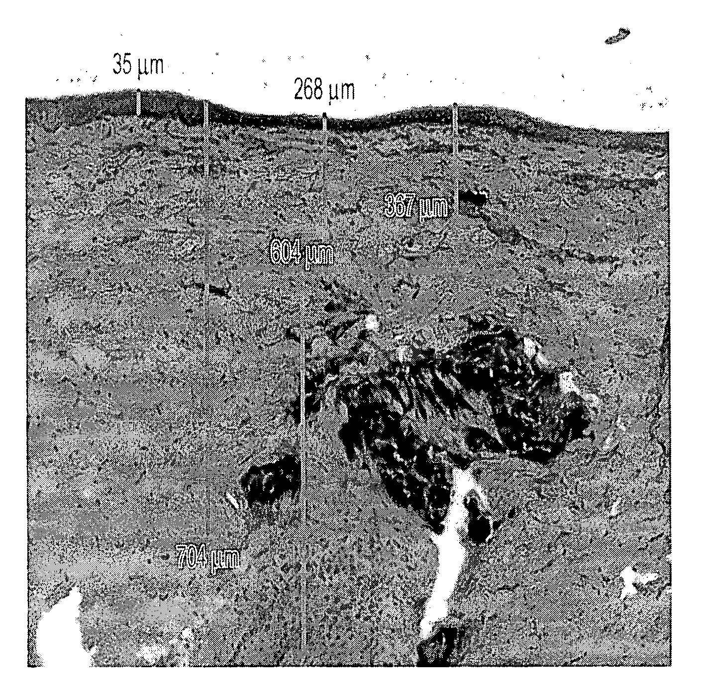
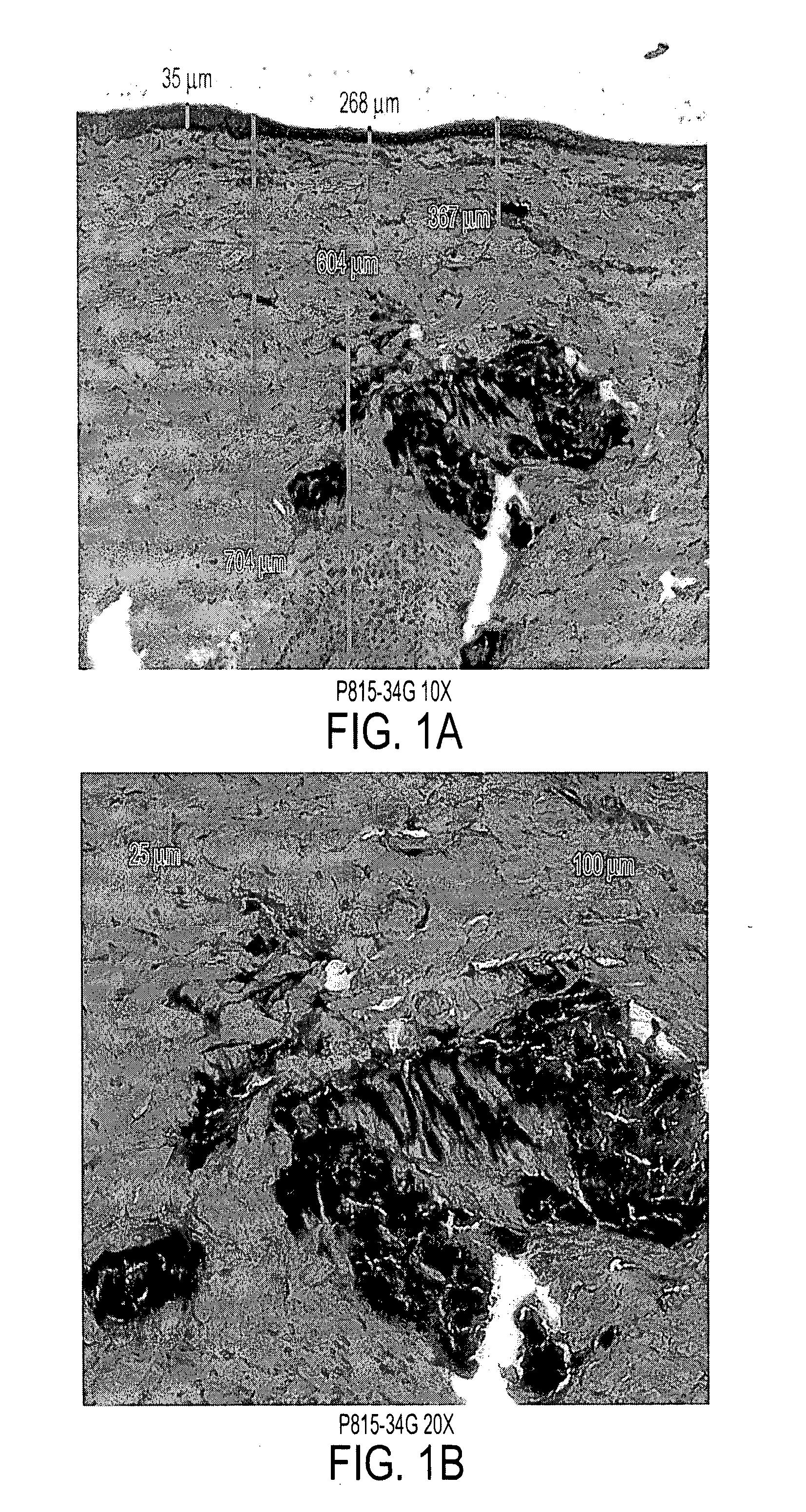
[0086] P815 cells were delivered intradermally by 34 gauge microneedles 1 mm in length. Cells were suspended to a concentration of 40×106 cells / ml. A total of 0.1 ml (4×10⁶ cells) was administered via bolus injection over a time period of approximately 1 minute.

[0087] 3. Evaluation of results.

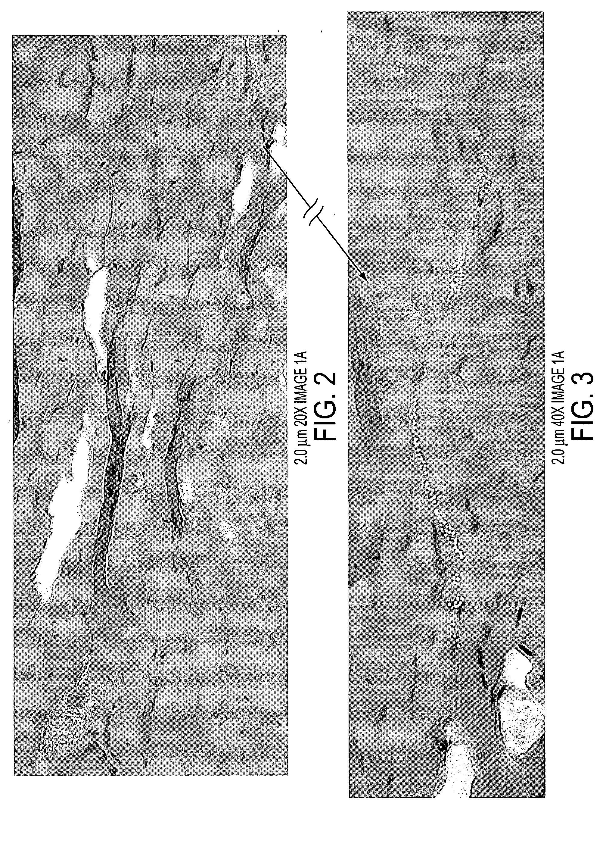
[0088] Immediately after allowing the bleb to resolve, full thickness skin biopsies were collected and processed for tissue sectioning and Haematoxylin & Eosin (H&E) staining. Pictures were taken from the light microscope observation.

[0089] 4. Results

[0090]FIG. 1 displays the distribution...


PUM

PropertyMeasurementUnit
depthaaaaaaaaaa
flow rateaaaaaaaaaa
flow rateaaaaaaaaaa

Abstract

A method for delivering cells into a subject by administering cells into the intradermal space of the skin of the subject by a microneedle. The cells are associated with cellular based therapeutics and vaccines and delivered by perpendicular insertion of the microneedle. The microneedle is a hollow needle having an exposed height of between about 0 and 1 mm, a total length of between about 0.3 mm to 2.5 mm, and a size of equal to or less than 30 gauge. An array of microneedles can also be used.

Description

[0001] This application is a continuation application of U.S. application Ser. No. 10 / 758,274, filed Jan. 16, 2004, which claims the benefit of U.S. Provisional Applications Nos. 60 / 440,348, filed Jan. 16, 2003, and 60 / 504,488, filed Sep. 19, 2003, all of which are incorporated by reference in their entirety.FIELD OF THE INVENTION [0002] The present invention relates to a method for delivering cellular based therapeutics and vaccines into subjects, particularly, for delivering dendritic cell or related cell type based therapeutics, islet cells, and vaccines into the intradermal space of the skin of the subjects by a microneedle. BACKGROUND OF THE INVENTION [0003] Cellular based therapeutics and vaccines refer to treatments that use cells and tissues as therapeutic agents to treat injury or disease. Examples of cellular based therapeutics include, but are not limited to, hematopoietic cell therapeutics, mesenchymal stem cell based therapeutics, immunotherapies, dendritic cell and rel...

Claims


Application Information

Patent Timeline
Patent Type & Authority Applications(United States)
IPC IPC(8): A61M5/31A01N63/00A61K35/12A61K35/14A61K35/15A61K39/00A61K45/00A61MA61M31/00A61M37/00
CPCA61K35/12A61K35/15A61K39/00A61M2037/0061A61K2039/5154A61K2039/54A61M37/0015A61K39/0005A61P37/04
Inventor BRITTINGHAM, JOHN M.MIKSZTA, JOHN A.WILLIAMS, DOMINIQUE J.DEAN, CHERYL H.
Owner BECTON DICKINSON & CO
Why Eureka
Try Eureka