Method for the Discrimination and Isolation of Mammary Epithelial Stem and Colony-Forming Cells
- Summary
- Abstract
- Description
- Claims
- Application Information
AI Technical Summary
Problems solved by technology
Method used
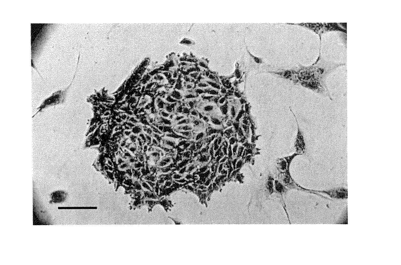
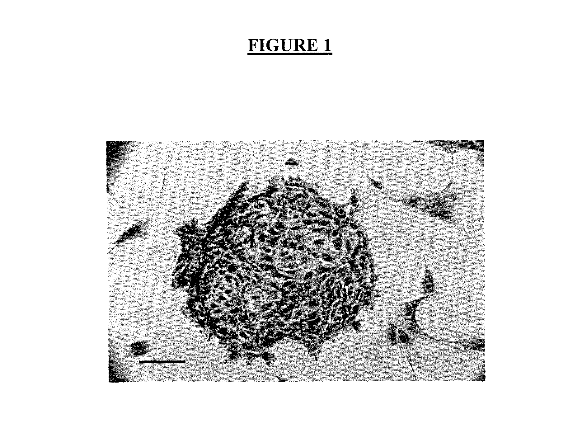
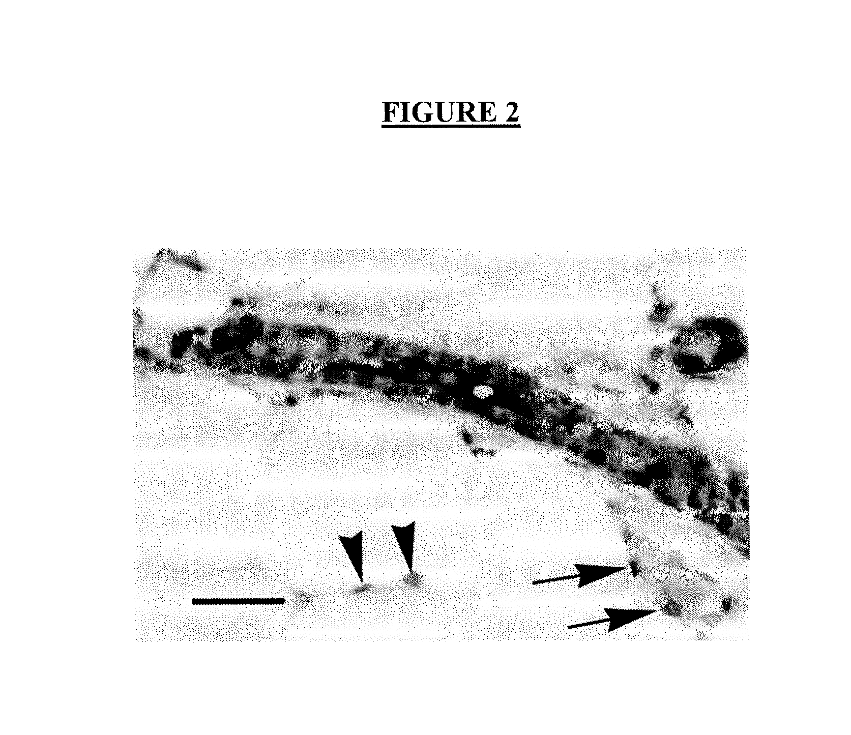
Image

Examples
example 1
Method for Separating Epithelial and Non-epithelial Cells in a Mammary Cell Sample
[0064] Mammary glands from young adult female mice were removed and digested for 8 hours at 37° C. in EpiCult-B™ (StemCell Technologies, Vancouver, BC, Canada) supplemented with 5% fetal bovine serum (FBS) and 300 U / mL collagenase and 100 U / mL hyaluronidase (StemCell Technologies). At the end of this time, the preparation was vortexed and centrifuged at 450 g for 5 minutes. The supernatant was discarded and contaminating red blood cells lysed with an ammonium chloride wash. Following centrifugation, the cells were suspended in 2 mL of 0.25% trypsin prewarmed to 37° C. and further dissociated by gentle pipetting for 1-2 minutes. The cells were then washed once with 10 mL of Hank's Balanced Salt Solution supplemented with 2% FBS (HF) and incubated with 5 mg / mL dispase II (StemCell Technologies) and 0.1 mg / mL deoxyribonuclease I (StemCell Technologies) for 2 min at 37° C. The resultant cell suspension wa...
example 2
Method for Isolating Mammary Stem Cells or Luminal-restricted Colony Forming Cells from a Suspension of Mammary Epithelial Cells
[0066] A suspension of mammary epithelial cells prepared as in Example 1 above, or prepared using flow cytometry, was labeled with antibodies recognizing epitopes of the cell surface proteins CD24 and CD49f. A suitable antibody clone specific for mouse CD24 is the M1 / 69 clone (BD Pharmingen). A suitable antibody clone specific for mouse CD49f is the GoH3 clone (BD Pharmingen). Both antibodies are used at a concentration of 1 μg / mL during the incubation process. The CD24 and CD49f antibodies are directly conjugated to different fluorochromes that are also distinct from that used to identify the CD45+ / Ter119+ / CD31+ / CD140a+ cells. Following treatment of the cell suspension with an agent to discriminate cells with non-intact plasma membranes (e.g., propidium iodide) the labeled cells were analyzed by flow cytometry and cells at any stage of differentiation fro...
example 3
Assessment of the Colony Forming Cell and Mammary Stem Cell Content of Enriched Cell Populations
[0067] The MaSC and CFC cell populations, isolated as described in Example 2, were then assessed for MaSC and CFC content by transplantation into cleared fat pads (Kordon E C, Smith G H. Development 1998; 12:1921-30) and by in vitro colony assays (Stingl J, Zandieh I, Eaves C J, Emerman J T. Breast Cancer Res Treat 2001:67:93-109), respectively. The MaSC fraction was found to be highly enriched from stem cells since approximately 80% of all stem cells present in the mouse mammary gland were in this subpopulation at a highly enriched frequency of 1 stem cell in 60 cells (from FVB mice) and 1 stem cell per 90 cells (from C57B1 / 6 mice). This represents an approximately 25-fold enrichment over non-sorted cells.
[0068] Self-renewal is the hallmark property of stem cells. To examine the self-renewal properties of MaSCs, 34 fat pads were transplanted with low numbers (11-42) of MaSC-enriched (C...
PUM
| Property | Measurement | Unit |
|---|---|---|
| Density | aaaaa | aaaaa |
Abstract
Description
Claims
Application Information
 - R&D Engineer
- R&D Manager
- IP Professional
- Industry Leading Data Capabilities
- Powerful AI technology
- Patent DNA Extraction
Browse by: Latest US Patents, China's latest patents, Technical Efficacy Thesaurus, Application Domain, Technology Topic.
© 2024 PatSnap. All rights reserved.Legal|Privacy policy|Modern Slavery Act Transparency Statement|Sitemap