Method for inducing differentiation into epithelial progenitor cell/stem cell population and corneal epithelial cell population from induced pluripotent stem cells

a technology of stem cell population and epithelial cell population, which is applied in the field of inducing differentiation into epithelial progenitor cells/stem cells and corneal epithelial cell population from induced pluripotent stem cells, can solve the problems of absolute donor shortage and rejection after transplantation, and the method of using corneal limbus epithelial cells cannot be adapted to patients with disease in both eyes, and achieves the effect of reducing rejection

Inactive Publication Date: 2012-06-07
TOHOKU UNIV +1
View PDF1 Cites 20 Cited by
  • Summary
  • Abstract
  • Description
  • Claims
  • Application Information

AI Technical Summary

Benefits of technology

[0043]Epithelial stem cells/progenitor cells or corneal epithelial-like cells of the present invention are derived from patient's own cells and thus are free from concerns about rejection. A layered corneal epithelial cell sheet prepared using the corneal epithelial-like cells of the present invention can be used as safe artificial cornea. Specifically, according to the present invention, the problems of donor shortage and rejection can be solved simultaneously in the field of regenerative medicine for corneal epithelial disease. Moreover, the cells of the present invention are not derived from ES cells but are obtained using, as a cell source, induced

Problems solved by technology

Keratoplasty based on eye donation has been carried out for intractable corneal epithelial disease and, however, has the problems of absolute donor shortage and rejection after transplantation.
However, the method using corneal limbus epithelial cells cannot be adapted to patients with disease in both eyes.
Also, since the oral mucosal epithelium does not differentiate into complete corneal epithelium, this method has the ri

Method used


Image

  • Method for inducing differentiation into epithelial progenitor cell/stem cell population and corneal epithelial cell population from induced pluripotent stem cells
  • Method for inducing differentiation into epithelial progenitor cell/stem cell population and corneal epithelial cell population from induced pluripotent stem cells
  • Method for inducing differentiation into epithelial progenitor cell/stem cell population and corneal epithelial cell population from induced pluripotent stem cells

Examples


example 1

Induction of Differentiation into Epithelial Cells from Mouse iPS Cells

1. Culture of Mouse iPS Cells:

[0134]Mouse iPS cells were kindly provided by Professor S. Yamanaka from the Kyoto University (Okita K et al., Nature (2007) 448: 313-317). SNL (SNL76 / 7) was kindly provided by Dr. Allan Bradley from the Bayer College of Medicine. The mouse iPS cells were maintained with this SNL (SNL76 / 7) as feeders using a medium for SNL feeders shown below.

[0135]SNL cells treated with mitomycin (MMC) were inoculated to a gelatin-coated culture dish and used as feeder cells. The mouse iPS cells were inoculated thereonto and maintained at 37° C. in a 5% CO2 atmosphere using a medium for iPS cell culture.

SNL feeder mediumDMEM (Nacalai Tesque)7% FBS (Daiichi Chemical)2 mM L-Glutamine (Invitrogen)1% Penicillin-Streptomycin (Invitrogen, 100x)

Medium for iPS cell cultureDMEM (Nacalai Tesque)15% FBS (Daiichi Chemical)2 mM L-Glutamine (100x, Invitrogen)1% Penicillin-Streptomycin (Invitrogen, 100x)1 μg / ml Pu...

example 2

Induction of Differentiation into Epithelial Cells from Human iPS Cells

[0161]1. Culture of Human iPS cells:

[0162]Human iPS cells were kindly provided by Professor S. Yamanaka from the Kyoto University (Takahashi K, Yamanaka S., et al. Cell, (2007) 131: 861-872). The human iPS cells were maintained with MEF cells (KITAYAMA LABES CO., LTD.) as feeders using a medium for MEF feeders shown below.

[0163]Specifically, MEF cells treated with mitomycin were inoculated to a gelatin-coated culture dish and used as feeder cells. The human iPS cells were inoculated thereonto and maintained at 37° C. in a 5% CO2 atmosphere using a medium for primate ES cells (Reprocell Inc.) supplemented with 4 ng / ml bFGF.

MEF feeder mediumDMEM (Nacalai Tesque)10% FBS (Daiichi Chemical)1% Penicillin-Streptomycin (Invitrogen, 100x)

2.1. Modified KCM (Keratinocyte Culture Medium) Method

(1) Culture on Collagen

[0164]The human iPS cells on the MEF feeders were treated with 0.25% trypsin / EDTA to disrupt the iPS cell colo...

example 3

Effect of Retinoic Acid on Induction of Differentiation into Epithelial Cells (Modified KCM Method)

[0171]The modified KCM method shown in Example 1 was examined for the influence of retinoic acid addition on the induction efficiency of differentiation into epithelial cells from mouse iPS cells or ES cells.

1. Differentiation Induction in Presence of Retinoic Acid

(1) Immunostaining Method

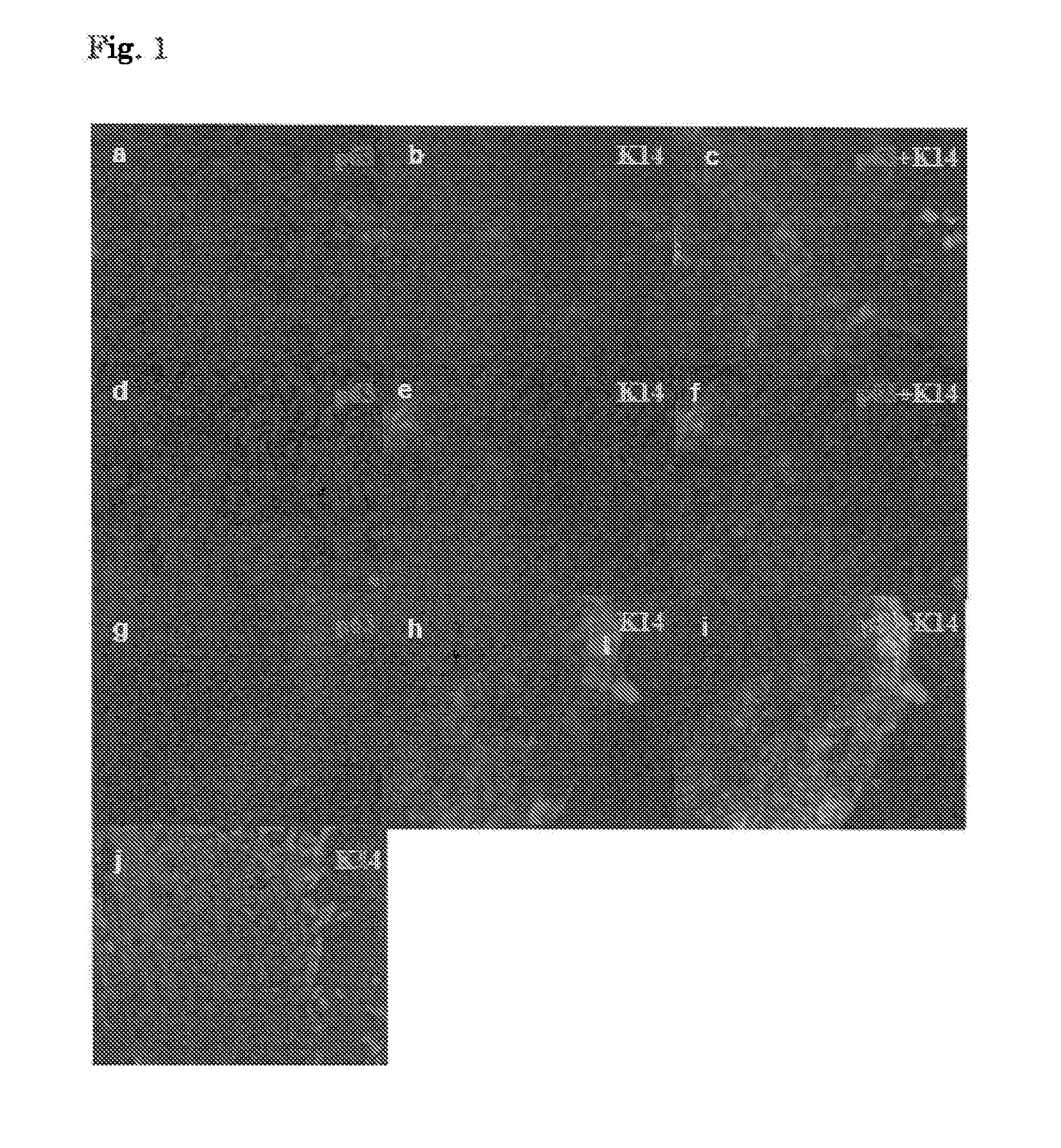
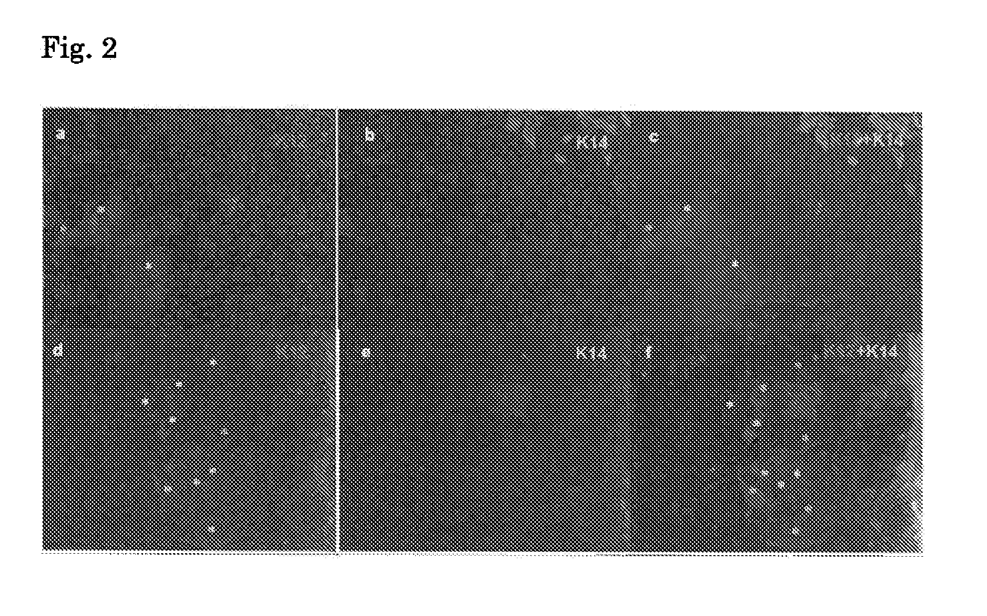
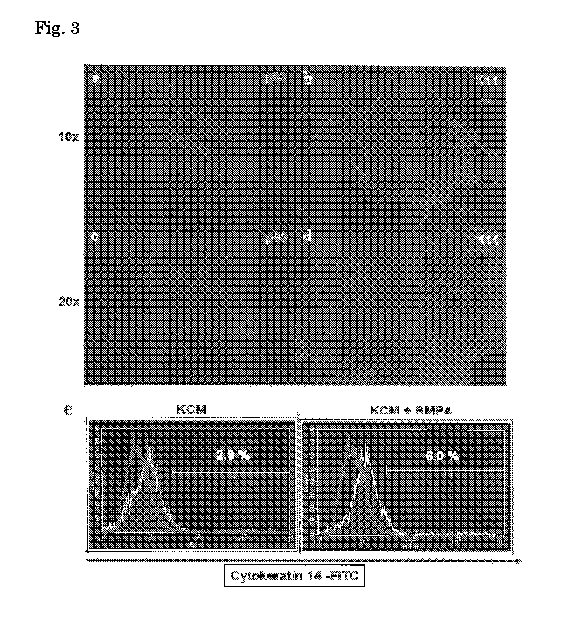
[0172]According to Example 1, mouse iPS cells were cultured on collagen using (i) a KCM medium, (ii) a KCM medium supplemented with 0.5 nM BMP4, or (iii) a KCM medium supplemented with 0.5 nM BMP4+1 μM retinoic acid.

[0173]Likewise, mouse ES cells (RF8; provided by Dr. Robert Farese, Jr. from the Gladstone Institute) were cultured on collagen using (i) a KCM medium, (ii) a KCM medium supplemented with 0.5 nM BMP4, or (iii) a KCM medium supplemented with 0.5 nM BMP4+1 μM retinoic acid.

[0174]The cells after 21-day culture (differentiation induction) were examined for their respective expressions of p63 ...


PUM


Abstract

The present invention relates to: a method for inducing differentiation into an epithelial progenitor cell/stem cell population or a corneal epithelial cell population by culturing, under particular conditions, induced pluripotent stem cells induced from mammalian somatic cells or undifferentiated stem cells; an epithelial progenitor cell/stem cell population or a corneal epithelial cell population obtained by the method; and a cell preparation for the treatment of epithelial disease and a cell sheet, which are prepared using these cell populations.

Description

TECHNICAL FIELD[0001]The present invention relates to: a method for inducing differentiation into an epithelial progenitor cell / stem cell population or a corneal epithelial cell population from induced pluripotent stem cells induced from mammalian somatic cells or undifferentiated stem cells; and a use of a cell population induced by said method in the treatment of epithelial disease.BACKGROUND ART[0002]Keratoplasty based on eye donation has been carried out for intractable corneal epithelial disease and, however, has the problems of absolute donor shortage and rejection after transplantation. To solve the problems, therapy has been developed using patient's own corneal limbus cells or oral mucosal epithelial cells. In this method, a cultured corneal epithelial cell sheet is prepared from corneal limbus cells of healthy eyes or oral mucosal epithelial cells and transplanted to an affected eye (Patent Literatures 1 and 2 and Non Patent Literature 1). However, the method using corneal...

Claims


Application Information

Patent Timeline
IPC IPC(8): C12N5/071
CPCA61L27/3834A61L27/3869A61L27/3895C12N5/0621C12N5/0629C12N2501/01C12N2506/45C12N2501/155C12N2501/385C12N2501/39C12N2501/395C12N2502/1323C12N2502/1394C12N2501/11
Inventor NISHIDA, KOHJIHAYASHI, RYUHEISAKURAI, MIHARUKAGEYAMA, TOMOFUMIYAMANAKA, SHINYAOKITA, KEISUKE
Owner TOHOKU UNIV
Why Eureka
Try Eureka