
Medical device for repair of tissue and method for implantation and fixation

a technology of medical devices and tissue, applied in the field of medical devices for tissue repair, can solve the problems of cartilage wear on the condylar or tibial plateau surface, fibrocartilage deterioration, and possible tear, and achieve the effect of securing the filamen

Inactive Publication Date: 2008-02-07
HOWMEDICA OSTEONICS CORP
View PDF95 Cites 57 Cited by
  • Summary
  • Abstract
  • Description
  • Claims
  • Application Information

AI Technical Summary

Benefits of technology

[0012]Advantageously, the present medical device allows for the provision of blood, biological factors, and cells from a vascular region of tissue to a torn or damaged area located in an avascular and / or semi-vascular region of tissue and provides for fixation of the tear walls or damaged area and the securement of a conduit in a desired position. Additionally, because the conduit itself anchors one side of the primary tear fixation, the conduit can be located with one end adjacent the plane of a tear, damaged area, or implant, allowing the conduit to efficiently deliver blood, biological factors, and cells thereto and increase the rapidity of the healing process. Moreover, in addition to facilitating the transfer of blood, biological factors, and cells from a vascular region to an avascular and / or semi-vascular region, the conduit can also provide for delivery of biological treatments, drugs, and other substances, such as blood, platelet rich plasma, growth factors, or cells, to the tear or defect area through the bore of the conduit. The desired substance can be delivered before, during, or after the conduit is inserted and positioned.

Problems solved by technology

Tears may occur due to the existence of prior defects in the fibrocartilage, shear loading of the fibrocartilage, and / or compounded loading resulting from repetitive compressive loading occurring over a period of time.
Additionally, fibrocartilage can deteriorate as a result of aging, resulting in hard and / or soft areas which further facilitate the creation of tears therein.
Additionally, these procedures may be performed when there is fibrillation and / or degeneration caused by defects in an avascular or limited vascular area, since these injuries are unlikely to heal.
However, cartilage wear on the condylar or tibial plateau surfaces and the eventual development of osteoarthritis may occur as a result of the meniscectomy.
Additionally, the onset of osteoarthritis may lead to more chronic conditions resulting in the need for a total knee replacement procedure.
While the device disclosed in Schwartz '926 is effective, the walls of the fibrocartilage tear may actually be pushed apart during implantation of the stent and prevent effective healing of the tear.
Additionally, even when the sides of the tear are properly aligned, the tear walls may loosen or migrate over time.
Further, the blood dissemination apertures in the stent may not be as effective in providing maximum blood flow to the area of interest as desired to effect healing.

Method used


Image

  • Medical device for repair of tissue and method for implantation and fixation
  • Medical device for repair of tissue and method for implantation and fixation
  • Medical device for repair of tissue and method for implantation and fixation

Examples


Embodiment Construction

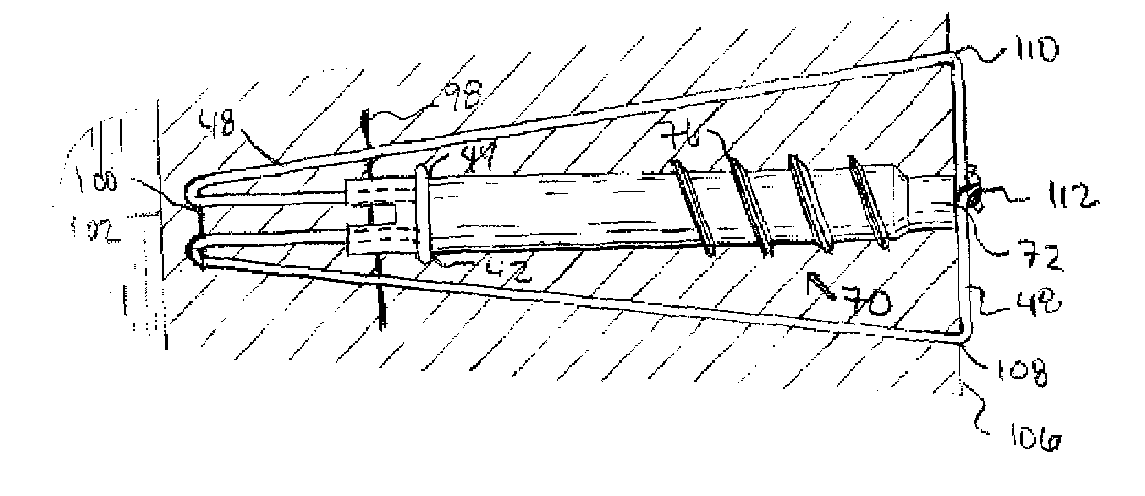
[0044]FIG. 7 shows conduit 30 according to one embodiment of the present invention. Conduit, as used herein, means only an elongate body and does not define any other structural features. Conduit 30 includes a first end 32, a second end 34, and through bore 36 extending from first end 32 to second end 34. In one embodiment, bore 36 has a non-circular cross-section. Body 30 can be manufactured from any biocompatible material. Body 30 has a length from first end 32 to second end 34 as small as 2 mm, 3 mm, or 4 mm and as large as 10 mm, 12 mm, or 15 mm. Additionally, conduit 30 may be coated with biocompatible substances to facilitate tissue regeneration, improve circulation, or achieve any other biologically desirable responses. For example, interior 38 of through bore 36 may be coated with an anti-coagulant to prevent coagulation of blood within through bore 36, thereby promoting the delivery of blood to a torn or damaged tissue area. Alternatively, bore 36 may contain a scaffold mat...


PUM


Abstract

The present invention relates to medical devices for repairing tissue and more specifically to devices which facilitate tissue regeneration and to surgical methods for the implantation and fixation of such devices. In one embodiment, the medical device is an elongate conduit that includes a longitudinal bore extending therethrough to facilitate the transfer of blood from a vascular region of tissue to a tear or damaged area located in an avascular and / or semi-vascular region of tissue. A filament and / or filaments are attached to the conduit and are positioned to secure the conduit and fixate the adjacent tear walls in mutual engagement. In another embodiment, a series of conduits are connected via a filament and / or filaments to facilitate the implantation of multiple conduits.

Description

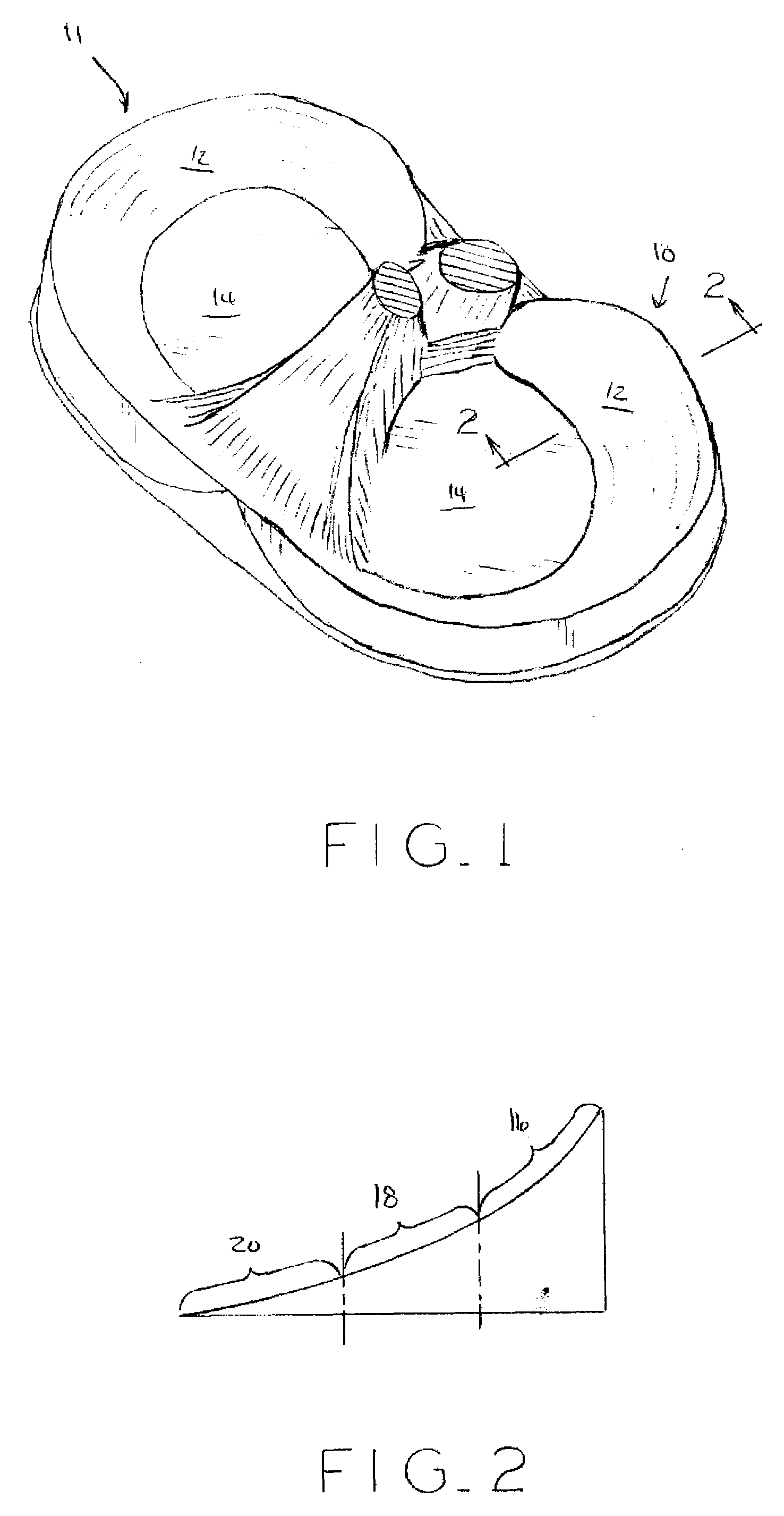
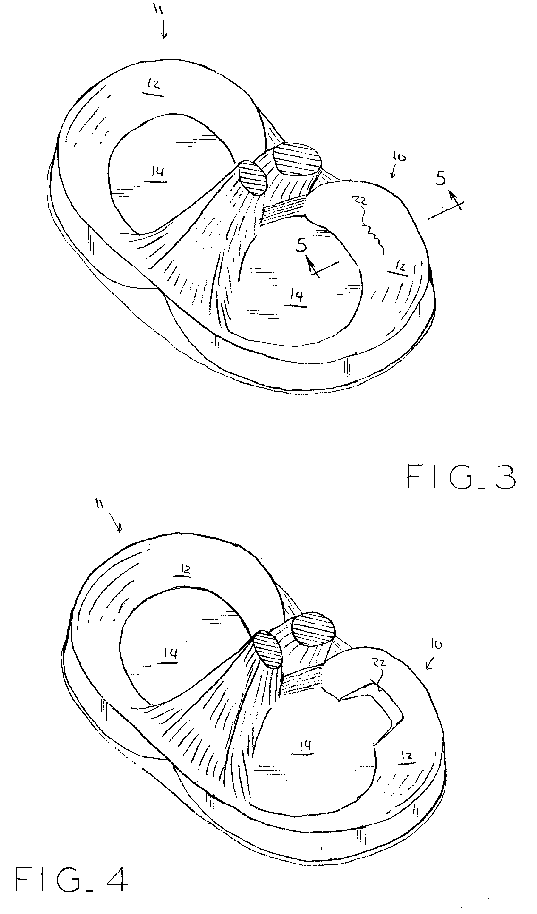
BACKGROUND OF THE INVENTION[0001]1. Field of the Invention[0002]The present invention relates to medical devices for repairing tissue and more specifically to devices which facilitate tissue regeneration and to surgical methods for the implantation and fixation thereof.[0003]2. Description of the Related Art[0004]Various parts of the human body are comprised of fibrocartilage. Fibrocartilage forms the disc, meniscus, and labrums, located in the spine and temporo-mandibular joint, knee, and shoulder and hip, respectively. Additionally, fibrocartilage is present in other parts of the human body, such as fingers, wrists, and ankles. Fibrocartilage is a resilient, compressive tissue capable of accepting and withstanding high loads imparted during bodily movement. Generally, fibrocartilage is found between two adjacent bones, such as the locations set forth hereinabove.[0005]The fibrocartilage of the knee forms menisci 10, 11, shown in FIG. 1. Menisci 10, 11 are semi-lunar, wedge-shaped ...

Claims


Application Information

Patent Timeline
Patent Type & Authority Applications(United States)
IPC IPC(8): A61B17/04
CPCA61B17/0401A61B17/0469A61B17/0487A61B17/064A61B2017/044A61B2017/0648A61B2017/0459A61B2017/0464A61B2017/0475A61B2017/0496A61B2017/0646A61B2017/0458A61B2017/0403A61B2017/0404A61B2017/0445
Inventor SCHWARTZ, HERBERT E.PROCH, FRANCIS S.
Owner HOWMEDICA OSTEONICS CORP
Learn More