Although exceptionally useful for the study of large groups of cells, multiwell plates are not suitable for the study of individual cells or even small groups of cells due to the large, relative to the cellular scale, size of the wells.
When cells float about in a well, specific individual cells are not easily found for observation.
Such variability in location makes high-
throughput imaging (for example for morphological studies) challenging as acquiring an individual
cell and focusing thereon is extremely difficult.
Such variability in location also makes high-
throughput signal processing (for example, detection of light emitted by a single cell through fluorescent processes) challenging as light must be gathered from the entire area of the well, decreasing the
signal to
noise ratio.
Further, a cell held in a well of a
multiwell plate well can be physically or chemically manipulated (for example, isolation or movement of a single selected cell or
single type of cell, changing media or introducing active entities) only with difficulty.
However, once a cell begins the
apoptosis process, the cell does not adhere to the bottom of the well: the cell detaches from the bottom and is carried away by incidental currents in the well.
An additional
disadvantage of multiwell plates is in the study of non-adhering cells.
The fact that the cells are not free-floating but are bound to the plate through some interaction necessarily compromises the results of experiments performed.
Further, the suction required to hold cells in picowells of a carrier of U.S. Pat. No. 4,729,949 deforms held cells and makes a significant portion of the cell membranes unavailable for contact, both factors that potentially compromise experimental results.
Study of cells with non-
fluorescence based methods generally gives poor results due to reflections of light from the carrier.
The fact that emitted light travels through an
optical fiber leads to loss of time-dependent and phase information.
One of the challenges of well-bearing devices known in the art for the study of single living cells, especially picowell-bearing devices, is of
information acquisition.
A disadvantage of using automatic
image analysis is that there is no easy way to sift through the massive amount of information acquired to identify important events from amongst all the images acquired.
One of the greatest challenges in both automatic image analysis and automatic signal analysis is the delineation of the borders of a single cell.
In both such cases, analysis of an image or of a signal gives completely wrong results.
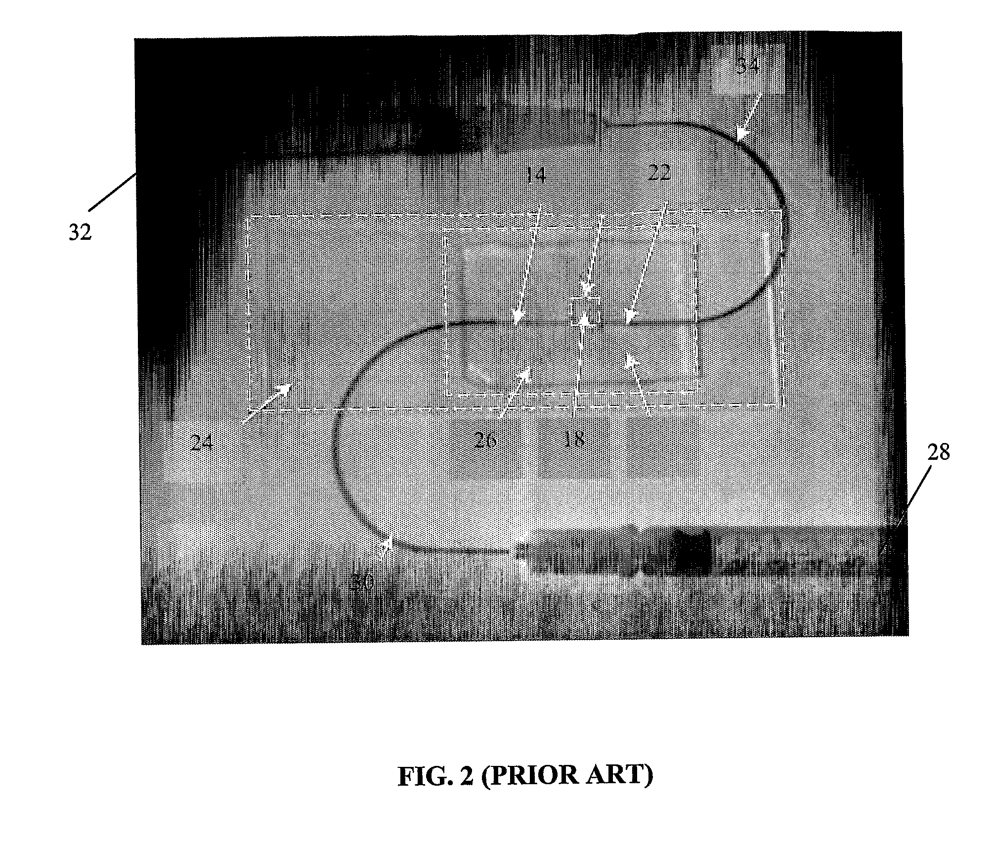
Further, due to the fact that the material from which wells are made is not invisible, distortions, reflections, diffractions and the image of the picowell walls often make delineation of cells difficult.
For example, differentiating cell 42 from cell 44 in FIG. 3 is a difficult task.
It is important to note that even the imperfect methods known in the art are
time consuming, expensive in terms of calculation resources, not robust and in general unsuited for high-throughput applications.
The problem of delineating the borders of a cell for automatic signal analysis is even greater.
In such applications, it is not practical to have a
time consuming cell-identification or picowell-identification step.
In addition, if the borders of the cell or picowells are not clearly delineated, the quality of the data is seriously compromised.
For example, when a cell is delineated conservatively, and only a portion of a signal emitted by a cell is acquired the values of the acquired signal will be inaccurate, especially in cases where signals are not emitted from all areas of a cell homogenously.
An additional problem arises when what is to be detected is not light emitted by a cell itself but rather light emitted by chromatogenic or fluorogenic entities in the medium in the immediate area of the cell, for example the medium held together with the cell in the same picowell.
As stated above, amongst other problems associated with the device of PCT
patent application US99 / 04473, the fact that the emitted light travels through an
optical fiber leads to loss of time dependent and phase information.
Further, the device of PCT
patent application US99 / 04473 is not suitable for acquiring high-resolution images.

