Method and apparatus that increases efficiency and reproducibility in immunohistochemistry and immunocytochemistry

a technology of immunohistochemistry and immunocytochemistry, applied in the field of methods and apparatuses, can solve the problems of uneven staining, unresolved problems, uneven staining, etc., and achieve the effects of improving consistency, reducing the sensitivity of assays, and facilitating transpor

Inactive Publication Date: 2008-04-03
PROHISTO
View PDF22 Cites 2 Cited by
  • Summary
  • Abstract
  • Description
  • Claims
  • Application Information

AI Technical Summary

Benefits of technology

[0012]The present invention relates to a container package partitioned to hold a plurality of individual standard laboratory slides and be secured with a removable lid. This design allows said slides to be rocked on laboratory equipment, and transported with ease. Said apparatus and technique results in no antibody pooling, nor tissue drying commonly encountered in standard sedentary immunohistochemical / immunocytochemical techniques. There is no risk for runoff with this combination of apparatus and method. This combination of apparatus and method provides stable transport for liquid immersed slides during techniques used for immunohistochemistry / immunocytochemistry. This results in better consistency, and increases the sensitivity of the assays. The base of the container package comprises partitions that divide said container package into individual compartments. Each individual compartment holds one standard laboratory slide, with a biological specimen affixed to the slide. The container package comprises a lid of the same or similar material as the base apparatus, so as to reduce evaporation, encourage stacking of the apparatus, and finally, give ease of storage and transport.

Problems solved by technology

This technique can lead to drying of tissues if the staining fluid does not cover all the specimens, and pooling of the stain resulting in uneven staining.
This technique can cause drying of tissues if the staining fluid does not cover all the specimens, and pooling of the stain resulting in uneven staining.
However, such inventions (e.g. U.S. Pat. No. 7,025,937) do not overcome issues such as tissue drying, antibody pooling, and inconsistent staining because of the sedentary techniques used during incubation.
Furthermore, the large volumes of liquid required for other automated techniques (e.g. U.S. Pat. No. 6,436,348; U.S. Pat. No. 6,017,495) make it financially infeasible to use for immunohistochemical and / or immunocytochemical staining, where expensive antibodies are used.
Although other inventions use low volumes, they do not overcome some problems present with systems requiring capillary action of fluid between two slides, especially when viscous reagents must be used.
Additionally, there is still risk for tissue drying, antibody pooling, and inconsistent staining associated with the sedentary nature of incubation (see U.S. Pat. No. 4,635,791; U.S. Pat. No. 5,002,736; U.S. Pat. No. 4,777,020; U.S. Pat. No. 4,985,206; and U.S. Pat. No. 5,192,503).
Although this method addresses issues such as tissue drying, and antibody pooling, it also relies on capillary action to stain tissues, which may be associated with uneven staining.
Finally, pulling the tissue up from the stain / liquid following incubation increases the risk of suctioning off part or all of the tissue, therefore compromising the integrity of the tissue.
The most frequent drawback of this technique is that the labeling reagent is pooled onto the slide, and can easily run off the slide, or stain unevenly.
This method also requires transporting slides, typically from an incubator or benchtop, to a working microscope, in order to analyze cellular staining visually, which dramatically increases the risk of reagent runoff.
Several other disadvantages of the Scharf invention include that only 2 slides can be stained per apparatus, that a relatively large quantity of expensive staining liquid is used for the immersion and staining of the two slides, and that the centrally extending ribs which separate the two slides can disturb a specimen affixed to the face of a slide.

Method used


Image

  • Method and apparatus that increases efficiency and reproducibility in immunohistochemistry and immunocytochemistry
  • Method and apparatus that increases efficiency and reproducibility in immunohistochemistry and immunocytochemistry
  • Method and apparatus that increases efficiency and reproducibility in immunohistochemistry and immunocytochemistry

Examples


Embodiment Construction

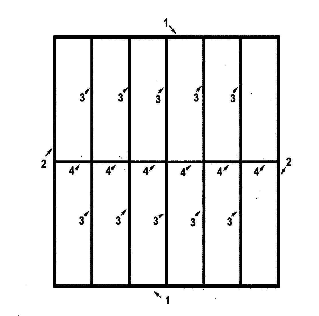
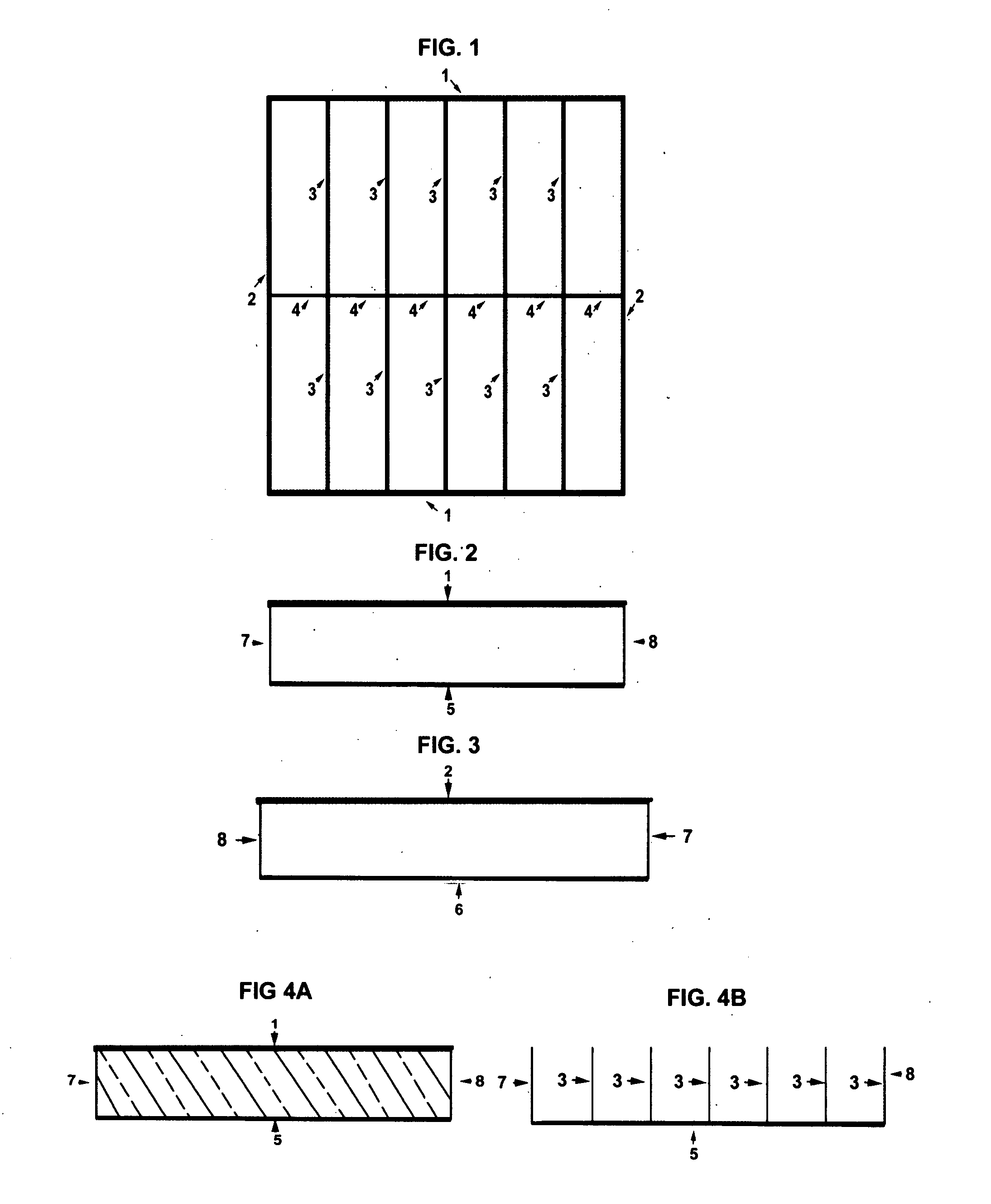
[0029]FIGS. 1, 2, 3 illustrates the top, side and end views of the container package base, without the container package lid. FIG. 4B is a cross sectional view of FIGS. 4A and 2. Said container package base has a bottom 5 and 7, that has walls outlined by 1, 5, 7, and 8 (FIG. 2), and 2, 6, 7, and 8 (FIG. 3). The container package base is partitioned into compartments by 3 and 4 (FIG. 1 and FIG. 4B). The compartments are preferably large enough to fit one standard laboratory microscope slide. FIG. 2 illustrates the side view of the container package base, with the wall outlined by 1, 5, 7 and 8. FIG. 4B illustrates a cross sectional view of FIG. 4B of the container package base, such that the compartments are visible.

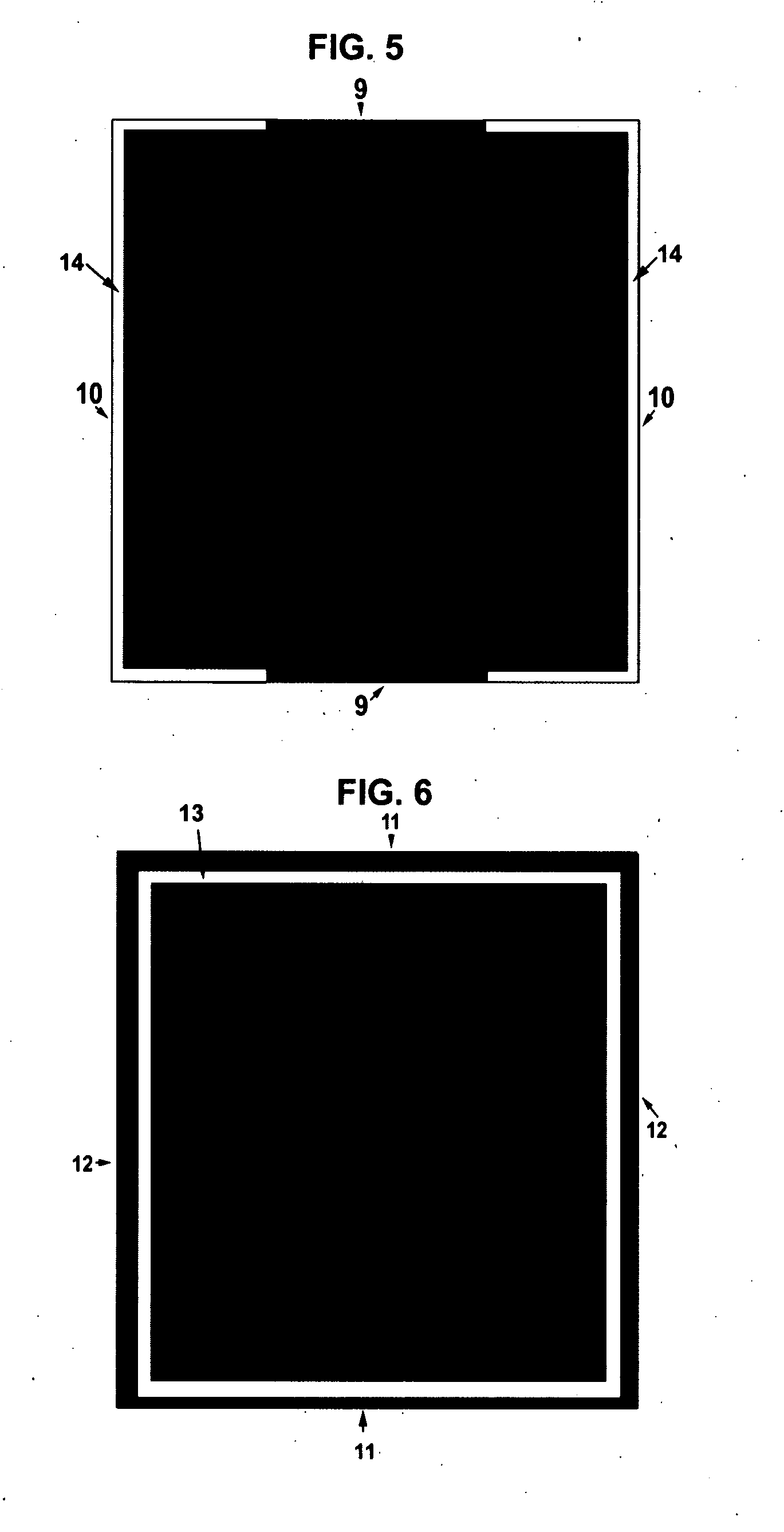
[0030]FIGS. 5 and 6 illustrate a top view of the top side, and the bottom side of the container package lid. The top side (FIG. 5) has an elevated lip 14 within which the bottom 5 and 7 fit into to allow for stacking of said container packages. The bottom side (FIG. 6) h...


PUM


Abstract

A method combined with its necessary container package that facilitates unique processing of microscope slides in a reagent solution is disclosed. This invention has not been described elsewhere. The container package is a partitioned box with each compartment fitting one standard microscope slide. The slides are immersed in a staining solution and the container package is covered with a lid. The container package and slides / staining solution are placed on a standard laboratory apparatus and rocked or agitated for an arbitrary incubation time. The method and its necessary container package provides a means for more reproducibility and sensitivity of results than the prior art that is critical for accurate scientific findings.

Description

CROSS-REFERENCE TO RELATED APPLICATIONS[0001]Not ApplicableSTATEMENT REGARDING FEDERALLY SPONSORED RESEARCH OR DEVELOPMENT[0002]Not ApplicableREFERENCE TO SEQUENCE LISTING, A TABLE, OR A COMPUTER PROGRAM LISTING COMPACT DISC APPENDIX[0003]Not ApplicableBACKGROUND OF THE INVENTION[0004]The staining and examining of biologic specimens occurs daily in great numbers in hospital and scientific laboratories, medical and veterinary clinics. Current techniques rely on a sedentary approach whereupon a plurality of staining fluid is placed over a biological specimen and incubated for a said period of time while remaining sedentary. This technique can lead to drying of tissues if the staining fluid does not cover all the specimens, and pooling of the stain resulting in uneven staining. To stain a biologic specimen fixed on a glass slide, it is necessary to bring a staining liquid or reagent into contact with the specimen on the slide. For the present purposes it will be understood that the ter...

Claims


Application Information

Patent Timeline
IPC IPC(8): C12N5/06C12M1/02
CPCG01N1/312
Inventor BAILEY, ANNE MARIEHOFSETH, LORNE JOHN
Owner PROHISTO
Why Eureka
Try Eureka