Device for tissue reinforcement having microporous and macroporous structures

a tissue reinforcement and micro-porous technology, applied in the field of tissue reinforcement devices, can solve the problems of vesicovaginal fascia injury, herniation of the bladder, and inconvenient reattachment or tying of tissue layers together, and achieve the effects of facilitating tissue regeneration, enhancing tissue repair, and eliminating or reducing potential scar contraction and formation

Inactive Publication Date: 2008-04-03
ETHICON INC
View PDF10 Cites 15 Cited by
  • Summary
  • Abstract
  • Description
  • Claims
  • Application Information

AI Technical Summary

Benefits of technology

[0012]The devices of the present invention may be incorporated with bioactives and therapeutic cells. The devices of the present invention have numerous advantages including enhanced tissue repair, facilitating tissue regeneration, eliminating or reducing potential scar contraction and formation, preventing or reducing the incidence of infection, and / or to preventing tissue adhesion.

Problems solved by technology

However, in some cases the simple repair by re-attachment or tying layers of tissue together is not appropriate to provide a satisfactory outcome.
This is a potentially serious medical condition that may occur, for example, during childbirth or from complications thereof, which can result in an injury of the vesicovaginal fascia.
Such an injury can result in a cystocele, which is a herniation of the bladder.
These conditions can be serious medical problems that can severely and negatively impact a patient both physicologically and psychologically.

Method used


Image

  • Device for tissue reinforcement having microporous and macroporous structures
  • Device for tissue reinforcement having microporous and macroporous structures

Examples


example 1

Formation of Tissue Reinforcement Device

[0046]A 10 weight percent solution of epsilon-caprolactone / glycolide copolymer in 1,4-dioxane was prepared as follows. 20.1 grams of 36 / 64 poly(epsilon-caprolactone-glycolic acid) copolymer (PCGA), obtained from American Polymer Incorporation (American Polymer Inc., Birmingham, Ala.), was added to 180 grams of 1,4-dioxane (Fisher Scientific, Raritan, N.J.) in a 250 milliliter Erlenmeyer screwed cap flask. The mixture was stirred for 4 hours in 60° C. water bath set on a temperature controlled heating plate. The polymer solution was filtered through an extra coarse thimble filter to remove any non-dissolved solids. This 10 weight percent solution was diluted to 5 weight percent by mixing 75.0 grams of 10 percent solution with 75.2 grams of 1,4-dioxane (Fisher Scientific, Raritan, N.J.).

[0047]A 3 weight percent solution was prepared by diluting 18.1 grams of 5 weight percent solution with 12.2 grams of 1,4-dioxane (Fisher Scientific, Raritan, N....

example 2

SEM Evaluation

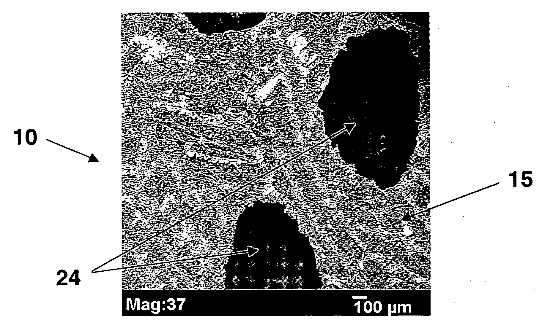
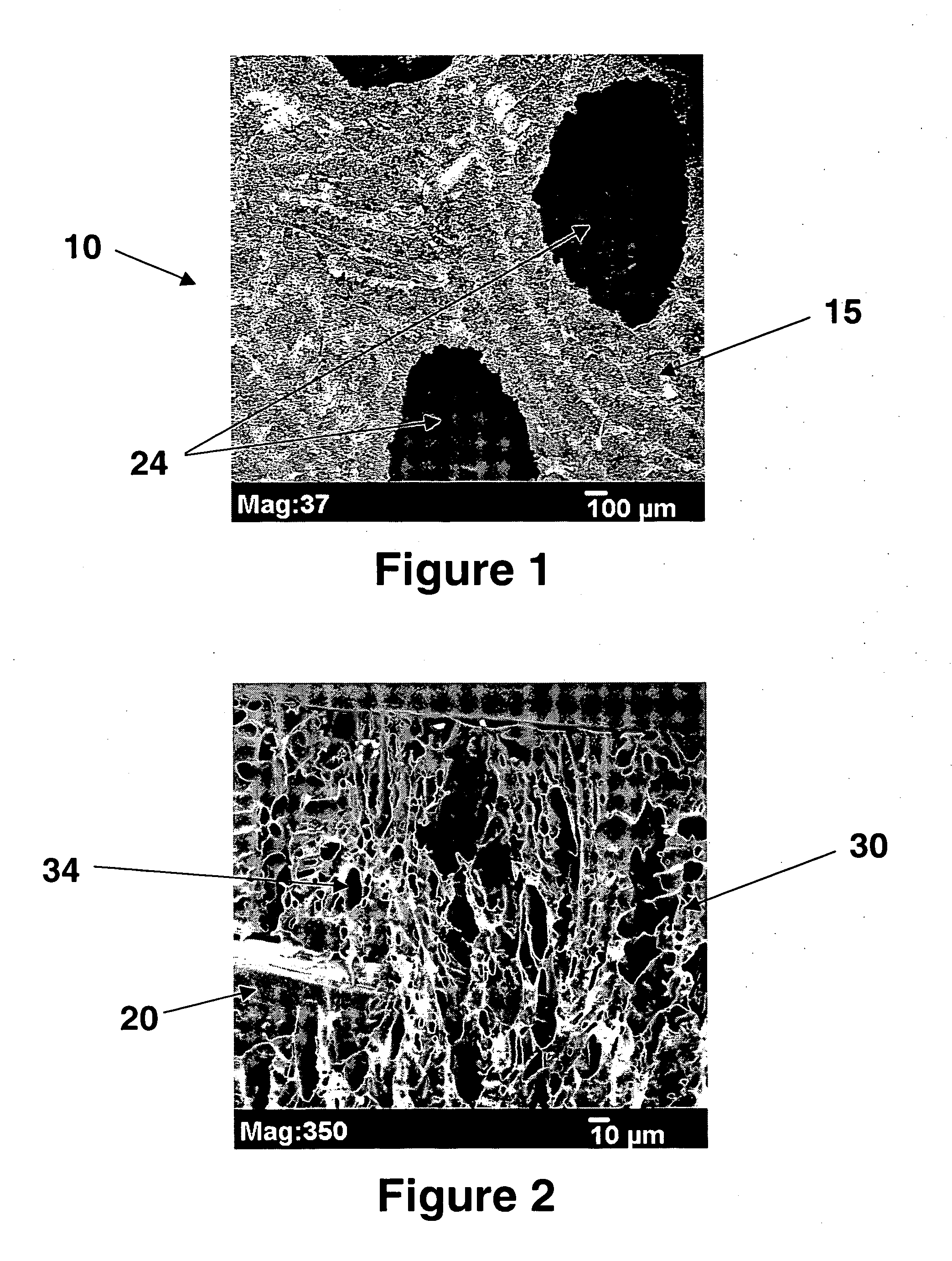
[0050]Samples of the tissue reinforcement device made in Example 1 were mounted on a microscope stud and coated with a thin layer of gold using a EMS 550 sputter coater. SEM analysis was performed using the JEOL JSM-5900LV SEM. The surfaces and cross-sectional areas were examined for each sample. FIGS. 1 and 2 shows tissue reinforcement device 10 where the individual components 20 of the macroporous structure 15 of the device 10 are polypropylene fibers. The fibers are coated with foam coating 30. Device 10 has macropores 24 and micropores 34 in coating 30.

example 3

Cell Attachment

[0051]Human fibroblasts were used for this experiment. Five samples of the tissue reinforcement device made in Example 1 were additionally coated with human fibroblast cells. As a control, five samples of polypropylene mesh were also coated with human fibroblast cells. The number of cells attached to the devices after overnight incubation were measured using a CyQuant kit from Molecular Probe Inc. A higher level of fluorescence indicates a higher level of cell attachment.

[0052]The tissue reinforcement device made in Example 1 had a fluorescence of 643.6+ / −333.6, while the fluorescence of the polypropylene mesh control was 174.6+ / −44.8. The mesh with individual fibers coated with polymer foam showed enhanced cell attachment.


PUM

PropertyMeasurementUnit
pores sizesaaaaaaaaaa
pore sizeaaaaaaaaaa

Abstract

A device for tissue reinforcement. The device has a macroporous structure. At least a section of the macroporous structure is coated with a microporous coating. The microporous and macroporous morphologies allow both cell in-growth and tissue integration.

Description

FIELD OF THE INVENTION[0001]The present invention relates to devices for tissue reinforcement, more specifically devices having both a macroporous and microporous structure to allow both cell in-growth and tissue integration.BACKGROUND OF THE INVENTION[0002]Individuals can sometimes sustain an injury to tissue (for example, trauma, stress induced, overuse, etc.), such as musculoskeletal tissue, that requires repair by surgical intervention. There are numerous surgical procedures in which a supportive or reconstructive technique is used to repair the injured tissue within the body. In many cases surgical sutures are employed to strengthen, support and repair weak tissue in these procedures. The sutures are especially useful in the repair of ligaments, muscles and fascia layers, which hold organs in place. However, in some cases the simple repair by re-attachment or tying layers of tissue together is not appropriate to provide a satisfactory outcome. In that case it may be required to...

Claims


Application Information

Patent Timeline
Patent Type & Authority Applications(United States)
IPC IPC(8): A61F2/02
CPCA61F2/0045A61L31/04A61L31/10A61L31/146A61L31/16C08L67/04
Inventor YANG, CHUNLINKAMMERER, GENE W.ARNOLD, KELLY R.DO, HIEP
Owner ETHICON INC
Why Eureka
Try Eureka