Retinal stem cell compositions and methods for preparing and using same

a technology of stem cells and compositions, applied in the field of retinal stem cell compositions, can solve the problems of slow disease progression, inability to replace lost retinal cells, and physical damage to retinal cells,

Inactive Publication Date: 2008-04-17
THE RES FOUND OF STATE UNIV OF NEW YORK
View PDF1 Cites 13 Cited by
  • Summary
  • Abstract
  • Description
  • Claims
  • Application Information

AI Technical Summary

Benefits of technology

[0022] In yet another aspect, the invention provides methods of repopulating one or more retinal cell types by providing a population having one or more non-retinal cell types, causing the cells to express or over-express one or more secreted activator or inhibitor o

Problems solved by technology

Current treatments can slow disease progression, but cannot replace lost retinal cells.
In addition to disease, physical damage to retinal cells may also occur through retinal detachment or other trauma to the eye.
Unfortunately, when transplanted to even the permissive environment of the embryonic mammalian eye, they differentiate into only three of the seven retinal cell types, suggesting restricted fates and a loss in multipotency.
Despite their obvious potential, endogen

Method used


Image

  • Retinal stem cell compositions and methods for preparing and using same
  • Retinal stem cell compositions and methods for preparing and using same
  • Retinal stem cell compositions and methods for preparing and using same

Examples


example 1

EFTFs Reprogram Primitive Ectoderm to Eyes

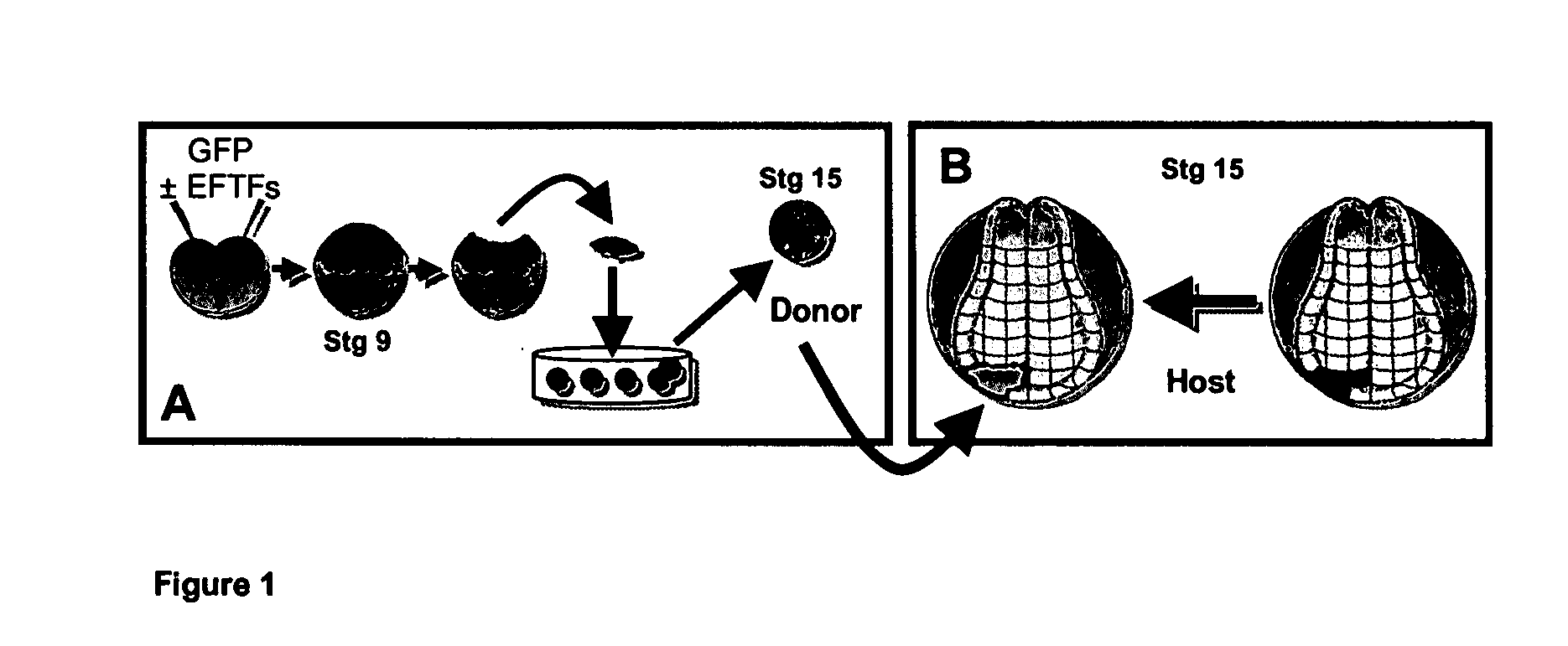
[0078] Seven eye field transcription factors (EFTFs) that are expressed in the retinal stem / progenitor cells of the early eye primordia are sufficient to induce the formation of ectopic eyes. An Animal Cap Transplant (ACT) assay makes it possible to detect the formation of retinal stem / progenitor cells. This method makes it possible to determine if non-retinal cells have been reprogrammed to retinal stem / progenitor cells based on their unique ability to generate retinal tissue when transplanted to the developing Xenopus embryo. The ACT assay is schematized in FIG. 1 and a description is detailed in Methods. This assay takes advantage of two strengths of the Xenopus system—the ectodermal explant assay and tissue transplantation assays. Both blastomeres of two-cell stage Xenopus embryos were injected with either EFTF RNA cocktail containing GFP RNA as a tracer or GFP RNA alone. Ectodermal explants (animal caps) are collected from injected emb...

example 2

EFTF-Induced Eyes are Morphologically and Molecularly Identical to Normal Eyes

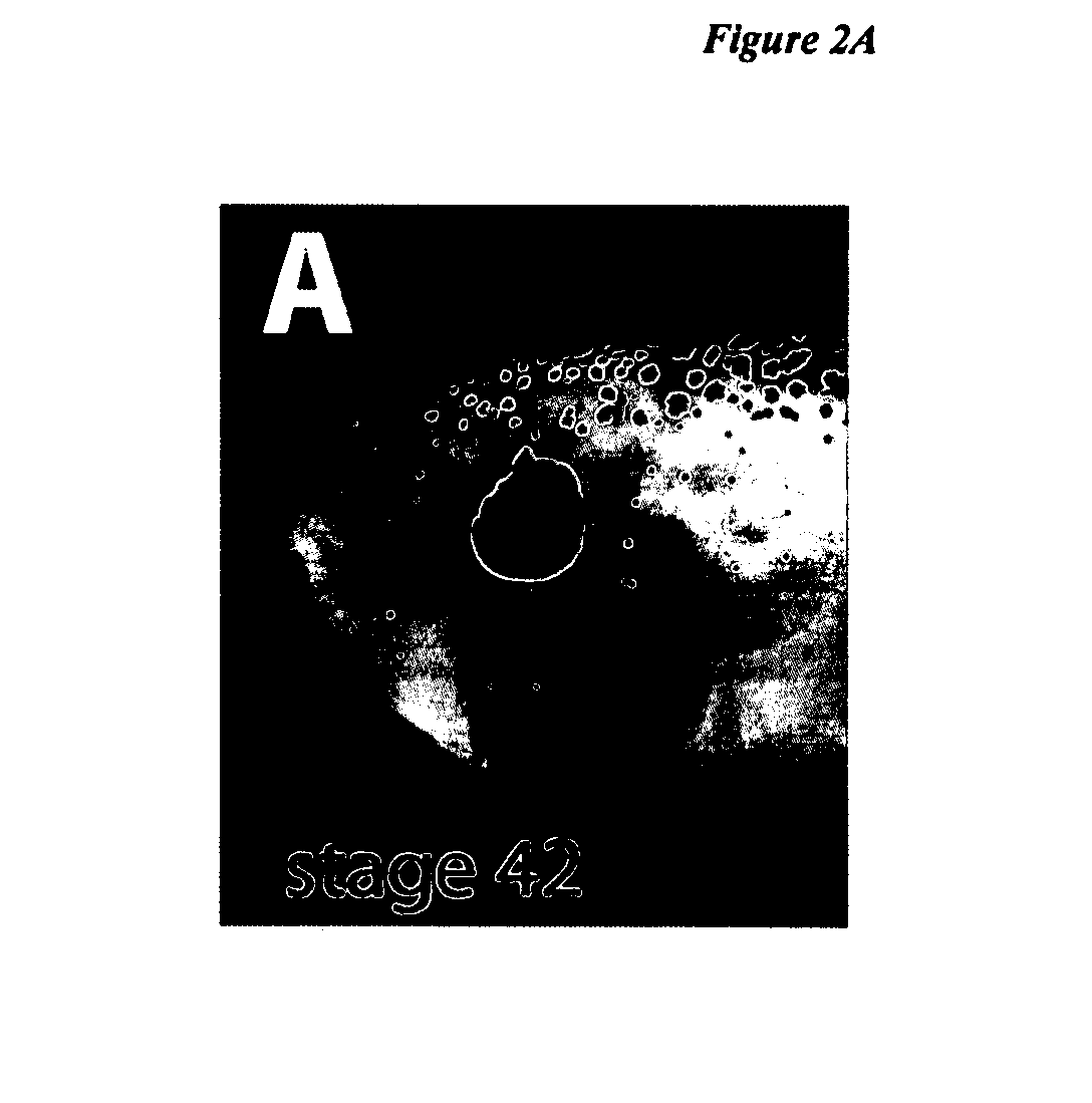
[0080] To better characterize the internal morphology and identify cell types present in EFTF-induced eyes, embryos with strongly fluorescent EFTF-induced eyes were fixed, cryostat sectioned and in situ hybridization or immunocytochemistry were used to identify retinal cell types. Induced eyes had internal morphology identical to normal eyes, containing the tri-layered structure of a normal retina and all the cell types that could be identified by morphology and available molecular markers. These including a lens, retinal pigment epithelium (RPE), rod and cone photoreceptors, and retinal ganglion cells (FIG. 2I-J). Retinal ganglion cell (RGC) axons, the only neural processes that leave the retina, exit the back of the eye as the optic nerve. When viewed using high contrast microscopy, axon tracts were observed exiting the back of induced eyes (opposite the lens), reminiscent of the path taken by RGC axons...

example 3

EFTF-Induced Eyes are Functionally Normal

[0082] In vertebrate eyes, the cornea and lens focus light reflected from images in the surrounding world onto the retina, which lines the back of the eyeball. Cells in the retina form complex circuits designed to convert light into electrical impulses that pass via RGC axons to the brain. An electroretinogram (ERG) can 1) detect additional retinal cell types not identifiable using molecular markers, 2) determine if the induced cells were functionally normal and 3) determine if they formed the intricate neural network necessary to detect and process a light stimulus. EFTF-induced eyes generated ERGs typical of normal eyes (FIGS. 2H& L). In the outer retinal layer, rod and cone photoreceptors use phototransduction to convert light into an electrical impulse. In the normal retina, photoreceptor initiated impulses pass through the inner nuclear layer via second order cell types. In induced eyes, brief light flashes with intensities as low as 0....


PUM


Abstract

Provided are cell compositions including non-retinal cell types that have been reprogrammed to form retinal stem cells, and methods for producing and using same. Such reprogrammed cells can be used to replace one or more retinal cell types that have been lost due to damage and/or disease and are thus useful in treating or preventing visual impairment.

Description

CROSS-REFERENCE TO RELATED APPLICATIONS [0001] This application claims priority from U.S. Provisional Application No. 60 / 795,404, filed Apr. 27, 2006, which is hereby incorporated by reference in its entirety.STATEMENT OF RIGHTS UNDER FEDERALLY SPONSORED RESEARCH [0002] This invention was made with government support under grant number 5R01EY015748-02 entitled “Retinal Stem Cell Culture and Characterization” awarded by the National Eye Institute of U.S. National Institutes of Health. Accordingly, the government has certain rights in the invention.FIELD OF THE INVENTION [0003] The present invention relates to retinal stem cell compositions and methods for reprogramming non-retinal cells to retinal stem progenitor cells. Such reprogrammed cells can be used to replace lost retinal cells and thus be used as a method of treating or preventing visual impairment caused by the loss of one or more retinal cell types. BACKGROUND OF THE INVENTION [0004] Nearly 10 million Americans are blind or...

Claims


Application Information

Patent Timeline
IPC IPC(8): A61K35/12A61P27/02C12N5/00C12N5/08A61K35/44C12N5/074C12N5/0797
CPCA61K35/12A61K35/44C12N5/0623C12N2506/02C12N2501/15C12N2501/155C12N2501/60C12N5/0687A61P27/02
Inventor ZUBER, MICHAEL EZRAVICZIAN, ANDREA SOPHIA
Owner THE RES FOUND OF STATE UNIV OF NEW YORK
Why Eureka
Try Eureka