Method and apparatus to produce ultrasonic images using multiple apertures

an ultrasonic image and multiple aperture technology, applied in the field of medical ultrasound, can solve the problems of poor lateral resolution, large lateral resolution, and small aperture, and achieve the effect of improving the signal-to-noise ratio

Active Publication Date: 2008-05-01
MAUI IMAGING
View PDF99 Cites 54 Cited by
  • Summary
  • Abstract
  • Description
  • Claims
  • Application Information

AI Technical Summary

Benefits of technology

[0017]There are numerous advantages to this approach, and these comprise the objects and advantages of the present invention. They include, among others:
[0022]Artificial scan lines can be considered as overlapping, and each pixel on an output image can be imaged from information from more than one omni line of data. Therefore the output pixel can be averaged from multiple data, thus improving the signal-to-noise ratio.
[0023]Omnidirectional probes can be placed in multiple or intercostal spaces, the suprasternal notch, the substernal window, multiple apertures along the abdomen and other parts of the body, and even on the end of a catheter. An advantage in using omnidirectional probes for catheter placement is that no steering is required of the probe.
[0024]Probes can be placed either on the image plane, off of it, or any combination. When placed away from the image plane, omni probe information can be used to narrow the thickness of the sector scanned.

Problems solved by technology

This design configuration lies at the heart of one of the most significant limitations in the use of ultrasonic imaging for medical purposes; namely, poor lateral resolution.
Theoretically the lateral resolution could be improved by increasing the aperture of the ultrasonic probe, but the practical problems involved with aperture size increase have kept apertures small and lateral resolution large.
For scanners intended for abdominal and other use, the limitation on aperture size is not so obvious, but it is a serious limitation nevertheless.
The problem is that it is difficult to keep the elements of a large aperture array in phase because the speed of ultrasound transmission varies with the type of tissue between the probe and the area of interest.
When the size of the aperture is increased to improve the lateral resolution, the additional elements of a phased array may be out of phase and may actually degrade the image rather than improving it.
In the case of cardiology, it has long been thought that extending the phased array into a second or third intercostal space would improve the lateral resolution, but this idea has met with two problems.
Second, the tissue speed variation described above, but not adequately addressed until this time, needs to be compensated.

Method used


Image

  • Method and apparatus to produce ultrasonic images using multiple apertures
  • Method and apparatus to produce ultrasonic images using multiple apertures
  • Method and apparatus to produce ultrasonic images using multiple apertures

Examples


Embodiment Construction

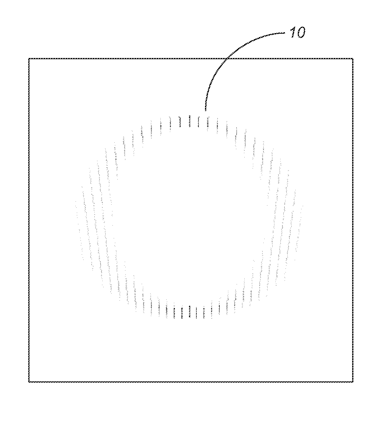
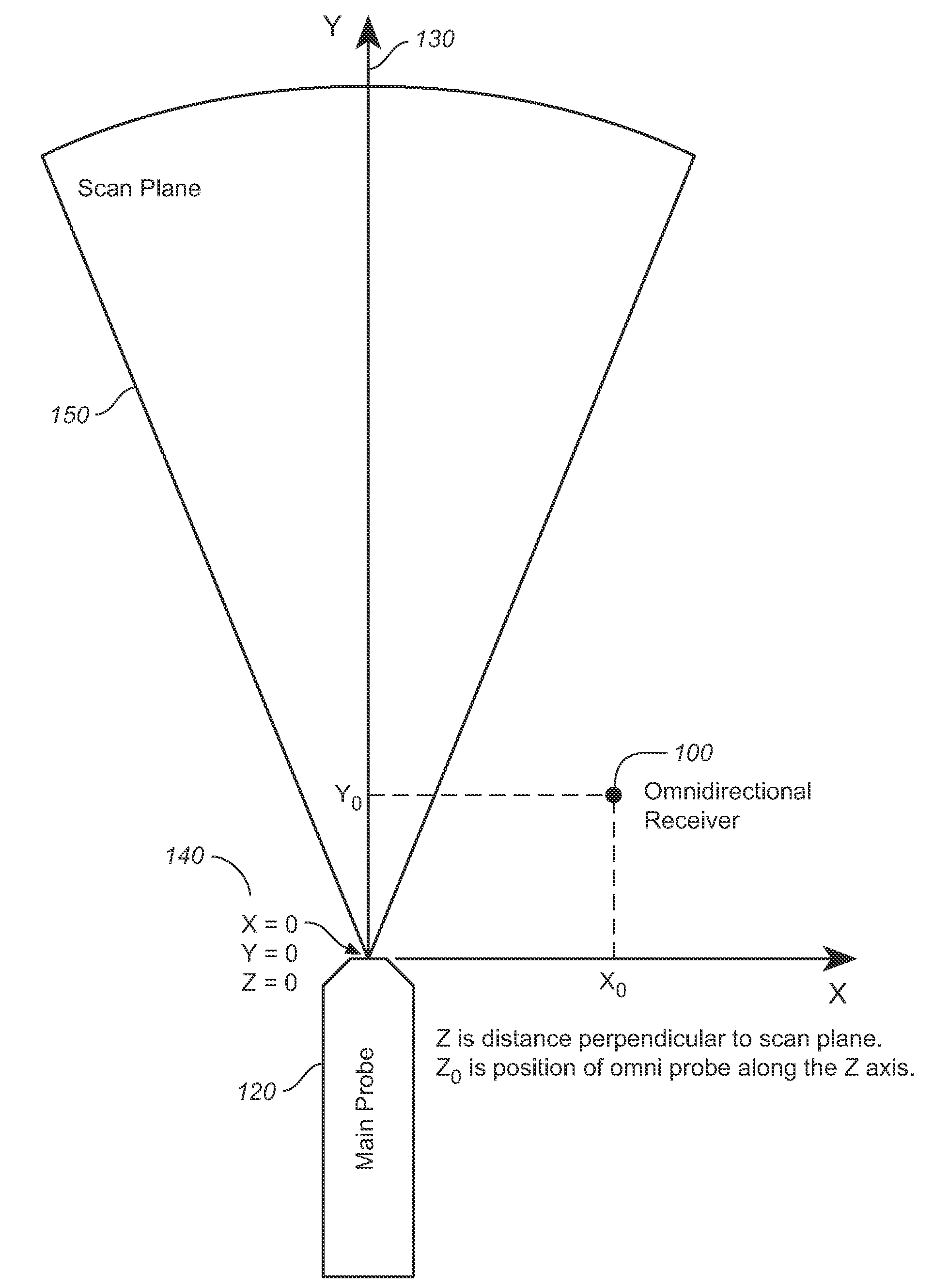
[0045]A key element of the present invention is that returned echoes in ultrasonography can be detected by a separate relatively non-directional receive transducer located away from the insonifying probe (transmit transducer), and the non-directional receive transducer can be placed in a different acoustic window from the insonifying probe. This probe will be called an omni-directional probe because it can be designed to be sensitive to a wide field of view.

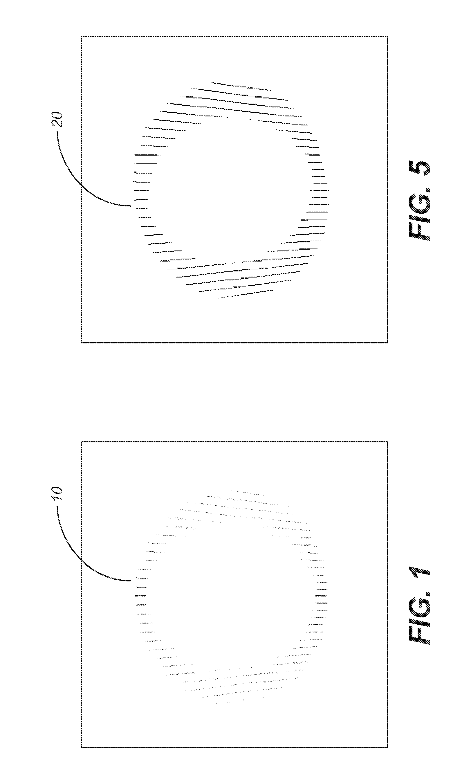
[0046]If the echoes detected at the omni probe are stored separately for every pulse from the insonifying transducer, it is surprising to note that the entire two-dimensional image can be formed from the information received by the one omni. Additional copies of the image can be formed by additional omni-directional probes collecting data from the same set of insonifying pulses.

[0047]A large amount of straightforward computation is required to plot the amplitude of echoes received from the omni. Referring now to FIG. 2, in which ...


PUM


Abstract

A combination of an ultrasonic scanner and an omnidirectional receive transducer for producing a two-dimensional image from the echoes received by the single omnidirectional transducer is described. Two-dimensional images with different noise components can be constructed from the echoes received by additional transducers. These can be combined to produce images with better signal to noise ratios and lateral resolution. Also disclosed is a method based on information content to compensate for the different delays for different paths through intervening tissue is described. Specular reflections are attenuated by using even a single omnidirectional receiver displaced from the insonifying probe. The disclosed techniques have broad application in medical imaging but are ideally suited to multi-aperture cardiac imaging using two or more intercostal spaces. Since lateral resolution is determined primarily by the aperture defined by the end elements, it is not necessary to fill the entire aperture with equally spaced elements. In fact, gaps can be left to accommodate spanning a patient's ribs, or simply to reduce the cost of the large aperture array. Multiple slices using these methods can be combined to form three-dimensional images.

Description

CROSS REFERENCES TO RELATED APPLICATIONS[0001]The present application claims the benefit of U.S. Provisional Patent Application Ser. No. 60 / 862,951, filed Oct. 25, 2006 (Oct. 25, 2006), and U.S. Patent Application Ser. No. 60 / 940,261, filed May 25, 2007 (May 25, 2007).SEQUENCE LISTING[0002]Not applicable.STATEMENT REGARDING FEDERALLY SPONSORED RESEARCH OR DEVELOPMENT[0003]Not applicable.THE NAMES OR PARTIES TO A JOINT RESEARCH AGREEMENT[0004]Not applicable.INCORPORATION-BY-REFERENCE OF MATERIAL SUBMITTED ON A COMPACT DISC[0005]Not applicable.BACKGROUND OF THE INVENTION[0006]1. Field of the Invention[0007]The present invention relates generally to imaging techniques used in medicine, and more particularly to medical ultrasound, and still more particularly to an apparatus for producing ultrasonic images using multiple apertures.[0008]2. Discussion of Related Art Including Information Disclosed Under 37 CFR § 1.97, 1.98[0009]In conventional ultrasonic imaging, a focused beam of ultraso...

Claims


Application Information

Patent Timeline
Patent Type & Authority Applications(United States)
IPC IPC(8): A61B8/00
CPCA61B8/42A61B8/5253A61B8/4281A61B8/4455A61B8/483A61B8/543G01S7/52046G01S7/5205G01S15/8977A61B5/725A61B8/085A61B8/145A61B8/4494A61B8/5207A61B8/4209A61B8/4218A61B8/4483A61B8/08A61B8/4245
Inventor SPECHT, DONALD F.
Owner MAUI IMAGING
Why Eureka
Try Eureka