

Selection of cells using biomarkers
a cell and biomarker technology, applied in the field of cell selection using biomarkers, can solve the problems of insufficient safety or effectiveness of obtaining the necessary sample component for conducting such analysis for a particular disease or condition, inaccurate, and potentially harmful to the mother and the fetus, and achieve high sensitivity and specificity.
- Summary
- Abstract
- Description
- Claims
- Application Information
AI Technical Summary
Benefits of technology
Problems solved by technology
Method used
Image

Examples
example 1
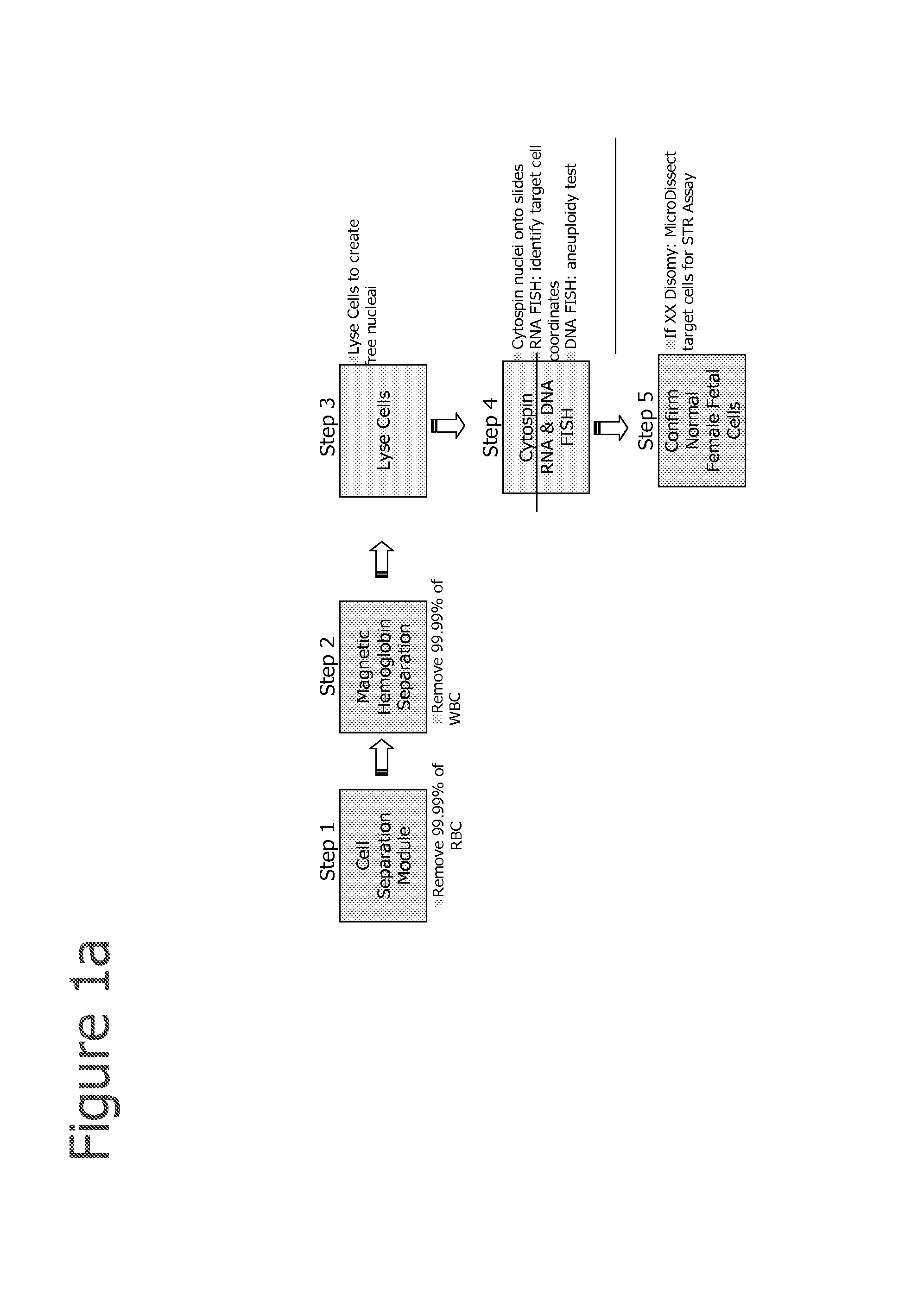
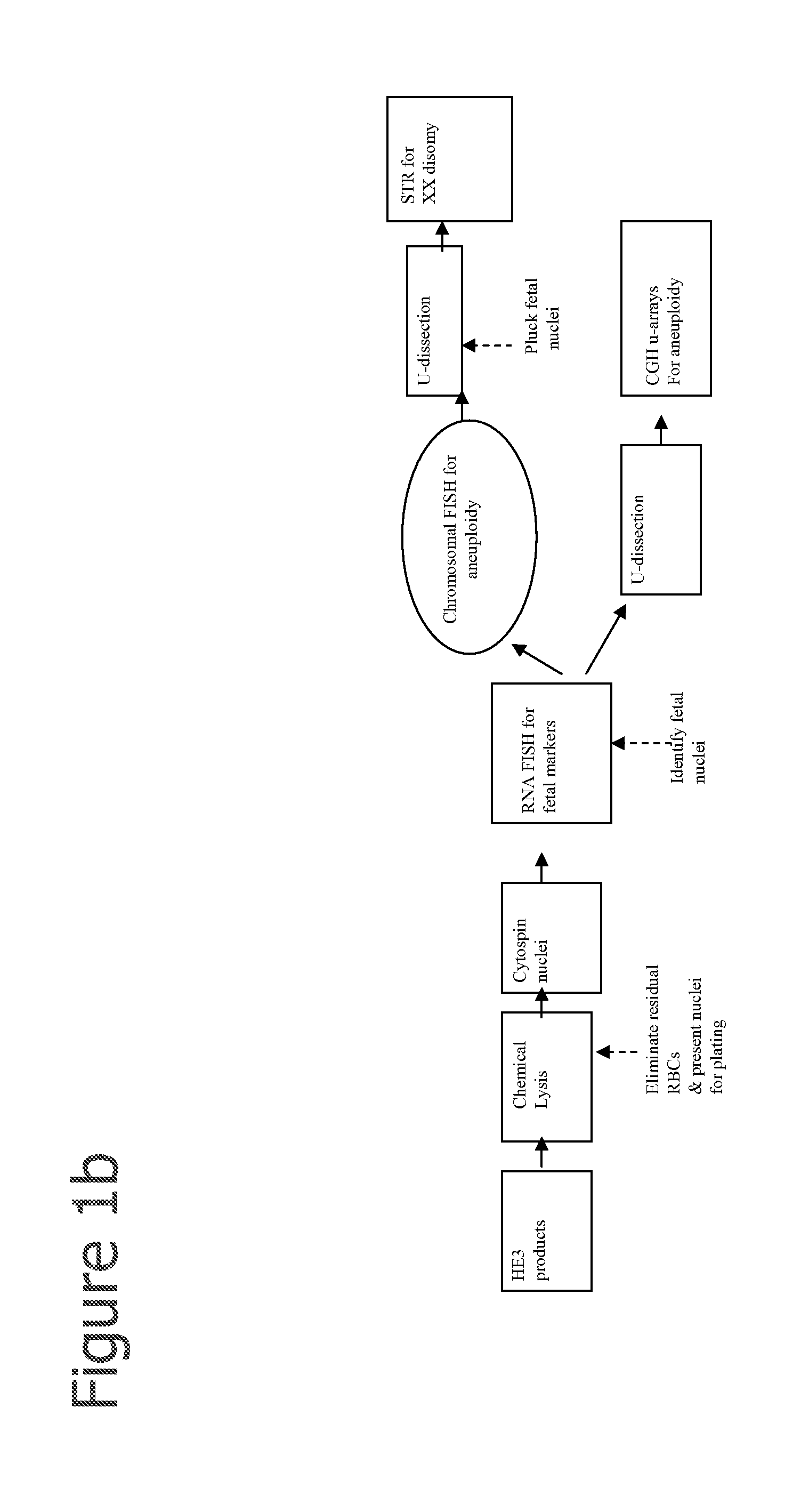
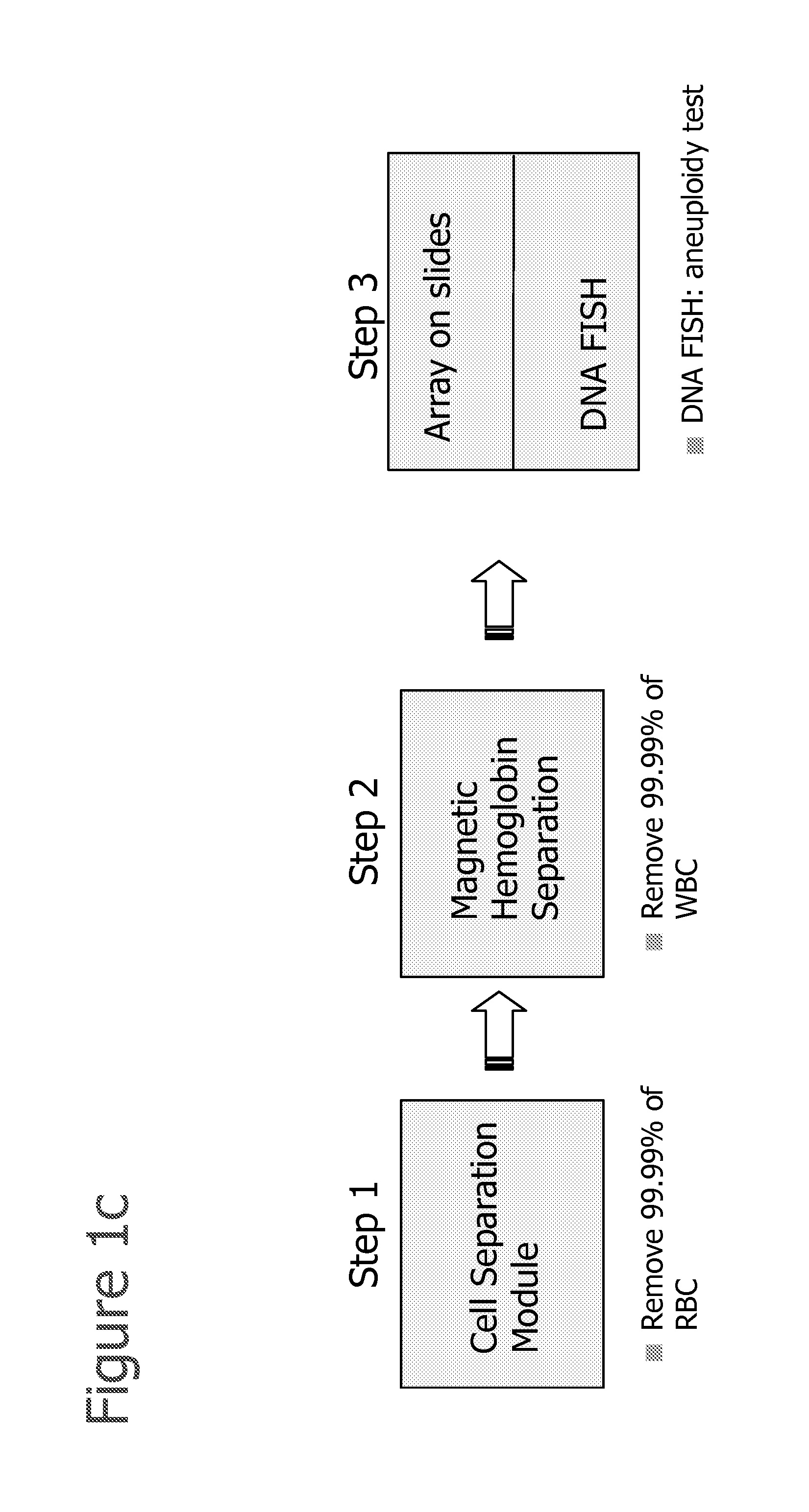
RNA FISH Analysis
[0210]After fetal cells are enriched by one or more of the methods described herein, RNA FISH analysis is performed using methods disclosed in Raamsdonk et al., 2001, NAR; 29:8:e42 or as described below:[0211]1. Cytospin the cells onto a glass slide (or cell grow on slide).[0212]2. Permeabilize the cells with cytoskeletal buffer (CB buffer 100 mM NaCl, 30 mM sucrose, 3 mM MgCl2, 10 mM PIPES, pH 6.8), 30 s (CB buffer can be replaced with 1×PBS).[0213]3. CB+0.5% Tx-100, 30 s[0214]4. CB buffer for 30 s.[0215]5. Fix in 4% paraformaldehyde in 1×PBS for 10 min[0216]6. 70% ethanol at 4 C for up to 2 weeks[0217]7. or dehydrate the slides with 70, 80, 90, 100% ethanol, and dried[0218]8. Hybridize with probes on at 37 C in 50% formamide, 6×SCC[0219]9. Slides wash in 50% formamide,[0220]10. 2×SSC at 39 C for 3×5 min,[0221]11. 2×SSC at 37 C for 3×5 min,[0222]12. Room temperature using 1×SSC 10 min[0223]13. 4×SSC 5 min
[0224]Preparation of Probe:[0225]1. Probes are prepared using...
example 2
In Situ rt-PCR Identification of Fetal Nucleated Red Blood Cells from Maternal Nucleated Red Blood Cells in Solution
[0260]A highly purified cells population with fnRBCs and mnRBCs, and maternal white blood cells is fixed in 2% paraformaldehyde solution in 1×PBS for 20 minutes at room temperature, then the cells are collected by a brief centrifugation, and resuspended in 1×RT-PCR reaction buffer with dNTP mix, reverse transcriptase, Hot-start Taq DNA polymerase, fluorescence dye labeled primers for beta and gamma globin. This is followed by a reverse transcription reaction step at 50° C. for 30 minutes to convert the mRNA into cDNA and then followed by 30 to 40 cycles of PCR. The amplified products can be visualized under a fluorescent microscope and images may be captured through a COD camera. Cells demonstrating a high gamma to beta globin expression ratio will be selected and further genetic information will be extracted.
example 3
In Situ rt-PCR Identification of Fetal Nucleated Red Blood Cell Nuclei from Maternal Nucleated Red Blood Cell Nuclei on Slide
[0261]A highly purified cells population with fnRBCs and mnRBCs, and maternal white blood cells is fixed in 2% paraformaldehyde solution in 1×PBS for 20 minutes at room temperature; the cells are then cytospin on to a slide. The cells are submerged in 1×rt-PCR reaction buffer with dNTP mix, reverse transcriptase, Hot-start Taq DNA polymerase, fluorescence dye labeled primers for beta and gamma globin. This is followed by a reverse transcription reaction step at 50° C. for 30 minutes to convert the mRNA into cDNA and then followed by 30 to 40 cycles of PCR. The amplified products can be visualized under a fluorescent microscope and images may be captured through a CCD camera. Cells demonstrating a high gamma to beta globin expression ratio will be selected and further genetic information will be extracted.
PUM
| Property | Measurement | Unit |
|---|---|---|
| volume | aaaaa | aaaaa |
| time | aaaaa | aaaaa |
| time | aaaaa | aaaaa |
Abstract
Description
Claims
Application Information