
Osteoblast Growth Factor
- Summary
- Abstract
- Description
- Claims
- Application Information
AI Technical Summary
Benefits of technology
Problems solved by technology
Method used
Image

Examples
example 1
Identification of a Novel Osteoclast-Derived Factor in Osteoclasts
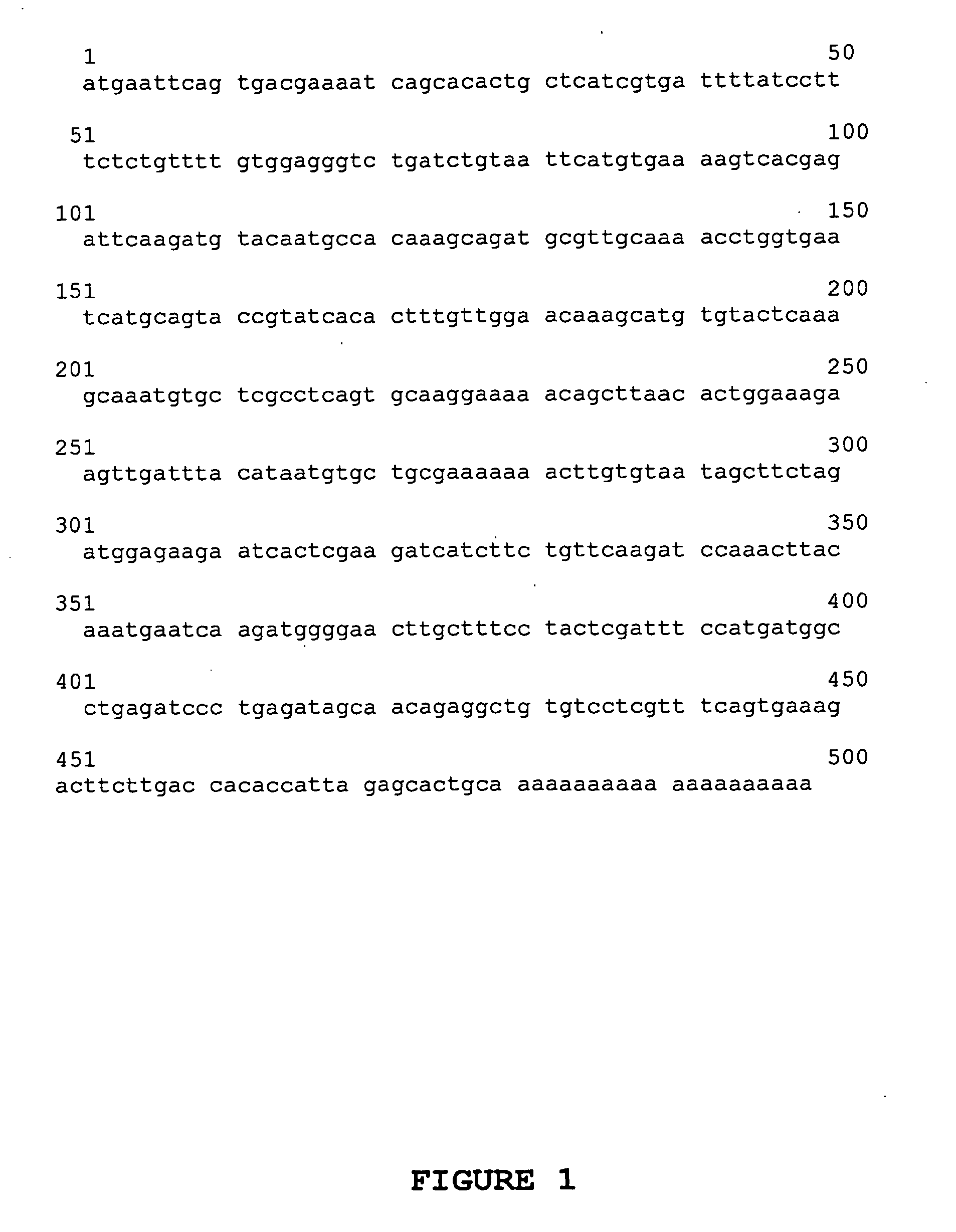
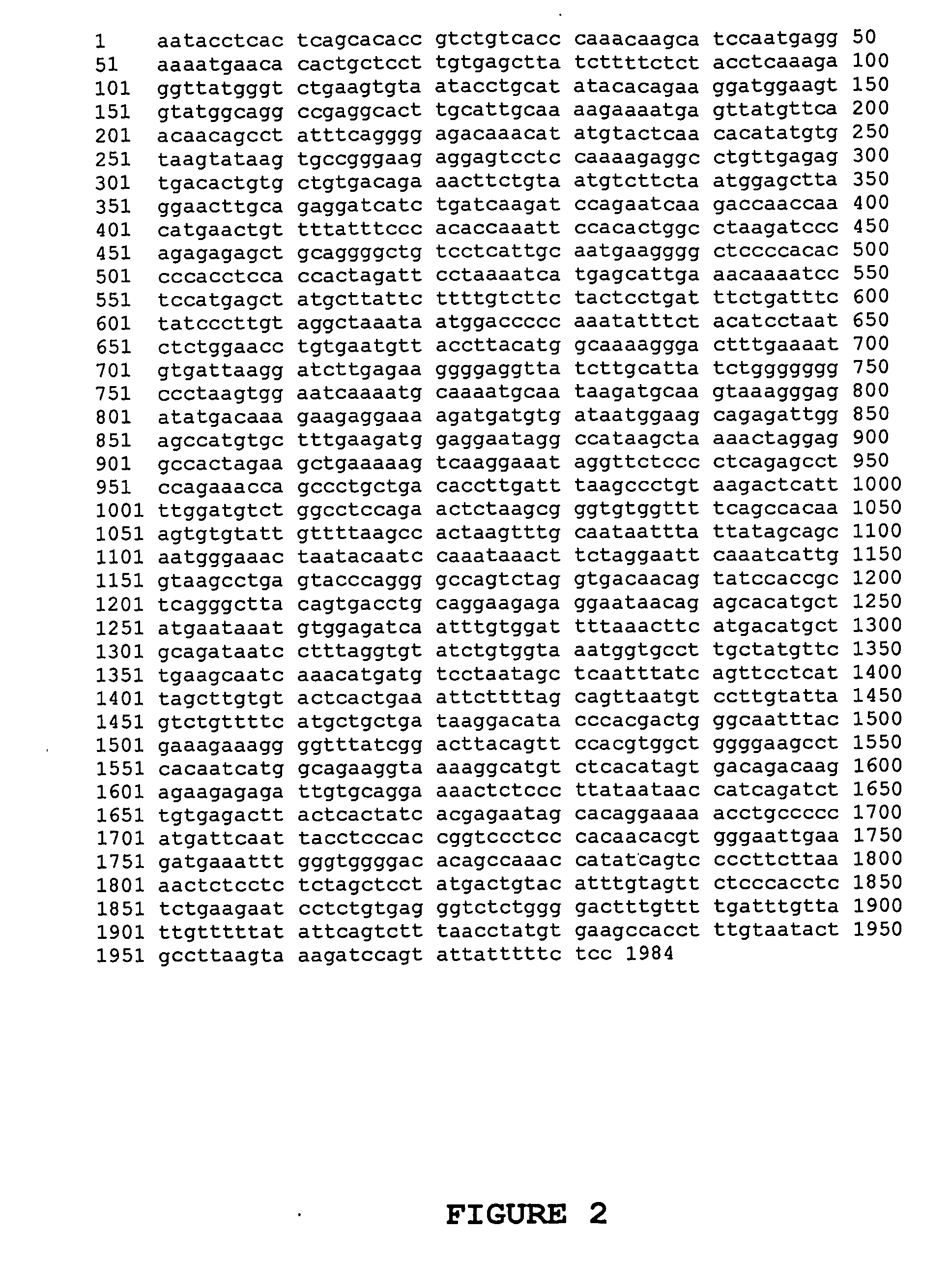
[0159]A cDNA-subtracted library was constructed according to the procedures of the ClonTech PCR-Select Subtraction Kit (California, USA). Briefly, double-stranded cDNA was prepared from 2 μg of poly(A)+ RNA obtained from the haematopoietic macrophage cell line RAW246.7 (Xu et al., 2000, J. Cell Biochem. 88, 1256-1264) treated with (tester) and without (driver) 100 ng / mL of RANKL for 5 days. Tester and driver cDNAs were digested with RsaI to generate shorter, blunt-ended double stranded cDNA fragments for optimal subtraction. The tester, but not the driver cDNA, was then treated with adaptors. Both tester and driver were subject to hybridisation, and primary PCR was employed to amplify differentially expressed sequences. The subtraction was also performed with RANKL-treated RAW246.7 cells as the driver, and untreated RAW246.7 cells as the tester to produce a reverse-subtracted cDNA pool. The forward-subtracted PCR prod...
example 2
Characterisation of the Expression Pattern of a Novel Caltrin
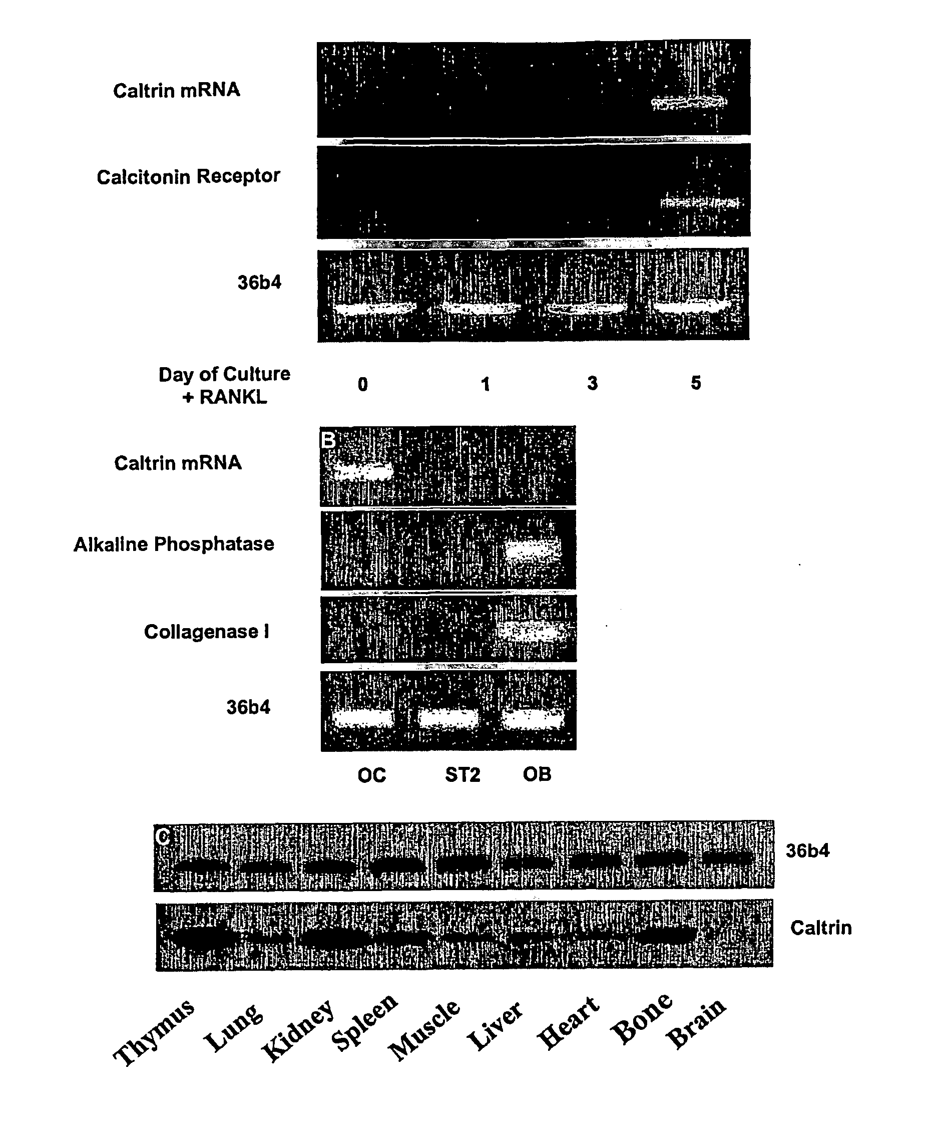
[0162]Once caltrin was identified in osteoclasts using subtractive hybridisation, the next step was to examine the level of caltrin mRNA in osteoclasts and other cells and tissues. To assess the level of caltrin mRNA expression during osteoclastogenesis, RAW246.7 cells were cultured in the presence or absence of soluble RANKL and subjected to semi-quantitative RT-PCR analysis.
[0163]Briefly, total RNA was isolated from cultured cells using RNAzol solution according to the manufacturer's instructions (Ambion Inc., Austin, Tex.). For RT-PCR, single stranded cDNA was prepared from 2 μg of total RNA using reverse transcriptase with an oligo-dT primer. Two microlitres of each cDNA was subjected to 30 cycles of PCR (94° C., 40 sec; 54° C., 40 sec; and 72° C., 40 sec) using mouse caltrin specific primers 5′ATGATTCAGTGACGAAAT3′; and 5′GAAGCTATTACACAAGTTTT3′. To examine whether caltrin was expressed in osteoblasts, total RNA was iso...
example 3
Expression of Recombinant Caltrin Using the Baculovirus Expression Vector System (BEVS)
[0166]In order to characterise the function of caltrin in bone, the BEVS (Kitts, 1995, Methods Mol Biol 39, 129-142) was utilised to produce active recombinant his-tagged proteins that can be used in subsequent in vitro and in vivo experiments.
[0167]Briefly, total RNA was isolated from RAW264.7 cell-derived osteoclasts. For RT-PCR, single-stranded cDNA was prepared from 2 μg of total RNA using reverse transcriptase and an oligo-dT primer. Two μl of cDNA was subject to 30 cycles of PCR amplification (94° C., 40 sec; 54° C., 40 sec; and 72° C., 40 sec) using the caltrin specific primers 5′ATGATTCAGTGACGAAAT3′ (forward) and 5′GAAGCTATTACACAAGTTTT3′ (reverse). The amplified product was gel purified on a 1% TAE-agarose gel and extracted using the QIAEX II gel purification kit (Qiagen Inc, Valenica, Calif.). The PCR products were cloned into the TOPO TA Cloning site of pcDNA3.1 / V5 / His-TOPO (Invitrogen, ...
PUM
| Property | Measurement | Unit |
|---|---|---|
| Fraction | aaaaa | aaaaa |
| Fraction | aaaaa | aaaaa |
| Length | aaaaa | aaaaa |
Abstract
Description
Claims
Application Information