Cell systems and methods for detecting proliferation acitvity

a cell system and proliferation factor technology, applied in the field of cell systems and cell systems for clinical assessment of proliferation factor activity, can solve the problems of undesirable clinical diagnostic use of radioactivity, loss of efficacy and risk of adverse side effects, and the immune system's ability to reduce or neutralize the effective concentration of administered biopharmaceuticals

Inactive Publication Date: 2008-06-05
AMGEN INC
View PDF0 Cites 16 Cited by
  • Summary
  • Abstract
  • Description
  • Claims
  • Application Information

AI Technical Summary

Problems solved by technology

However, despite the use of human polypeptides and safeguards in the preparation and approval process, the patient's own immune system can reduce or neutralize the effective concentration of an administered biopharmaceutical.
Loss of biopharmaceutical functionality through host immune mechanisms can lead to loss of efficacy and risk of adverse side effects.
One method of measuring DNA synthesis uses radioactive precursors such 3H-thymidine and, although this method can provide good sensitivity, the use of radioactivity in a clinical diagnostic setting is undesirable.
The BrdU incorporation method is often cumbersome and, therefore, is limited in assay throughput.
Therefore, use of this method as a reliable indicator of cell proliferative activities has not been fully established in clinical settings.

Method used


Image

  • Cell systems and methods for detecting proliferation acitvity
  • Cell systems and methods for detecting proliferation acitvity
  • Cell systems and methods for detecting proliferation acitvity

Examples


example i

Cell Proliferation Determination by Luciferase Expression

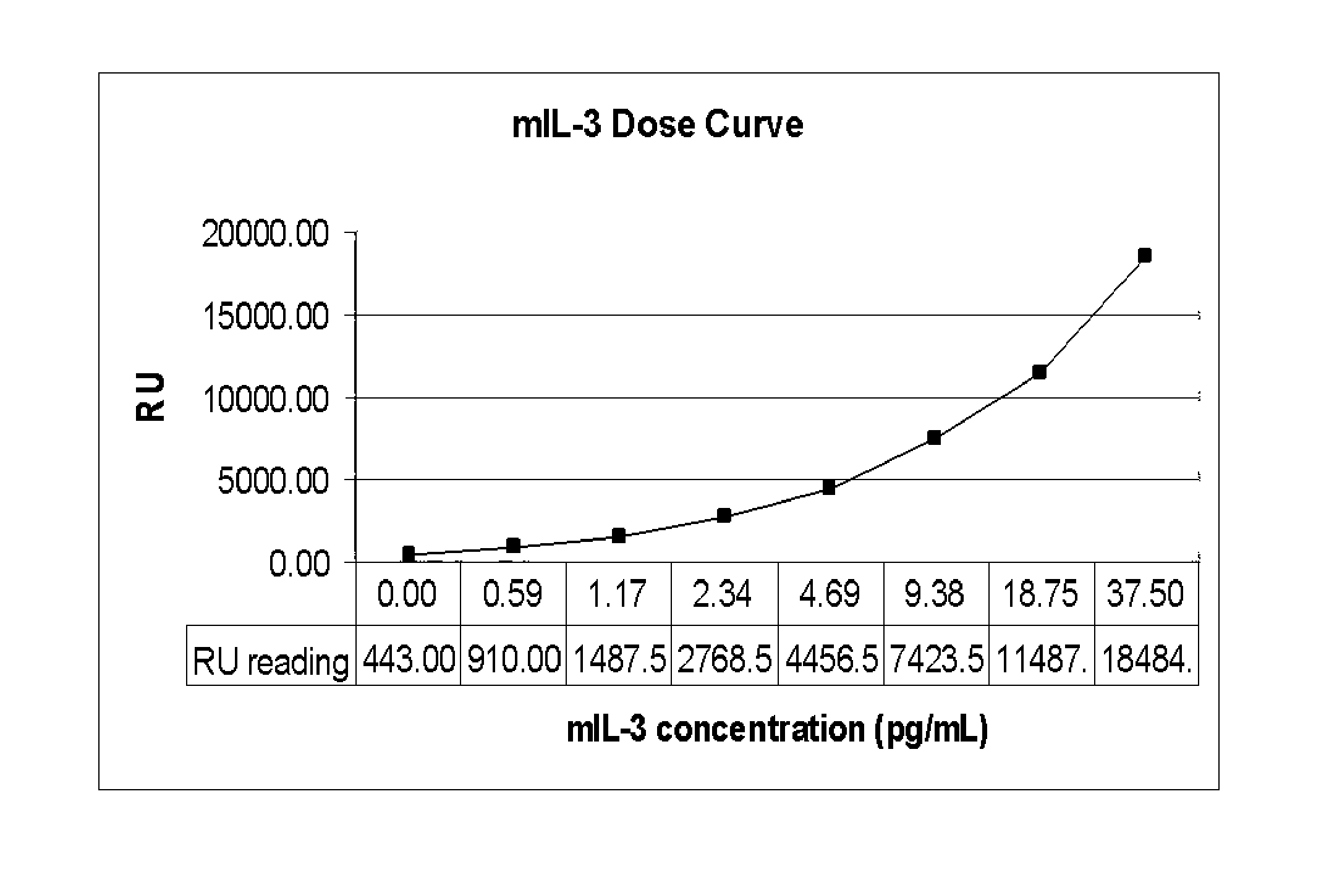
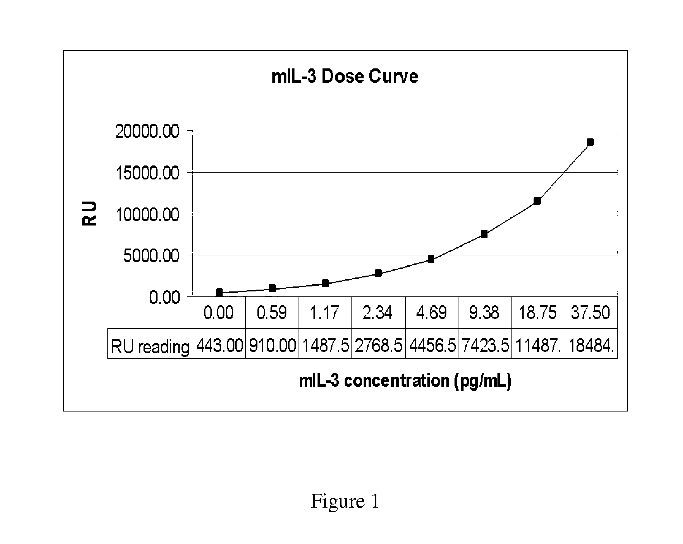
[0092]This Example shows that cell proliferation and / or cell viability can be assessed by luciferase activities constitutively expressed in stably transfected cells.

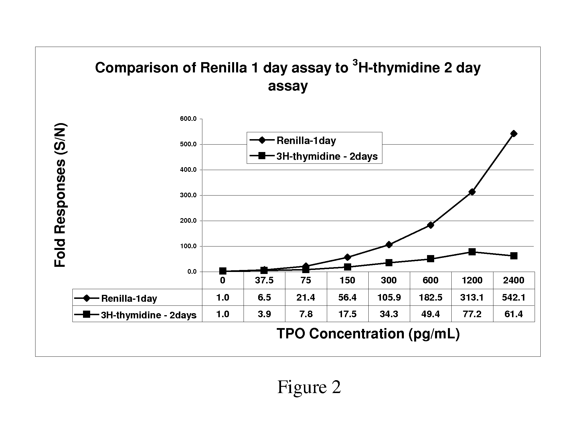
[0093]The cell line 32Dcl23 is a stably transfected 32D cell line that expresses human mpl gene encoding the receptor for Thrombopoietin (TPO). These 32D clone 23 cells are dependent on recombinant murine interleukin-3 (IL-3) for routine culturing and growth and respond to both TPO and mIL-3 with cell proliferation. Responses to cytokine induced proliferation was determined by light emission from constitutively expressed luciferase and compared to tritiated thymidine and ATP content proliferation assays.

[0094]The TPO receptor-expressing stable cell line constitutively expressing Renilla luciferase, termed 32Dcl23pRL-F11, was generated by cotransfection of pRL-CMV (Promega, Madison, Wis.) and pBK-CMV (Stratagene, San Diego, Calif.) plasmids into 32Dcl23 cells. pRL-C...


PUM

PropertyMeasurementUnit
timeaaaaaaaaaa
timeaaaaaaaaaa
concentrationaaaaaaaaaa

Abstract

The invention provides proliferative response indicator cell having a vertebrate cell having a luciferase encoding nucleic acid and a heterologous proliferation factor receptor encoding nucleic acid, wherein each of the encoding nucleic acids are operationally linked to expression elements for co-expression of a luciferase polypeptide and a heterologous proliferation factor receptor. The invention also provides a method of determining a cell proliferative response to a proliferation factor. The method includes: (a) contacting a vertebrate cell expressing luciferase and a proliferation factor receptor with a proliferation factor for sufficient time for the proliferation factor to bind to the proliferation factor receptor; (b) culturing the contacted cell expressing luciferase for at least one generation, and (c) measuring the amount of light emission, wherein the luciferase expression is driven from a promoter non-responsive to the proliferation factor and the light emission directly correlates with proliferation factor-mediated cell proliferation. The proliferation factor can be a growth factor, a cytokine or a hormone or an agonist or antagonist thereof. The methods of the invention additionally include determining the effect of an inhibitor of the proliferation factor. Cells used in the method are contacted with a proliferation factor in the presence of a sample suspected of containing an inhibitor of the proliferation factor. The inhibitor can be neutralizing antibody, or binding fragment thereof, to the proliferation factor. The methods of the invention also are applicable as an indicator of cell health or viability. The invention further provides a diagnostic system. The diagnostic system includes a plurality of different vertebrate cell lines each encoding a luciferase gene and a different proliferation factor receptor, the luciferase gene being operationally linked to a promoter non-responsive to a proliferation factor bound by the proliferation factor receptor, wherein light emission from each of the different cell lines being characterized as directly correlating with proliferation factor-mediated cell proliferation.

Description

[0001]This application is based on, and claims the benefit of, U.S. Provisional Application No. 60 / 832,313, filed Jul. 21, 2006, entitled CELL SYSTEMS AND METHODS FOR DETECTING PROLIFERATION ACTIVITY, and is incorporated herein by reference.BACKGROUND OF THE INVENTION[0002]This invention relates generally to cellular proliferation factors and, more specifically to non-radioactive methods and cell systems for clinical assessment of proliferation factor activity.[0003]With the advent of recombinant DNA technology, polypeptide-based therapeutics have become continually and increasingly commonplace in the repertoire of drugs available to medical practitioners for the treatment of a wide range of diseases from cancer to autoimmune diseases. Along with the scientific and technical advances that have occurred in the production of recombinant proteins, another reason for the success of protein therapeutics is their high specificity towards target molecules. The ability to employ biological ...

Claims


Application Information

Patent Timeline
Patent Type & Authority Applications(United States)
IPC IPC(8): C40B40/02C12N5/06G01N33/567
CPCC12N2503/00C12N2510/00C12Q1/66G01N33/5091G01N33/5011G01N33/5017C12Q1/6897
Inventor ZHUANG, YAOHU, ZHENG
Owner AMGEN INC
Why Eureka
Try Eureka