Long length imaging using digital radiography

a technology of digital radiography and long-length body parts, applied in the field of system and method for imaging a long-length body part, can solve the problems of insufficient imaging of longer-length body parts, workflow and data collection problems, and the difficulty of both cr and dr systems to perform long-length body parts imaging, so as to reduce the need for over-size film and simplify the long-length imaging process

Inactive Publication Date: 2008-06-26
CARESTREAM HEALTH INC
View PDF19 Cites 89 Cited by
  • Summary
  • Abstract
  • Description
  • Claims
  • Application Information

AI Technical Summary

Benefits of technology

[0017]The present invention can provide an automated method for long length imaging that is suited to DR imaging apparatus.
[0018]The present invention

Problems solved by technology

This makes long length imaging more difficult for both CR and DR systems.
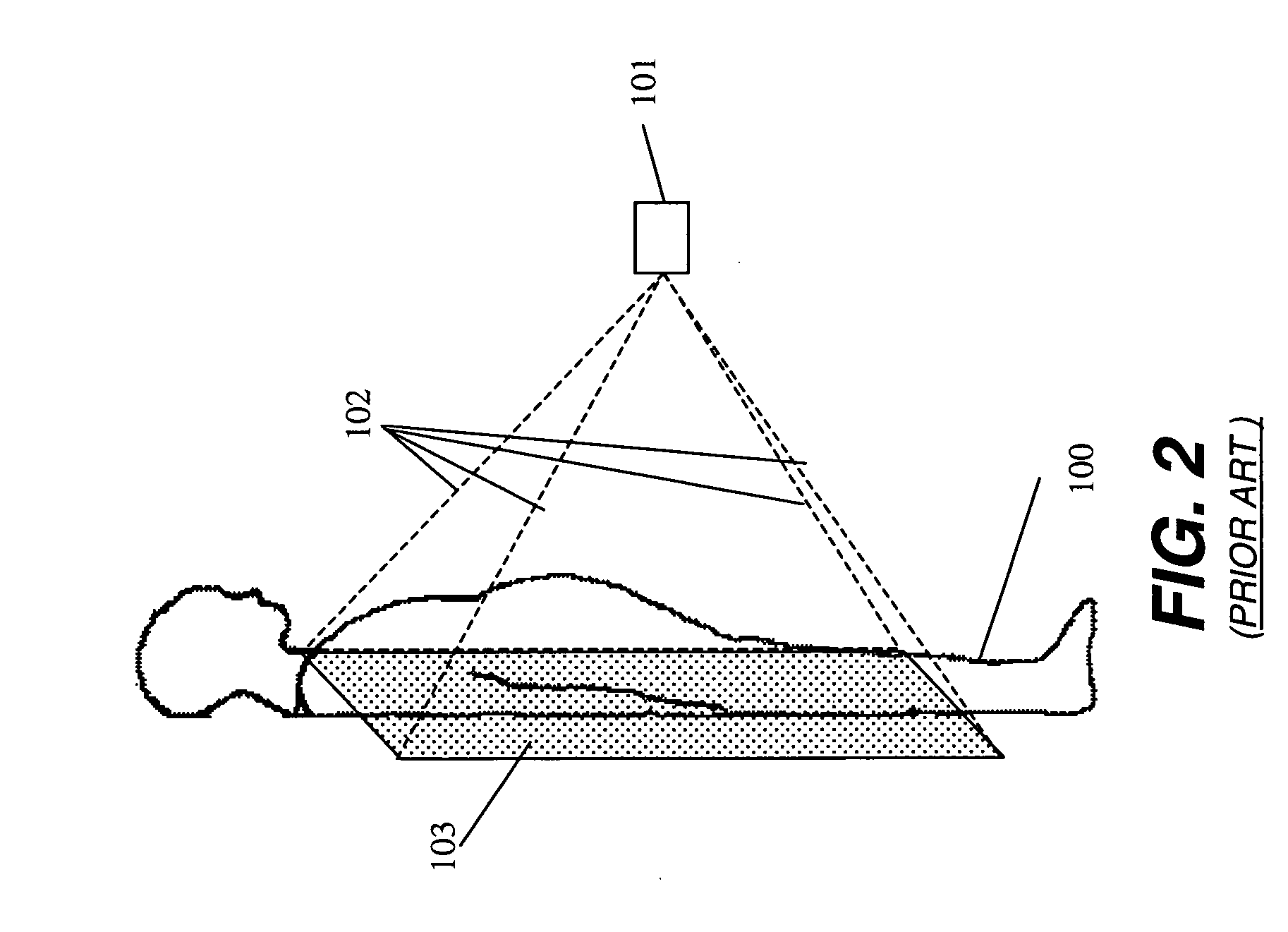
A detector of this size can image only a portion of the body part at a time and so is inadequate for performing imaging exams of longer length body parts such as the full spinal column or full leg.
The handling and identifying of the unprocessed cassette data that is not yet scanned creates workflow and data collection issues at some sites.
However, the cost and technology constraints of DR panels limit detector size and complicate the task of long length imaging.
For example, an apparatus providing movement of both the detector and the x-ray tube can be mechanically complex.

Method used


Image

  • Long length imaging using digital radiography
  • Long length imaging using digital radiography
  • Long length imaging using digital radiography

Examples


Embodiment Construction

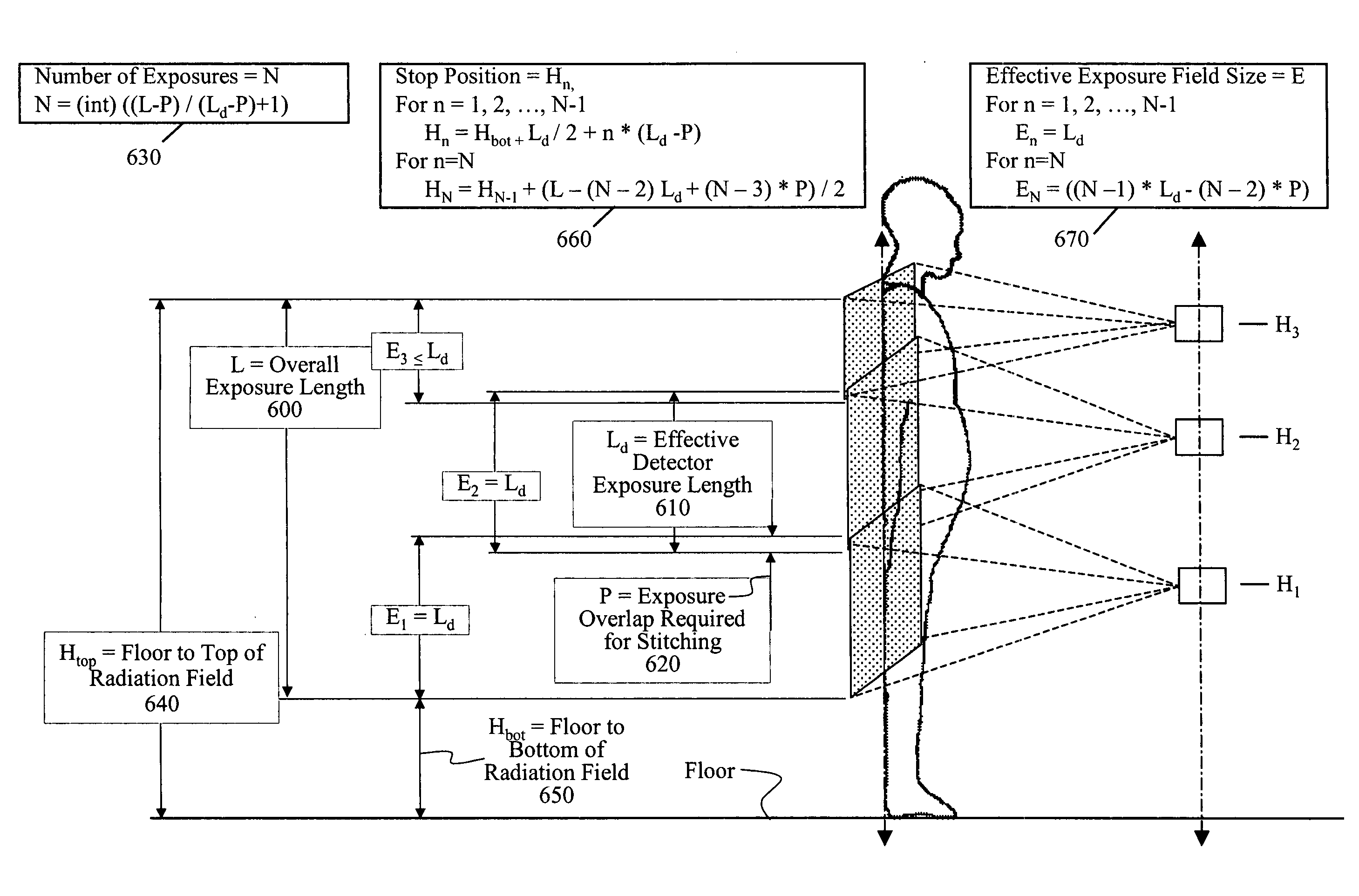
[0035]The present invention provides a method and apparatus that automate long length imaging using a DR imaging apparatus and minimize operator interaction during the imaging sequence. The present invention provides for a long length imaging sequence to be executed once set up and initiated by the operator, and provides automatic adjustment of exposure controls during the long length imaging session. It is to be understood that elements not specifically shown or described herein may take various forms well known to those skilled in the art.

[0036]FIG. 14 shows an imaging apparatus 50 suitable for practicing the method of the present invention. A collimator 62 is provided which can change it opening size by an actuator 66 that is in communication with a control logic processor 54. A sensor 64 indicates the position of collimator 62 to control logic processor 54. Processor 54 can be in communication with a display 56. Other sensing apparatus (not shown in FIG. 14) provide information ...


PUM


Abstract

A method for long length imaging with a digital radiography apparatus. Setup instructions are obtained for the image and a set of imaging positions is calculated for an exposure series according to the setup instructions. An operator command is obtained to initiate an imaging sequence. The imaging sequence is executed for each member of the set of imaging positions in the exposure series by automatically repeating the steps of positioning a radiation source and a detector at a location corresponding to the specified member of the set of imaging positions and obtaining an image from the detector at that location and storing the image as a partial image. The long length image is generated by combining two or more partial images.

Description

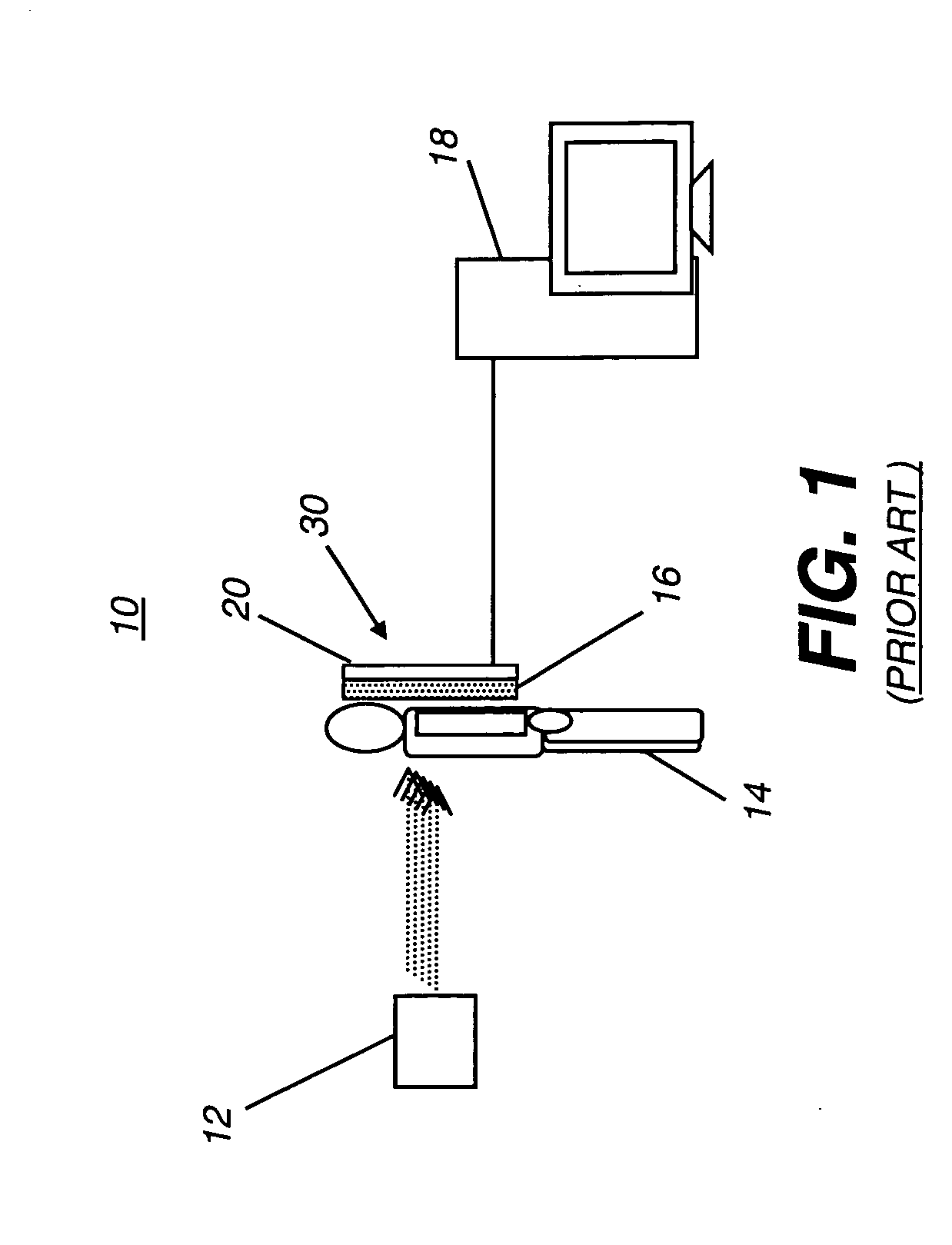
FIELD OF THE INVENTION[0001]The invention generally relates to the integration of digital radiography projection x-ray hardware and software systems, and more particularly relates to a system and method for imaging a long length body part.BACKGROUND OF THE INVENTION[0002]Digital Radiography (DR) systems are being employed in medicine and industry, with particular value as clinical imaging tools. As shown in the simplified block diagram of FIG. 1 of a prior art system, radiation from a radiation source 12 in a DR imaging apparatus 10 is directed through a subject 14 and impinges on a radiation detector 30 that includes a scintillator screen 16 for converting the energy from ionized radiation into light radiation having a different frequency, typically within the visible spectrum, and an image sensing array 20. Image sensing array 20, typically mounted on the backplane of scintillator screen 16 or otherwise optically coupled with scintillator screen 16, forms a digital image from the ...

Claims


Application Information

Patent Timeline
IPC IPC(8): H05G1/64
CPCA61B6/02A61B6/06A61B6/4441A61B6/4476H04N5/32A61B6/542A61B6/545G03B42/02H04N5/2624A61B6/5241
Inventor WANG, XIAOHUIRUSCIO, RICHARD
Owner CARESTREAM HEALTH INC
Why Eureka
Try Eureka