
Apparatus and methods for early stage peritonitis detection and for in vivo testing of bodily fluid

a peritonitis and in vivo testing technology, applied in the field of medical diagnostic testing, can solve the problems of only being able to do tests in the lab, end the patient's ability to stay on apd and capd therapy, and vomiting, so as to facilitate the cleaning of the inner wall of the effluent, the effect of increasing the turbulence of the effluent flow

Inactive Publication Date: 2008-07-31
FRESENIUS MEDICAL CARE HLDG INC
View PDF18 Cites 63 Cited by
  • Summary
  • Abstract
  • Description
  • Claims
  • Application Information

AI Technical Summary

Benefits of technology

[0028]Still further aspects of the invention provide apparatus as described above wherein the chamber comprises a deflector disposed in the flow path. The deflector can increase turbulence of effluent flowing in the chamber, increasing the Reynold's number of the fluid, e.g., to a value of about 100 or greater and, more preferably, of about 250 or greater. In these and other aspects of the invention, turbulence introduced into the flow as a result of the deflector facilitates cleaning its inner walls—particularly, for example, removing from them white blood cells, red blood cells and other components of the effluent.

Problems solved by technology

Often caused by introduction of bacteria (e.g., from the tubing, connectors and other apparatus that make up the PD transfer set) to the peritoneum during dialysis, this swelling of the peritoneum can cause vomiting, abdominal tenderness and a host of other symptoms.
Although responsive to antibiotics, peritonitis can end a patient's ability to stay on APD and CAPD therapies.
In extreme cases, it can be be fatal.
Largely, these tests can only be done in the lab, after a patient has presented with symptoms.
By that time, the peritonitis may well have set in, resulting in undue patient distress and potentially necessitating more extensive treatment.
However, test strips have a limited time window of utility and have generally not been successful in early stage detection.
This can be late-developing, unfortunately, and is further compounded if the PD solution remains in the body for a long period before expulsion (as is the case, for example, during daytime dwells of APD patients).
Detection of turbid effluent is further complicated in APD equipment with long drain lines, since patients may only see the effluent lines and not the effluent bag (where the turbidity is more readily apparent).
Implementations of this technique have generally not proven reliable because of poor signal-to-noise.

Method used


Image

  • Apparatus and methods for early stage peritonitis detection and for in vivo testing of bodily fluid
  • Apparatus and methods for early stage peritonitis detection and for in vivo testing of bodily fluid
  • Apparatus and methods for early stage peritonitis detection and for in vivo testing of bodily fluid

Examples


Embodiment Construction

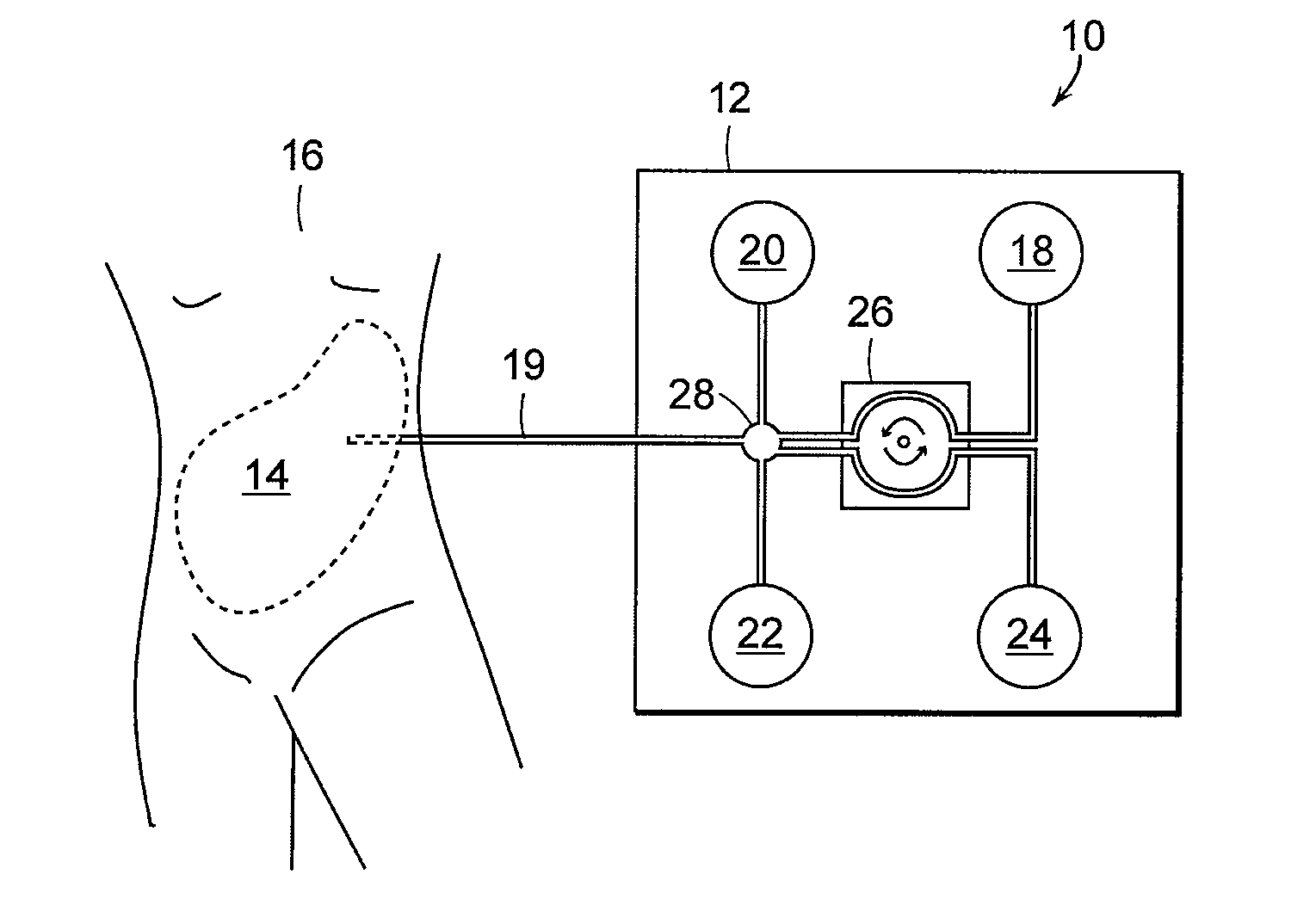
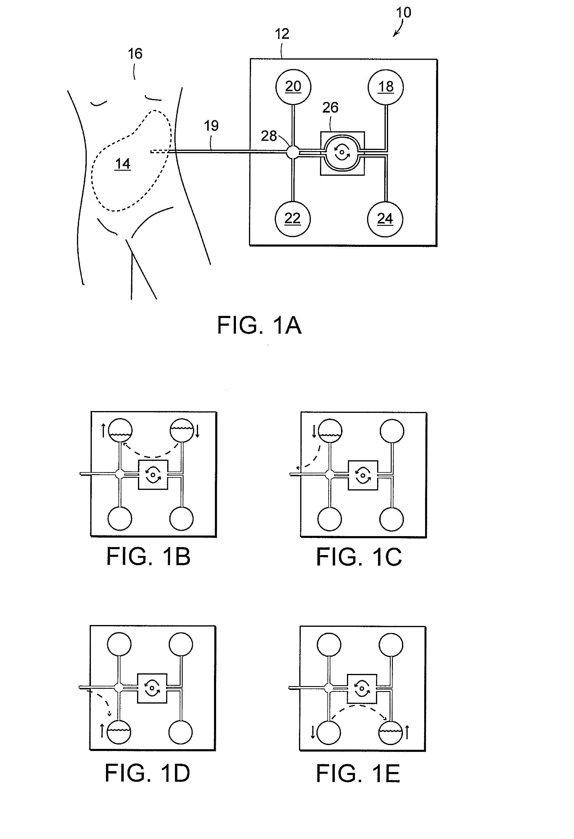
[0053]FIG. 1A depicts an automated peritoneal dialysis (APD) treatment system 10 according to one practice of the invention and of the type with which the invention can be practiced. The system 10 includes a cycler 12 or other apparatus to facilitate introducing fresh peritoneal dialysis (PD) solution into, and removing spent PD solution from, the peritoneum 14 of a patient 16.

[0054]The system 10 includes a PD solution supply chamber 18, a heating chamber 20, a weigh chamber 22, and a disposal chamber 24, all constructed an operated in the conventional manner known in the art (albeit as adapted for inclusion of PD effluent test apparatus as discussed elsewhere herein). Thus, PD supply chamber 18 holds a supply of fresh PD solution for delivery to the patient 16; heating chamber 20 brings the fresh PD solution to an appropriate temperature for delivery to the peritoneum; weigh chamber 22 hold spent PD solution expelled from the peritoneum, e.g., for weighing; and, disposal chamber 24...


PUM


Abstract

The invention provides, inter alia, automated medical methods and apparatus that test PD effluent in a flow path (e.g., with an APD system or CAPD setup) to detect, for example, the onset of peritonitis, based on optical characteristics of the effluent resolved at cellular scales of distance. For example, according to one aspect of the invention, an APD machine includes, in an effluent flow path, apparatus for early stage peritonitis detection comprising an illumination source and a detector. The source is arranged to illuminate peritoneal effluent in a chamber that forms part of the flow path, and the detector is arranged to detect illuminant scattered by the effluent. The detector detects that reflected or scattered illuminant at a cellular scale of resolution, e.g., on a scale such that separate cellular-sized biological (or other) components in the effluent can be distinguished from one another based on scattering events detected by the detector. Other aspects of the invention provide automated medical testing methods and apparatus that detect the onset of peritonitis and other bodily conditions by testing fluids in the body in vivo, e.g., the patient's peritoneum. Such apparatus and methods utilize a first fiber optic bundle to carry illuminant from a source of the type described above into a bodily organ or cavity, and a second fiber optic bundle to carry illuminant scattered by fluid in that organ or cavity to a detector as described above.

Description

[0001]This application is a continuation in part of U.S. patent application Ser. No. 11 / 880,656, filed Jul. 23, 2007, entitled “Early Stage Peritonitis Detection Apparatus and Methods,” which claims the benefit of U.S. Provisional Patent Application Ser. No. 60 / 833,763, filed Jul. 27, 2006, entitled “Early Stage Peritonitis Detection Apparatus and Methods,” the teachings of which are incorporated herein by reference.BACKGROUND OF THE INVENTION[0002]The invention relates to apparatus and methods for medical diagnostic testing. It has application, inter alia, in detecting the onsite of peritonitis, for example, during continuous ambulatory peritoneal dialysis (CAPD) and automated peritoneal dialysis (APD) procedures.[0003]Peritoneal dialysis (PD) is a medical procedure for removing toxins from the blood that takes advantage of the semi-permeable membrane surrounding the walls of the abdomen or peritoneal cavity. During a PD procedure, a solution is introduced into the patient's abdome...

Claims


Application Information

Patent Timeline
IPC IPC(8): A61M1/28
CPCA61B5/0059A61M2205/331A61M1/28A61M1/285
Inventor LANDHERR, FRANK J.LAN, JAY M.
Owner FRESENIUS MEDICAL CARE HLDG INC
Learn More