

Quantitation of cellular DNA and cell numbers using element labeling
a cell number and element labeling technology, applied in the field of rapid and sensitive assays, can solve the problems of relatively low fluorescence enhancement of reagents upon binding nucleic acids, low extinction coefficient, and high intrinsic background fluorescen
- Summary
- Abstract
- Description
- Claims
- Application Information
AI Technical Summary
Problems solved by technology
Method used
Image

Examples
experiment 1
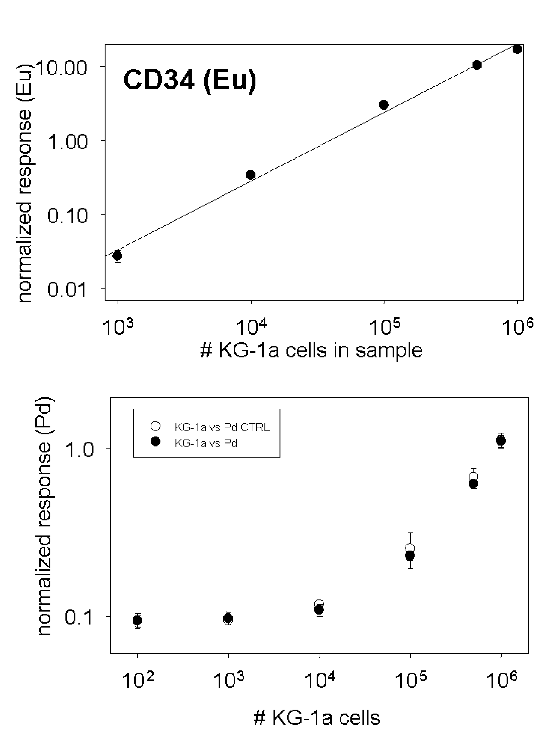
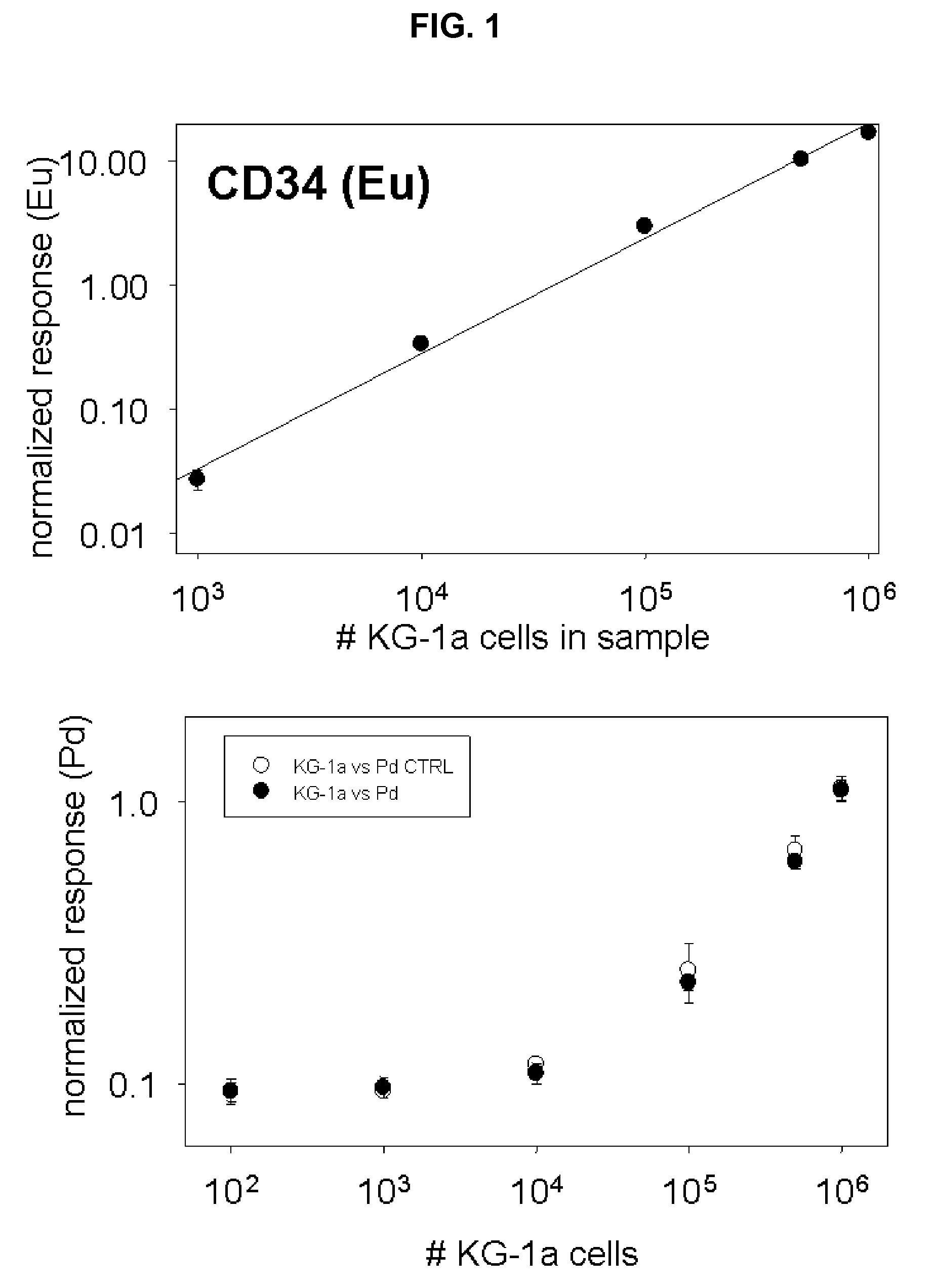
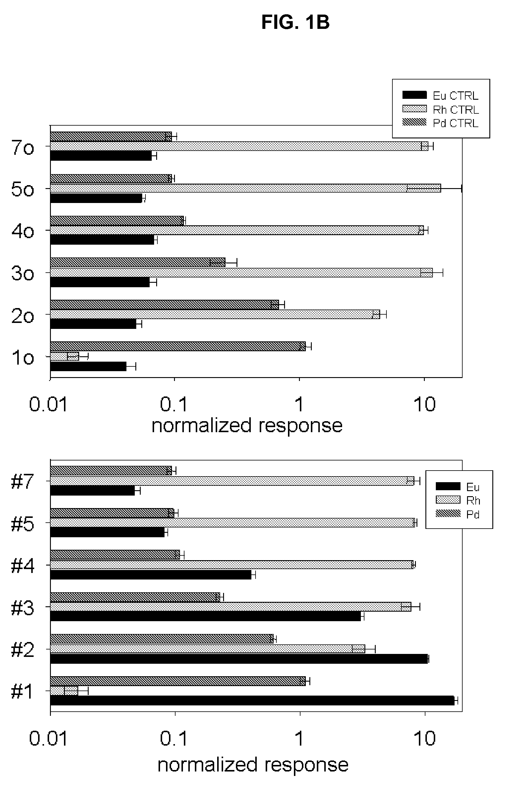
[0043]One embodiment can be demonstrated by example of rhodium (Rh) and palladium (Pd) in mixed cell populations with surface marker identified by europium (Eu). The experiment was set up to investigate if metals passively accumulated by cells could be indicative of cell number and cell type. A work flow chart is presented in FIG. 7. For this purpose two different leukemia cell lines were cultured for 72 hours in growth media supplemented with very low amounts of rare earth metals: 100 ppb Rh was added to MBA-1 cells, and 500 ppb Pd was added to KG-1a cells which express the cell surface marker. During the three days of cell culture, aliquots of cells were tested for viability with trypan blue. No excessive cell death was recorded due to the presence of metals in media. Cells were collected by centrifugation (1200 rpm, 10 minutes), washed once in PBS / 10% FBS and counted in a hemocytometer. Serial dilutions were prepared to give 1×10⁶, 0.5×10⁶, 1×10⁵, 1×10⁴, 1×10³, and 1×10² KG-1a ce...
experiment 2
[0046]Yet another embodiment can be demonstrated by example of rhodium-containing (Rh(III)) metallointercalator binding to cells. Binding of an intercalator to DNA is governed by the law of mass action. Assuming that a mammalian cell in G1 phase has 3.3×10⁹ DNA base pairs of which only 10-70% are free from protein interaction (nucleosomes) and available for binding with the intercalator (one molecule per two base pairs at saturation), the number of molecules of dye needed is approximately 1-10×108. Thus for [Rh(phi)2bpy]Cl3, with 671 MW, 1 ml of 200 uM solution should be 100-fold excess of intercalator over DNA binding sites for 1×10⁶ cells. The binding of various amounts of the Rh(III) intercalator with cultured cells was assessed. MBA-1 cells were fixed in 3.7% formaldehyde / PBS for 15 minutes, followed by permeabilization with 0.3% TritonX-100 / PBS; washed once with PBS and incubated for 45 minutes with 1 ml of increasing concentrations of [Rh(phi)2bpy]Cl3 from 2.5 nanoM to 250 mic...
experiment 3
[0048]Yet another embodiment can be demonstrated by example of DNA labeling with Rh(III) metallointercalator and antigen CD33 surface labeling using solution and particle elemental analysis as shown in FIG. 4 and FIG. 9.
[0049]MBA-1 cells were fixed / permeabilized with 3.7% formaldehyde and 0.3% Triton X-100 and labeled with Rh(III) intercalator (250 microM). Triplicate samples with 3×10⁴, 3×10⁵, and 3×10⁶ cells per tube were prepared. Cells were then incubated with the primary anti-CD33 antibody, followed by secondary Eu-labeled anti-mouse antibody. Cells were washed in 0.65 um centrifugal spin filters (Durapore). After the final wash, filter units were placed over new eppendorf tubes and 80 microL of concentrated HCl were added. Overnight digestion of cellular material was followed by centrifugation and the filtrate was collected, combined with equal volume of Ir internal standard and subjected to ICP-MS. FIG. 4 shows simultaneous identification of Rh (representing cell numbers) and...
PUM
| Property | Measurement | Unit |
|---|---|---|
| Fraction | aaaaa | aaaaa |
| Fraction | aaaaa | aaaaa |
| Concentration | aaaaa | aaaaa |
Abstract
Description
Claims
Application Information