Anti-restenotic therapeutic device

a technology of anti-restenotic and therapeutic devices, which is applied in the direction of prosthesis, surgery, blood vessels, etc., can solve the problems of ischemic events, significant clinical problems, and restnosis after percutaneous coronary intervention, and achieve the effect of promoting beneficial cell growth and enhancing the growth of a type of cells

Inactive Publication Date: 2008-08-21
NFOCUS NEUROMEDICAL
View PDF14 Cites 72 Cited by
  • Summary
  • Abstract
  • Description
  • Claims
  • Application Information

AI Technical Summary

Benefits of technology

[0005]An anti-restenotic device is provided for repairing a tissue, particularly an arteriosclerosed blood vessel or a damaged wall of a luminal or chambered organ. In some embodiments, the device comprises a structure having a first surface and a second surface. A bioactive layer is disposed on the first surface, wherein the bioactive layer enhances growth of a type of cells thereon. And an anti-restenosis layer is disposed on the second surface, wherein the anti-restenosis layer inhibits growth of another type of cells thereon. Thus, positioning of the structure within a blood vessel (so that the first surface faces the lumen) promotes beneficial cell growth, such as endothelial cell growth, to form a more blood-compatible lining. At the same time, the second surface inhibits ingrowth of undesirable cells which lead to restenosis, such as smooth muscle cells.
[0006]Such an anti-restontic device can also be used to repair other tissues, such as hernias, hepatic ducts, meninges, lung passageways, patent foramen ovale, atrial septal defects, and tracheal bronchial strictures, to name a few. For example, when repairing a hernia, the device may have the form of a patch which is positionable against the herniated muscle layer. The first surface which contacts the herniated muscle layer promotes beneficial cell growth, such as muscle cell ingrowth. This assists in holding the patch in place. The second surface which faces away from the herniated muscle layer inhibits growth of cells which lead to adhesions.
[0009]In some embodiments, the structure further comprises at least one security ring configured to secure the sleeve to the support frame. In such embodiments, the second surface may be disposed on at least one surface of the at least one security ring. Further, the first surface is typically disposed on an inner surface of the sleeve. A single security ring may be used, or multiple security rings may be spaced along the device allowing greater flexibility of the device. The rings could also align with specific portions of the underlying expandable support frame, such as alternating gaps in the internal member cells or separate sections, to provide even greater flexibility. Thus, in some embodiments, the security rings serve two purposes, to hold the sleeve in place and to deliver the anti-restenotic agent. Because of this, the rings may have very low radial strength, which would lead to a more flexible / desirable device.

Problems solved by technology

The material accumulates over time and can eventually impede the flow of blood.
When disturbed, this material can become dislodged and occlude blood flow thereby causing an ischemic event.
However, the cells can allow the friable diseased material to extrude into the vessel lumen and may break off to cause harmful emboli.
However, restenosis after percutaneous coronary intervention is a significant clinical problem, occurring after 15% to 30% of angioplasty procedures or intracoronary stenting.
This is particularly problematic in saphenous vein grafts used in coronary bypass surgery.

Method used


Image

  • Anti-restenotic therapeutic device
  • Anti-restenotic therapeutic device
  • Anti-restenotic therapeutic device

Examples


Embodiment Construction

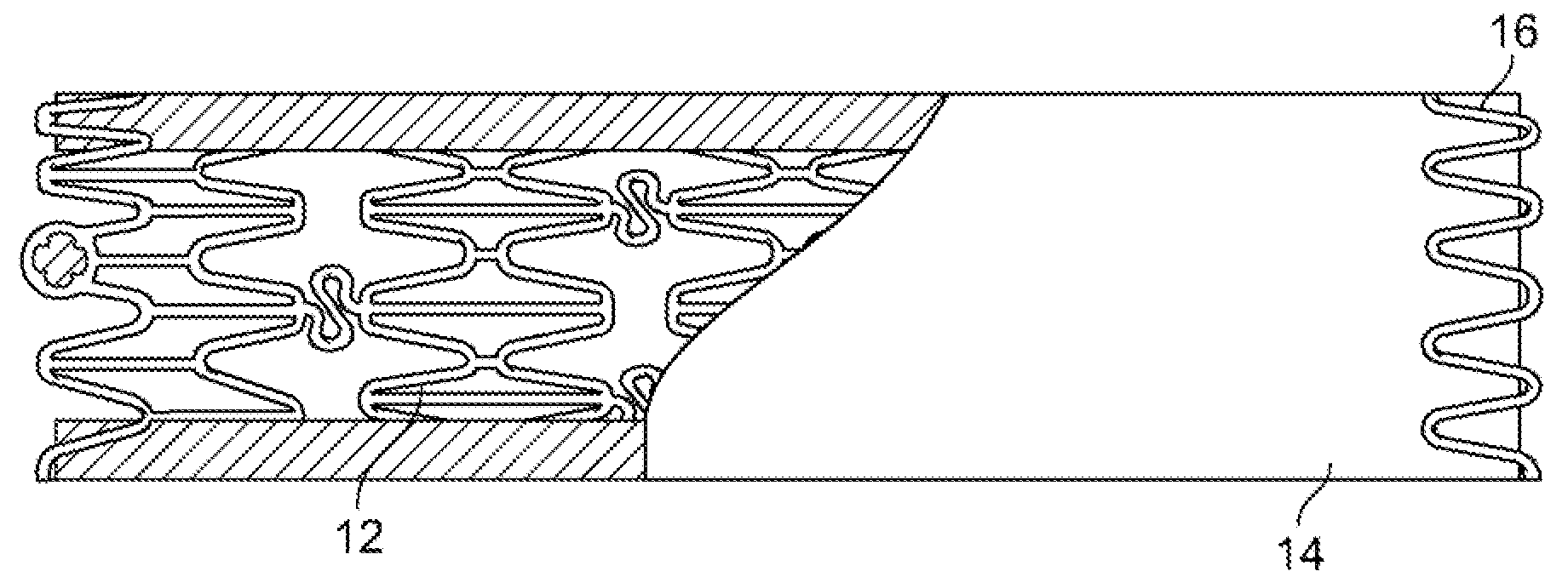
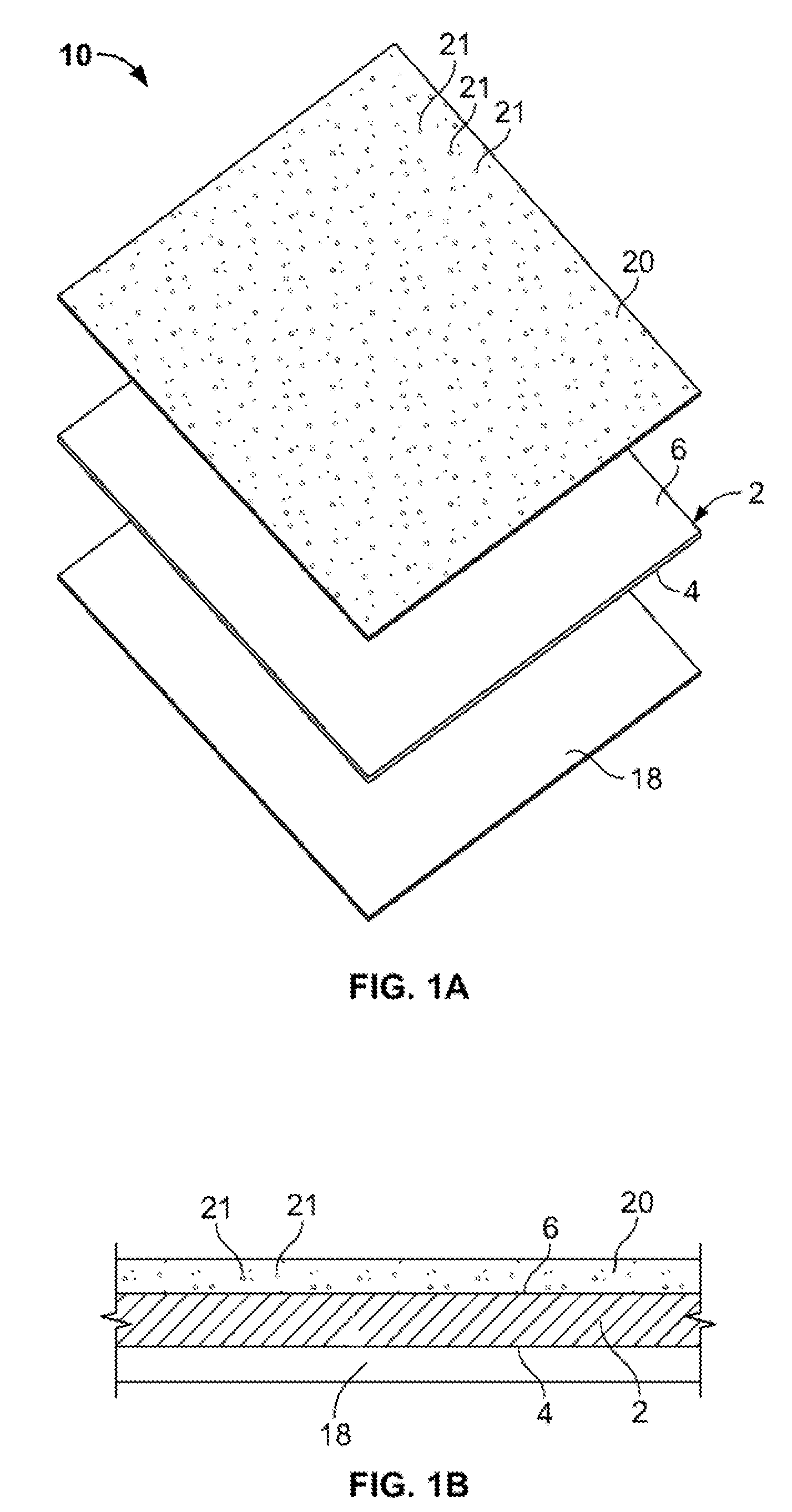
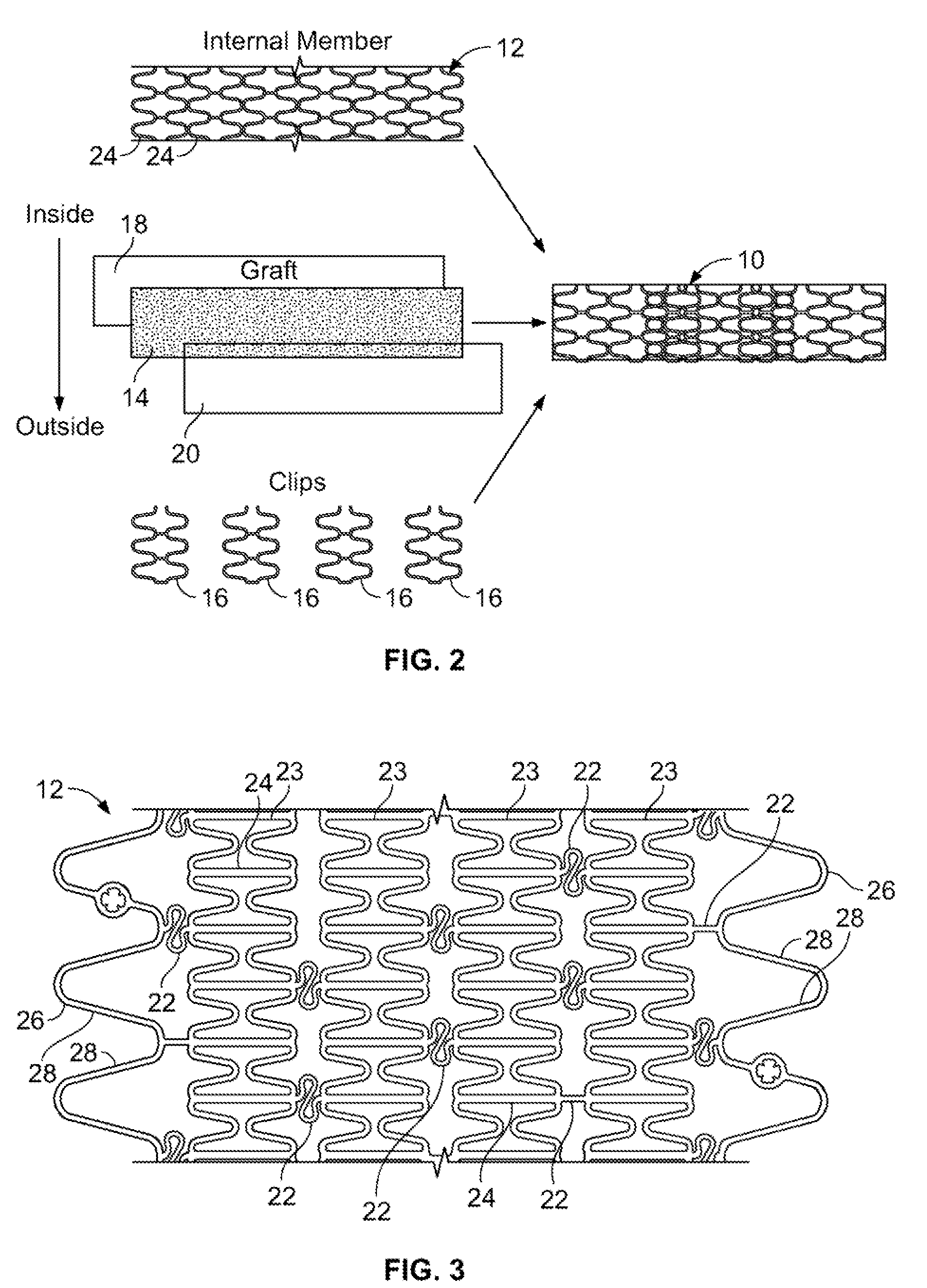
[0027]An anti-restenotic device is provided for repairing a tissue. Such a device may take a variety of forms. Examples include a patch, a sheet, a tube, a pocket, a sleeve, a stent, a graft-stent, and, particularly, a composite expandable device. FIGS. 1A-1B illustrate an example of a device 10 having the form of a patch or sheet. Here the device 10 comprises a structure 2 having a first surface 4 and a second surface 6. The device 10 further comprises a bioactive layer 18 disposed on the first surface 4, wherein the bioactive layer 18 enhances growth of a type of cells thereon. The device 10 further includes an anti-restenosis layer 20 disposed on the second surface 6, wherein the anti-restenosis layer inhibits growth of another type of cells thereon. The anti-restenosis layer includes an anti-restenosis agent 21 which is eluted therefrom as will be discussed in later sections. The structure 2 and layers 4, 6 are shown separated for illustration purposes. FIG. 1B provides a cross ...


PUM

PropertyMeasurementUnit
thicknessaaaaaaaaaa
timeaaaaaaaaaa
bioactiveaaaaaaaaaa

Abstract

An anti-restontic device is provided for repairing a tissue, particularly an arteriosclerosed blood vessel or a damaged wall of a luminal or chambered organ. In some embodiments, the device comprises a structure having a first surface and a second surface. A bioactive layer is disposed on the first surface, wherein the bioactive layer enhances growth of a type of cells thereon. And an anti-restenosis layer is disposed on the second surface, wherein the anti-restenosis layer inhibits growth of another type of cells thereon.

Description

CROSS-REFERENCE TO RELATED APPLICATIONS[0001]This application is a continuation of co-pending International Application No. PCT / US2006 / 031059 filed Aug. 10, 2006 which claimed benefit of priority to U.S. Provisional Application No. 60 / 707,296 filed Aug. 10, 2005, both applications of which are incorporated herein by reference in their entirety.BACKGROUND OF THE INVENTION[0002]Vascular disease often presents as friable material on the inner lumens of blood vessels throughout the body, particularly the coronary arteries. The material accumulates over time and can eventually impede the flow of blood. When disturbed, this material can become dislodged and occlude blood flow thereby causing an ischemic event. The primary minimally invasive method of treating this disease is to open the vessel mechanically while preventing the embolic material from being dislodged causing further harm.[0003]Two types of mechanical implants are typically used to open the vessels, stents and stent-grafts. S...

Claims


Application Information

Patent Timeline
Patent Type & Authority Applications(United States)
IPC IPC(8): A61F2/06
CPCA61F2/0077A61F2/07A61F2/91A61F2002/075A61F2/915A61L31/16A61L2300/252A61L2300/412A61L2300/416A61F2250/0051
Inventor DIECK, MARTIN S.DEBEER, NICHOLAS C.
Owner NFOCUS NEUROMEDICAL
Why Eureka
Try Eureka