Biomolecule immobilization on surface via hydrophobic interactions

a technology of hydrophobic interactions and biomolecules, applied in the field of genetic analysis, can solve problems such as difficult tasks

Inactive Publication Date: 2008-10-30
APPL BIOSYSTEMS INC
View PDF13 Cites 22 Cited by
  • Summary
  • Abstract
  • Description
  • Claims
  • Application Information

AI Technical Summary

Problems solved by technology

However, the complexity of the human genome and the interrelated functions of genes often make this task difficult.

Method used


Image

  • Biomolecule immobilization on surface via hydrophobic interactions
  • Biomolecule immobilization on surface via hydrophobic interactions
  • Biomolecule immobilization on surface via hydrophobic interactions

Examples


example 1

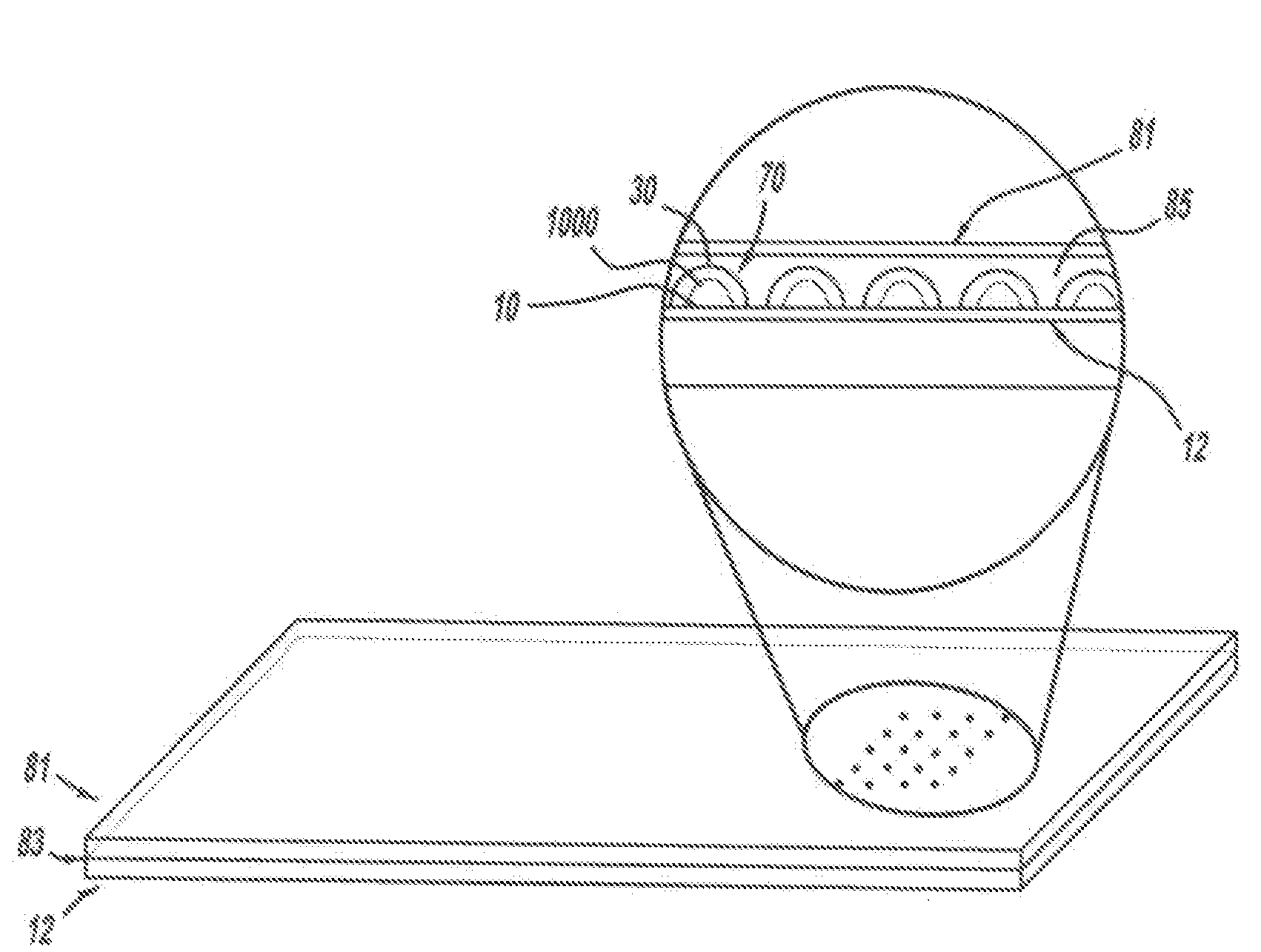
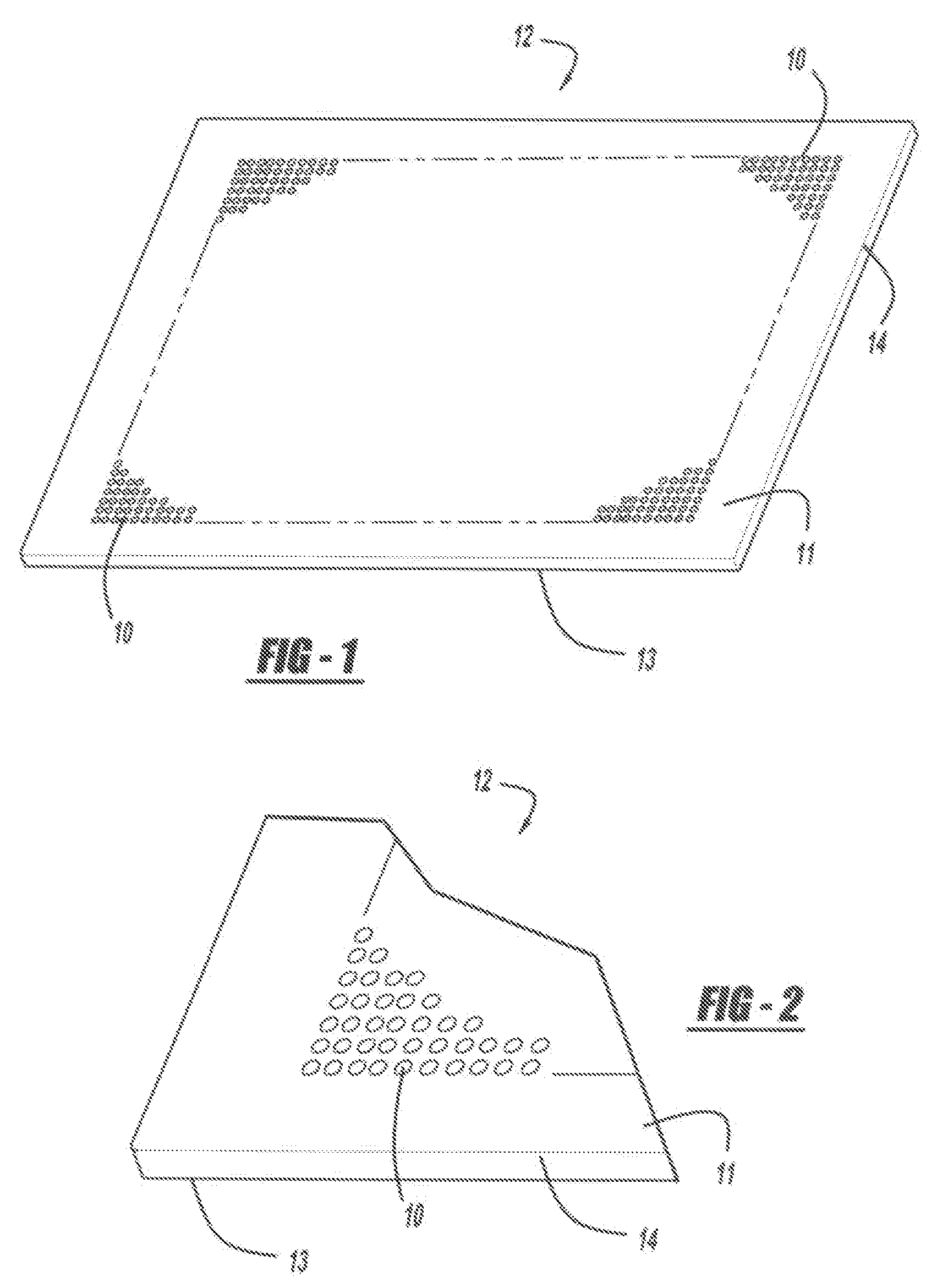
[0123]An exemplary amplification method of these teachings is performed using a surface-treated microscope slide, supplied by Scienion AG (Berlin, Germany), on which discrete reaction spots comprising hydrophilic areas are created. Each reaction spot is essentially circular in shape, having a diameter of about 160 μm. An array of 30,000 reaction spots is formed on the surface of the slide. Sets of PCR primers and detection probes, for hybridizing with known oligonucleotides, such as, for example, polynucleotide targets, are then deposited on the hydrophilic areas of the reaction spots and covalently linked to the reaction spots through a cleavable disulfide linker, forming reaction spots. A unique set of primers and detection probes is deposed on each reaction spot.

[0124]A sample containing a mixture of polynucleotide is then flooded across the surface of the slide, contacting the reaction spots. The sample is allowed to incubate for about twelve hours, after which excess sample is ...

example 2

[0125]A microplate is made according to these teachings by applying discrete reaction spots of agarose onto a polycarbonate plastic substrate. A solution is made comprising 3% (by weight) of agarose having a melt point≦65° C., supplied as NuSieve GTG, by FMC BioProducts (Rocland, Me., USA). The solution is then spotted onto the surface of the substrate in an array comprising 15,000 reaction spots. The microplate is then used in a method according to Example 1. In this method, high resolution blend agarose 3:1, and monoclonal anti-biotin-agarose, supplied by Sigma (St. Louis, Mo., USA) can be substituted for the low melt agarose, with substantially similar results. In some embodiments, biotinylated polynucleotides such as primers and detection probes are used.

example 3

[0126]A microplate is made according to these teachings, by cutting an optical adhesive cover comprising a plastic material, to the size of a standard glass microscope slide, and pasting the cover to the standard glass microscope slide. Heat and pressure is applied while smoothing the cover over the glass surface in order to expel air bubbles between the cover and glass surface. 2 uL droplets of 1% low melting agarose are delivered onto the plastic surface of the cover at a 4500 μm pitch in a matrix and dried at low heat on a hot plate to create a plurality of reaction spots. The plastic surface is rinsed with deionized water. A matrix of water droplets is retained on the reaction spots on the plastic surface when the excess of water was removed. 2 uL of RNase P TaqMan® reaction mix, supplied by Applied Biosystems (Foster City, Calif., USA) with human genomic DNA is then added onto each reaction spot and covered with mineral oil to seal the reaction spots and create reaction chamber...


PUM

PropertyMeasurementUnit
volumeaaaaaaaaaa
lengthaaaaaaaaaa
widthaaaaaaaaaa

Abstract

A method, apparatus, or system for generating a pattern of polynucleotides on a substrate. The method includes providing a substrate having a hydrophobic surface. The method further includes conjugating a polystyrene moiety to a polynucleotide and applying a polystyrene-polynucleotide conjugate to create a plurality of reaction spots on the hydrophobic surface of the substrate. An apparatus includes a substrate with at least one polystyrene-polynucleotide conjugate on a surface of the substrate. A system can analyze a polystyrene-polynucleotide conjugate and the system may perform PCR.

Description

INTRODUCTION[0001]Currently, genomic analysis, including that of the estimated 30,000 human genes, is a major focus of basic and applied biochemical and pharmaceutical research. Such analysis can aid in developing diagnostics, medicines, and therapies for a wide variety of disorders. However, the complexity of the human genome and the interrelated functions of genes often make this task difficult. There is a continuing need for methods and apparatus to aid in such analysis.DRAWINGS[0002]The skilled artisan will understand that the drawings, described herein, are for illustration purposes only. The drawings are not intended to limit the scope of the present teachings in any way.[0003]FIG. 1 is a top perspective view illustrating a plurality of reaction spots on a hydrophobic substrate in accordance with some embodiments;[0004]FIG. 2 is an enlarged perspective view illustrating a plurality of reaction spots on a hydrophobic substrate in accordance with some embodiments;[0005]FIG. 3 is...

Claims


Application Information

Patent Timeline
Patent Type & Authority Applications(United States)
IPC IPC(8): C12Q1/68C12M1/34
CPCB01J19/0046C12Q1/686B01J2219/00529B01J2219/00596B01J2219/00608B01J2219/00619B01J2219/00635B01J2219/00637B01J2219/00659B01J2219/00722B01L3/50851B01L3/5088B01L7/52B01L2300/0636B01L2300/0819C40B50/18C40B80/00C12N15/1093B01J2219/00387
Inventor LIU, TIMOTHY Z.CHEN, JER-KANGGRAHAM, RONALD J.
Owner APPL BIOSYSTEMS INC
Why Eureka
Try Eureka