

Microneedles and Methods for Microinfusion
a technology of microneedles and microinjection, which is applied in the field of transdermal therapies, can solve the problems of limited use of infusion pumps outside the clinical setting, inconvenient intradermal injection, and limited self-administration by patients or other uses outside the clinic or laboratory, and achieves the effect of being easily visibl
- Summary
- Abstract
- Description
- Claims
- Application Information
AI Technical Summary
Benefits of technology
Problems solved by technology
Method used
Image

Examples
example 1
Fluid Delivery Through Hollow Microneedles Into Human Cadaver Skin
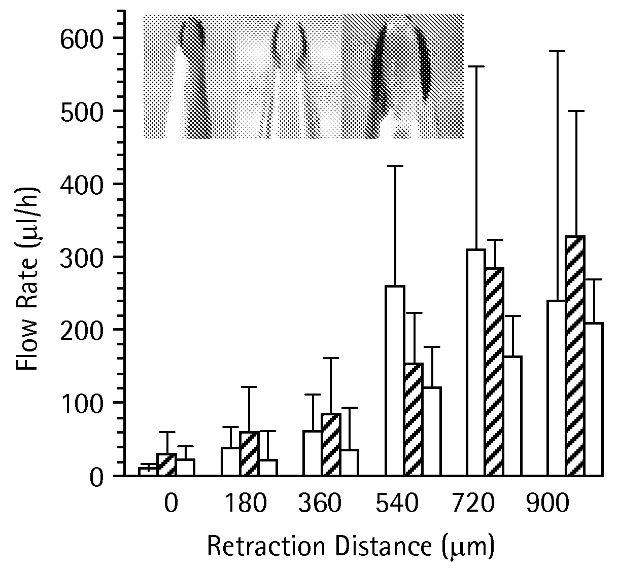
Effect of Partial Retraction Following Insertion
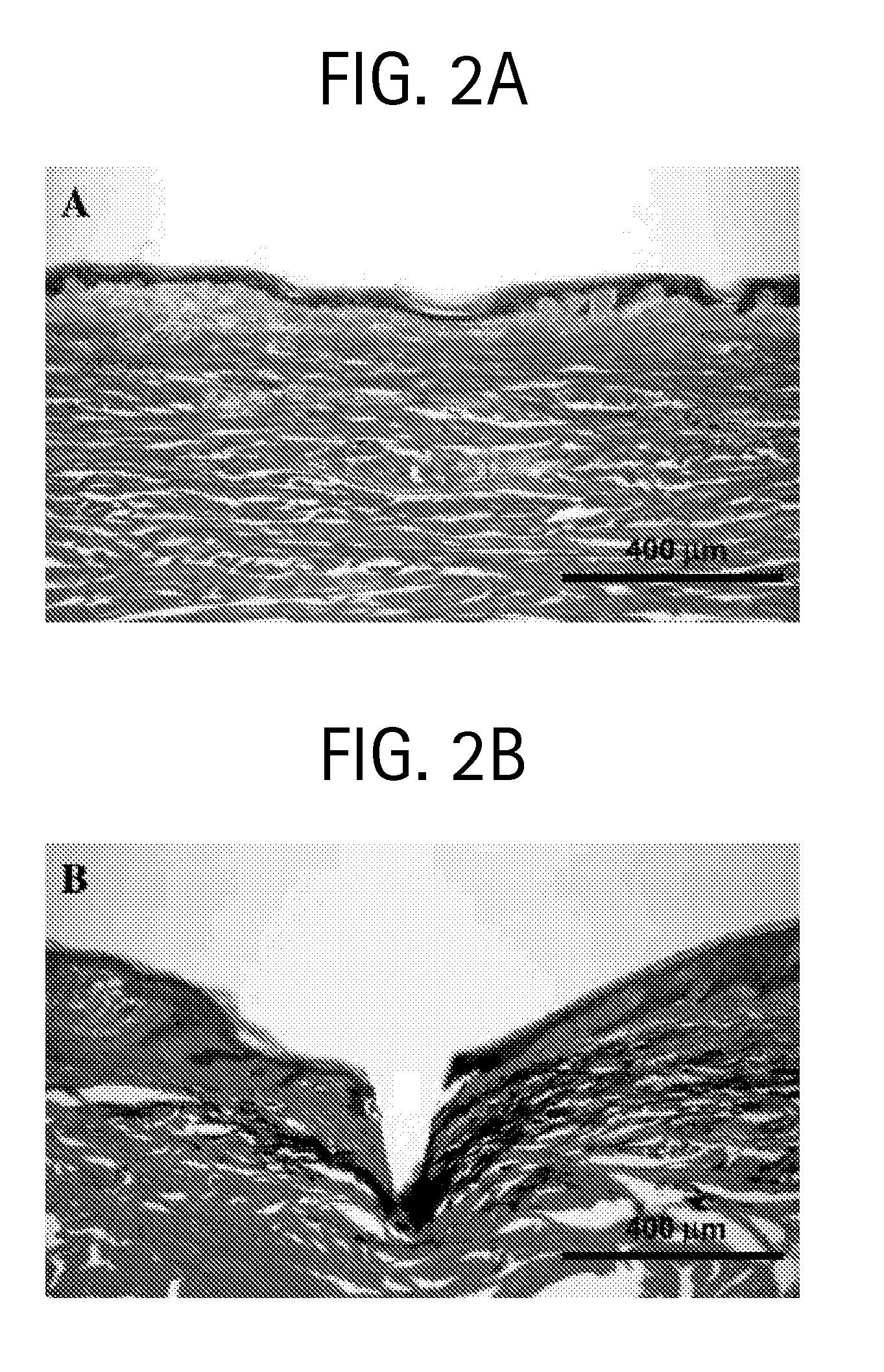
[0093]Single microneedles were inserted into human cadaver skin in vitro to a controlled depth and then, sometimes, partially retracted.
Microneedle Fabrication
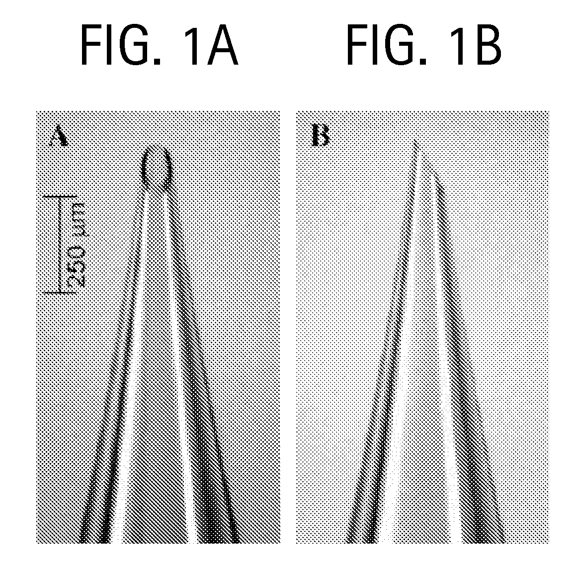
[0094]Glass microneedles were fabricated by pulling fire-polished borosilicate glass pipettes (o.d. 1.5 mm, i.d. 0.86 mm, BF150-86-15, Suffer Instrument, Novato, Calif.) using a micropipette puller (P-97, Sutter Instrument). In most cases, the resulting blunt-tip microneedles were then beveled (BV-10, Sutter Instrument) and cleaned using chromic acid (Mallinckrodt, Hazelwood, Mo.), followed by filtered DI water and acetone (J. T. Baker, Phillipsburg, N.J.) rinses. Microneedle geometries were determined by bright-field microscopy (Leica DC 300; Leica Microsystems, Bannockburn, Ill.) and image analysis (Image Pro Plus, version 4.5, Media Cybernetics, Silver Spring, Md.). The microfabricated microneedles typically had...
example 2
Effect of Hyaluronidase on Microinfusion Flow
[0110]Hyaluronidase is known to reduce flow resistance in the skin by rapidly breaking down hyaluronan, a glycosaminoglycan within skin collagen fibers (Kreil, Protein Sci 4:1666-69 (1995); Bruera, et al., Annals of Oncology 10:1255-58 (1999); Meyer, “Hyaluronidases” in The Enzymes, vol. 5, pp. 307-20 (Boyer, ed) Academic Press, New York, N.Y. (1971)). This enzyme might similarly break down the resistance of dermal tissue compressed during microneedle insertion. To test this prediction, microneedle infusion was carried out using the sulforhodamine solution described in Example 1 mixed with a purified ovine testicular hyaluronidase preparation (Vitrase™) that is commercially available and FDA approved for human use to facilitate injection when simultaneously co-injected using a hypodermic needle (Hyaluronidase (Vitrase)—ISTA. Drugs in R&D 4:194-97 (2003)).
[0111]The effect of hyaluronidase was determined by comparing to an infusion flui...
example 3
Tissue Resistance to Microinfusion Flow
[0112]The resistance to fluid flow into tumor tissue at constant pressure has been shown to decrease over time, probably due to flow-induced changes in tissue microstructure. To address this possibility during infusion using microneedles as described in Example 1 above, the flow into skin was measured continuously for 100 min for needles inserted and left in place and for needles inserted and retracted (FIG. 9). The microneedles were inserted into skin to a depth of 1080 μm and then retracted 720 μm to a final insertion depth of 360 μm (solid line) and microneedles inserted to a depth of 1080 μm without retraction (dashed line). Infusion was carried out at 138 kPa using microneedles having 35-38° beveled tips with 30-32 μm effective radius openings. Data are expressed as mean values (n≧3) with average standard deviation of 40% for both curves (not shown). In both cases, the cumulative volume of fluid infused into skin increased linearly with ti...
PUM
| Property | Measurement | Unit |
|---|---|---|
| Fraction | aaaaa | aaaaa |
| Fraction | aaaaa | aaaaa |
| Depth | aaaaa | aaaaa |
Abstract
Description
Claims
Application Information