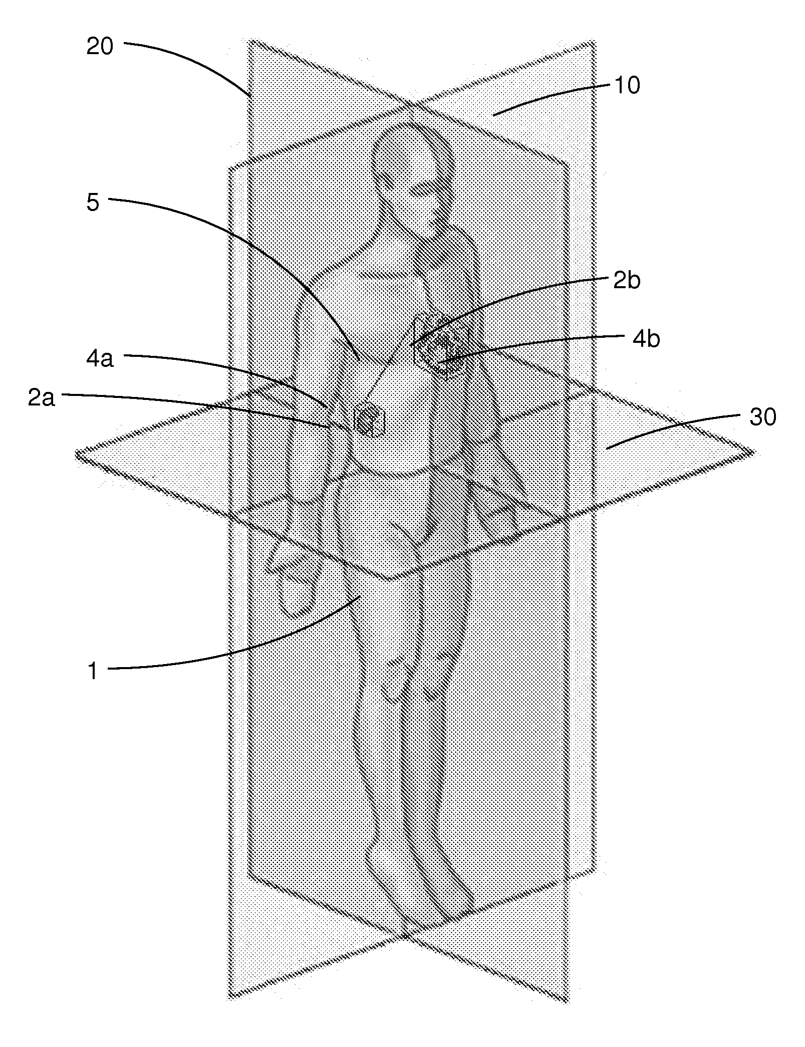
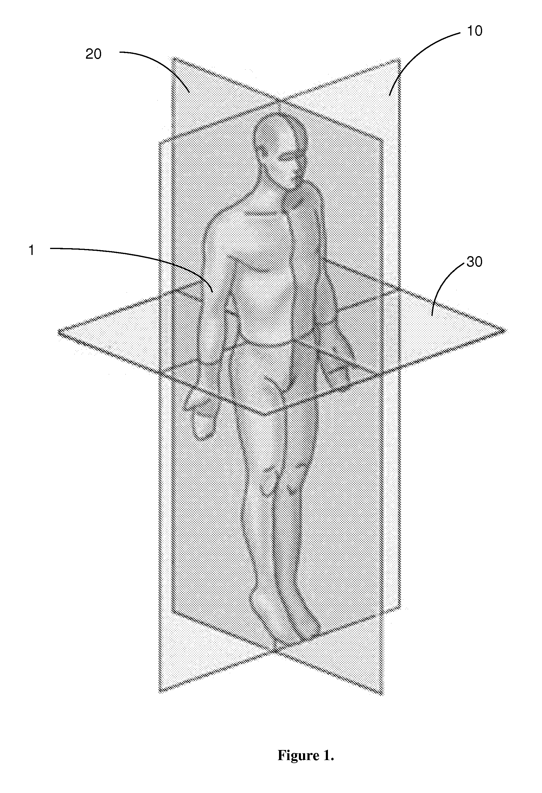
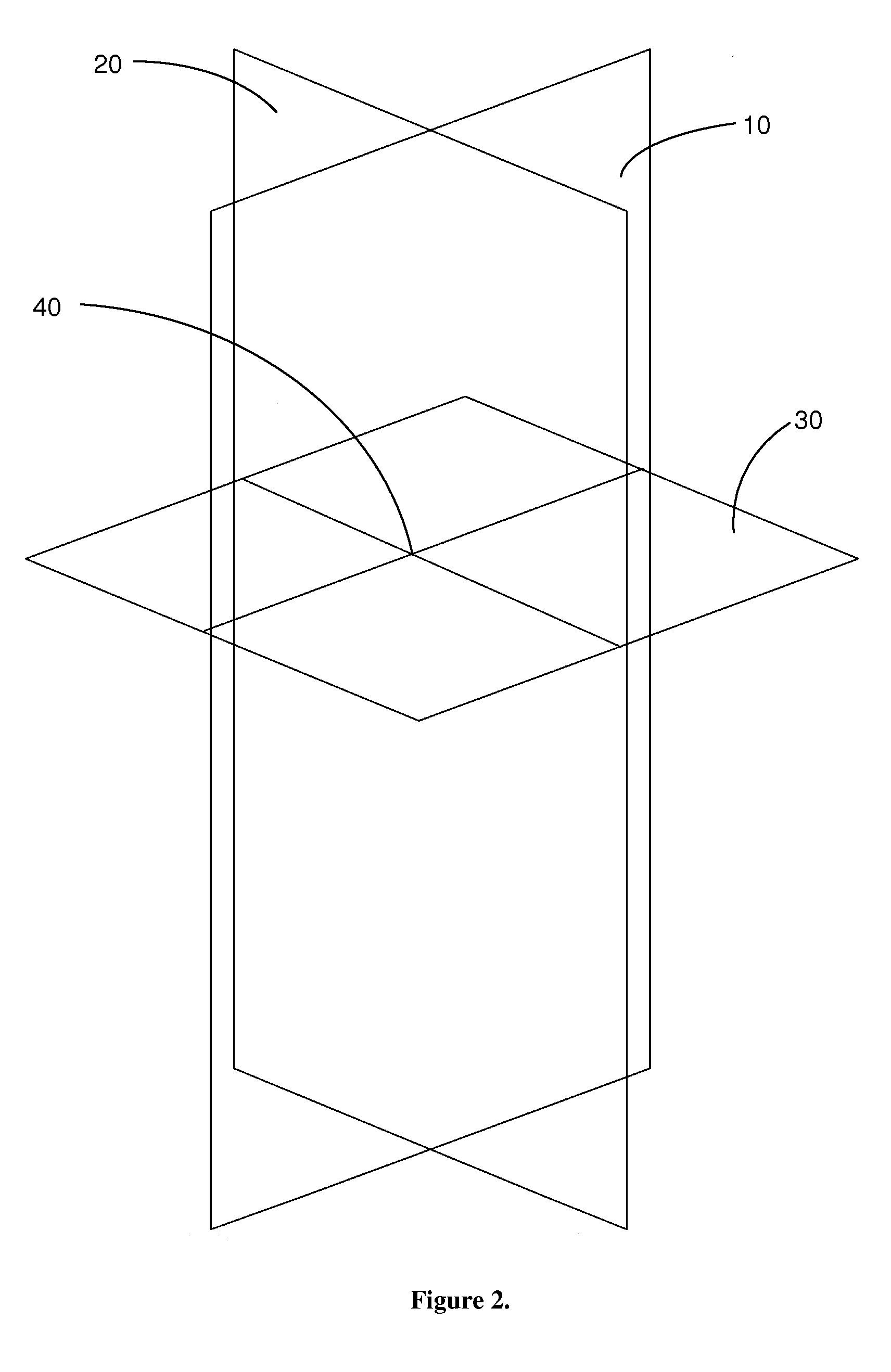
[0011]Use of a defined three dimensional coordinate
system to align and register volumetric
medical imaging data of the human body within
three dimensional space makes possible the
objective analysis of the morphometric organization of any individual human as well as the ability to compare and statistically define the morphological characteristics of populations of humans. This permits description of quantitative data of the morphometric features of the body of any individual human, relative to any set of three dimensional coordinates. Further, changes in any morphological feature over time that occur as a result of normal development, growth, aging, acute insult or progressive changes related to
disease processes may be described using the three dimensional coordinate system. Moreover, patterns related to normal development, growth, aging, acute insult or
disease processes may be documented, described, analyzed, and diagnosed using the present invention. Statistically-
derived data sets of morphometric changes, patterns or changes in patterns of
digital image data characteristics are also useful in the present invention.
[0017]Volumetric medical images are composed of arrays of rows and columns of voxels. Regardless of how that voxels are obtained (CT, MR, PET, etc) the final representation is organized in a 3D array. One of the features provided by the bounding box data approach is that the 3D
grey scale voxel array patterns can be defined for
normal conditions as well as for any and all variants of
pathological conditions. With sufficient validated data sets of
voxel grey scale array patterns, the scanning device computer may track the
anatomical structures it is actively scanning and compare the active-scan structures to a validated
database of voxel grey scan array patterns. Comparing the patient's array patterns with known patterns allows the computer to perform a real-time,
first pass differential diagnosis of the image while the patient is still on the scan table. The results of the scan may be analyzed and, if the computer requires more information to make a decision, it has the opportunity to rescan the patient with an appropriate protocol the patient is still in the
scanner.
[0020]A normative human morphology
database may be developed for all relevant structures for a
large population of normal
healthy individuals to describe complete array of statistical descriptors of the morphological features.
Medical imaging of millions of patients is performed each year. For each of these scans a radiologist provides a medical opinion as to whether the morphology is normal or abnormal. When abnormal, the
pathology is described. Using normal and validated abnormal morphology, a
database or series of databases may be developed to define the array patterns of normal
anatomical structures and
disease conditions for which
medical imaging is utilized as a diagnostic tool. This database then can be used to provide a measure of limits between normal (healthy) and abnormal (diseased /
pathological) morphological structure. The information is used to develop
software for a
first pass differential diagnosis and more efficient and accurate scanning protocols. This will ultimately reduce the number of scans a person may require, reduce unnecessary
radiation exposure, and will result in faster, cheaper and more accurate imaging processes.
[0021]Data may be stored in any format known in the art, including Picker SPECT, GE MR SIGNA—including SIGNA 3, 5, and
Horizon LX—Siemans Magnatom Vision, CTI, CTI ECAT 7, SMIS MRI, and ASI / Concorde MicroPET. The standard format for volumetric medical image data to be captured and stored is in the
DICOM format. Images in
DICOM format can be viewed, modeled and measured on a wide range of public domain and
commercial software available today. Any and all volumetric medical image data oriented as described above to a defined human morphometric coordinate system can be data mined to provide precise and comparable measurements for any and all relationships of anatomical features.
Software plug-ins for several
software packages have been developed to permit efficient mining of data from the
DICOM image sets oriented within the coordinate system. The plug-ins permit the point and click identification and storage of the 3D coordinate of specific anatomical features. Line distant length between two anatomical features can be determined. Any 2D area or 3D volume can be
user defined by a point and click approach and the volume and 3D coordinate location recorded.
[0022]If and when
volumetric image data of human morphology is placed in registration within a three dimensional coordinate system, a wide range of quantitative measurements can be made to better document the structural features of the body. When fully implemented
computer based technology will be able to determine if all internal structures are normal, and if not what are the likely medical problems. Use of the coordinate system permits quantitative description and analysis of structural features includes but are not limited to: location, volume, orientation, length and
diameter of all individual structure as well as the spatial relationships of any combination of morphological feature of the human body relative to a any three dimensional coordinate system. If a similar process is applied to a
population of humans with image data all registered with the same
three dimensional space relative to a consistent coordinate system, then detailed
statistical analysis can be conducted to establish mathematical descriptors for all levels of normal
human anatomy as well as each and every known
pathological condition that has, as one of its features, an alteration in the patient's anatomical structure. The resulting human morphometric data sets can be used in the development of
computer software for the automated analysis of medical images. The ability to quantitatively describe the structure of the human body will significantly increase the value of medical imaging in 21st century clinical
medicine. This approach will ultimately provide faster, cheaper and more accurate initial stages of differential diagnosis of a wide range of medical conditions.


