
Modification of properties and geometry of heart tissue to influence function

Active Publication Date: 2008-12-18
JAYARAMAN SWAMINATHAN
View PDF63 Cites 0 Cited by
  • Summary
  • Abstract
  • Description
  • Claims
  • Application Information

AI Technical Summary

Benefits of technology

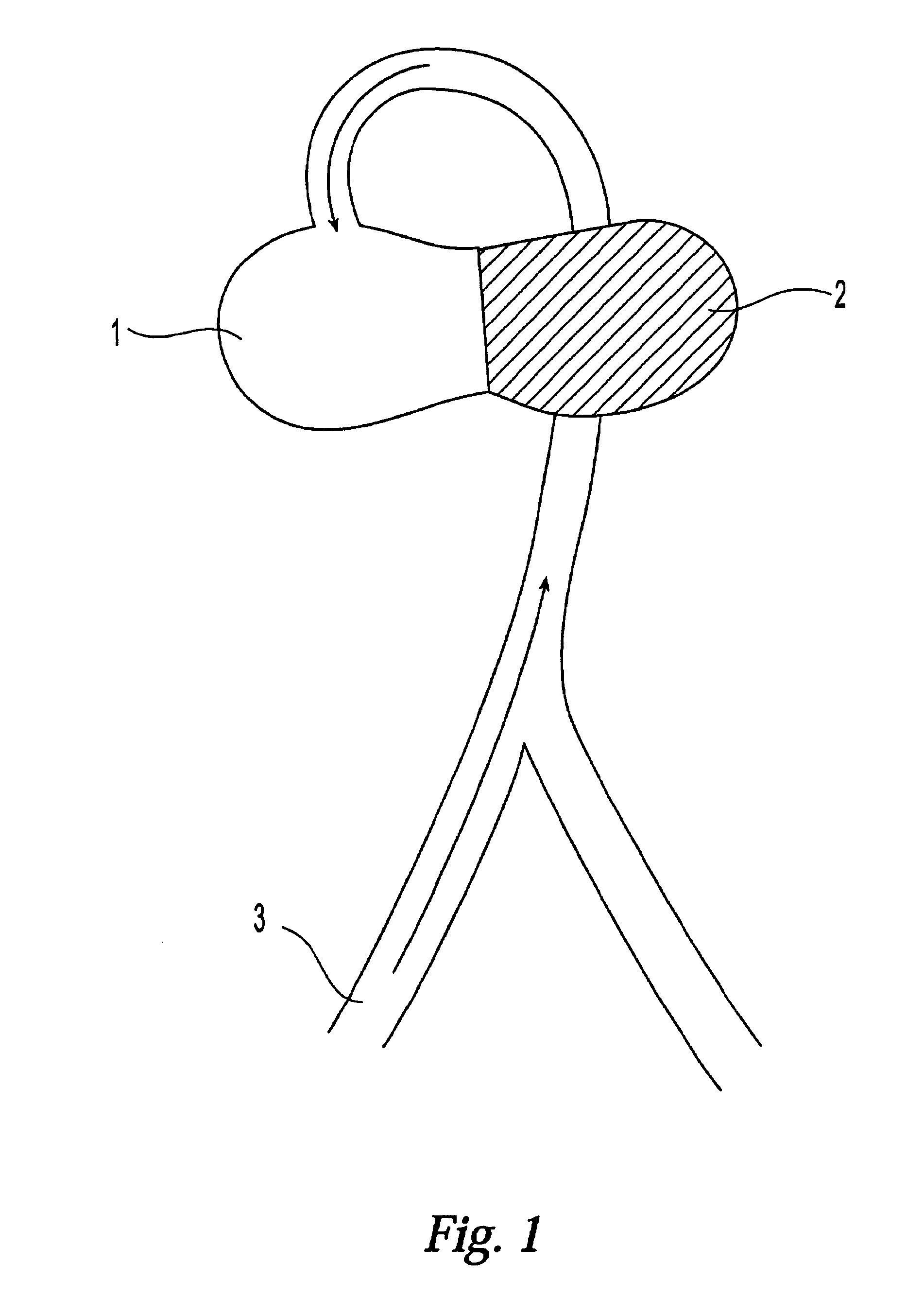
[0029]This present invention relates to devices, materials and methods and is directed toward treatment of heart failure by physically modifying the diseased or damaged heart tissue in such a manner that the internal volume of the damaged left ventricle is reduced, thereby improving the pumping efficiency of the diseased heart and ameliorating the symptoms of heart failure.
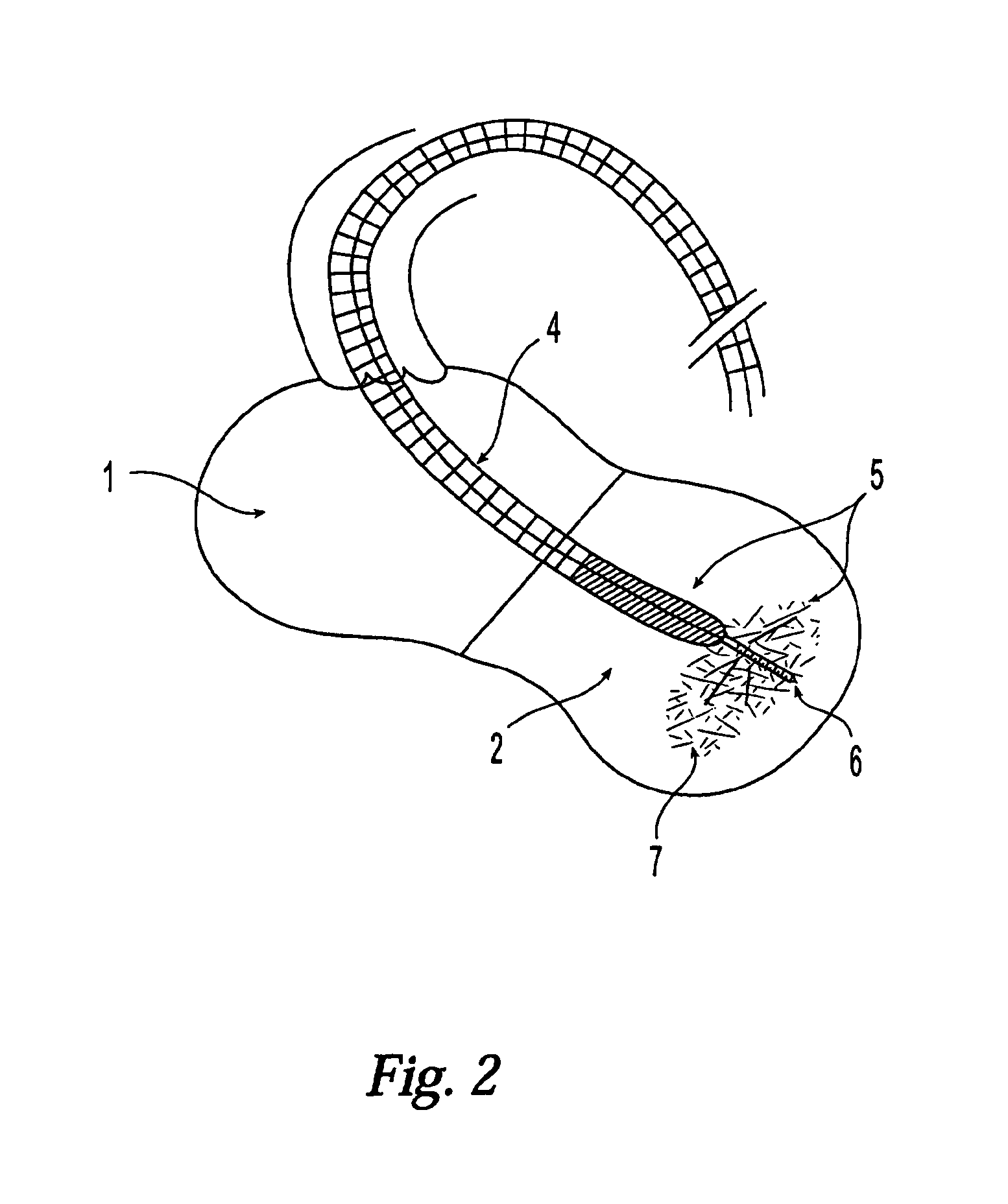
[0030]One embodiment of this invention comprises a catheter-based, minimally invasive procedure that will introduce biocompatible materials into the left ventricle of the heart. Part of the left ventricular cavity will be filled with biocompatible material which will be applied and attached to the left ventricle using catheter-guided techniques and equipment rather than conventional cardiosurgical procedures. The biocompatible filling materials introduced will decrease the volume of the left ventricle and improve the hemodynamics of the heart, thereby alleviating the symptoms of heart failure.
[0031]A second embodiment, which may be used either alone or in combination with the first embodiment, comprises direct injection of suitable, substantially non-compressible biocompatible materials into the wall of the left ventricle. This procedure will increase the bulk of the wall and thereby diminish the interior volume of the left ventricle. These materials may also strengthen and reinforce the wall as well, diminishing the risk that the ventricle might rupture.
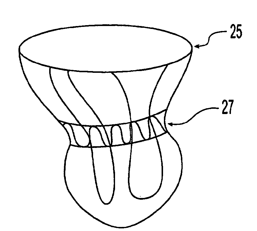
[0033]A third embodiment of the method relates to a method for treating heart failure comprising attaching at least one band to the surface of a human heart comprising a left atrium, a right atrium, a left ventricle and a right ventricle, thereby compressing the heart, whereby cardiac performance is improved. The internal volume of at least one of the left atrium, right atrium, left ventricle, and right ventricle can be decreased by this method, preferably by attaching a plurality of elastic bands to the surface of the heart. If desired, cross-links between a plurality of circular elastic bands attached to the surface of the heart can be provided. If desired, the circular elastic bands can be substantially horizontally positioned and substantially parallel to one another, wherein the bands are of varying diameter arranged in order of descending size from the atria toward the ventricles in order to form a conical structure on the outer surface of the heart.
[0044]The tightening element may be made of wire. Wire is very inelastic compared to elastomeric bands. Therefore, a tightening element of wire will prevent the heart from expanding beyond a given volume. Tightening elements of wire can be tied (i.e. knotted) easily. In addition, the strength of wires allows the tightening element to be cinched under enough force that would break other materials.

Problems solved by technology

This procedure will increase the bulk of the wall and thereby diminish the interior volume of the left ventricle.
These materials may also strengthen and reinforce the wall as well, diminishing the risk that the ventricle might rupture.

Method used


Image

  • Modification of properties and geometry of heart tissue to influence function
  • Modification of properties and geometry of heart tissue to influence function
  • Modification of properties and geometry of heart tissue to influence function

Examples


example 1

[0120]An angiogram is used to determine the heart rate of a subject, and it is found that D=180, S=100, and F is 44.4%. Computer modeling of the heart is used to image the heart and determine where dead or non-functioning tissue is located.

[0121]A biocompatible filler material of a hydrogel is placed in the heart in the area of the non-functioning tissue. After the material is placed, another angiogram is taken to find that D=170, S=100 and F is 42%. The preceding steps are repeated until F is increased into the range of 60 to 75% and preferably, as close to 65% as possible. The final values of D=150, S=60 and F=60% are acceptable and the procedure is terminated.

example 2

[0122]A left ventricular angiogram was done with a LV catheter and the ejection fraction was determined using the dye outflow technique. Later, a Noga catheter was used to map the ventricular wall motion to determine the areas of the ventricular wall where the tissues are ischemic.

[0123]A biocompatible filler material was introduced in the areas of the ventricular wall where the tissues were found to be ischemic. The ventricular wall mapping was repeated to notice any changes in wall motion. The wall mapping was repeated after 4 weeks, one month and six months to monitor any changes.

example 3

[0124]The experiment of Example 3 was repeated after a filling material consisting of a biological material and a filler material was introduced inside the ventricular wall.


PUM


Abstract

Materials, devices and methods for the treatment of congestive heart failure are disclosed. In these methods, the volume of the left ventricle is reduced, thereby increasing the efficiency of the pumping action of the heart. Volume reduction is accomplished by introduction of biocompatible materials into the wall of the left ventricle, or into the ventricle itself. Suitable biocompatible materials include those that undergo a phase transition within the ventricle or within the wall of the ventricle, and are thereby converted from a substantially liquid state to a substantially solid state. Such materials also include those which increase in volume during the transition from the liquid state to the solid state. Also disclosed is a method for ventricular geometry reduction wherein flexible, elastic bands are attached to the external surface of the heart to effect a decrease in the volume of the left ventricle. Finally, disclosed are devices including catheters and elastic bands that are usable in these treatments

Description

CROSS-REFERENCE TO RELATED APPLICATION[0001]This application is a continuation in part of U.S. application Ser. No. 10 / 770,266, which will be U.S. Pat. No. 7,390,293, which is a continuation of pending U.S. application Ser. No. 09 / 988,293 filed Nov. 19, 2001, now U.S. Pat. No. 6,685,627, which is a continuation-in-part of pending U.S. application Ser. No. 09 / 414,708 filed Oct. 8, 1999, now U.S. Pat. No. 6,360,749, and claims the benefit of U.S. Provisional application No. 60 / 103,824 filed Oct. 9, 1998, the contents of which are expressly incorporated herein by reference thereto.FIELD OF THE INVENTION[0002]This invention relates to treatment of heart failure and more particularly, treatment of heart failure by reducing the internal volume of a dilated and diseased left ventricle.BACKGROUND OF THE INVENTION[0003]Congestive heart failure is a chronic, degenerative condition that impairs the heart's ability to pump blood at normal filling pressures to adequately meet the energy requirem...

Claims


Application Information

Patent Timeline
IPC IPC(8): A61F2/02
CPCA61B17/00234A61B17/12022A61B17/12122A61B17/12186A61B17/1219A61B2017/00044A61B2017/00243A61F2/2481A61M2210/125
Inventor JAYARAMAN, SWAMINATHAN
Owner JAYARAMAN SWAMINATHAN
Learn More