Endoscopic imaging device

a technology of endoscope and imaging device, which is applied in the field of endoscope, can solve the problems of limiting the operation of the endoscope and limiting the spatial confocal resolution, so as to reduce the need for biopsy, reduce the time of examination, and reduce the discomfort of the patien

Active Publication Date: 2009-01-08
PURDUE RES FOUND INC
View PDF11 Cites 22 Cited by
  • Summary
  • Abstract
  • Description
  • Claims
  • Application Information

AI Technical Summary

Benefits of technology

[0006]An improved endoscope, offering the ability to image tissues in vivo on both a gross and a cellular level is provided. Embodiments of the present invention allow the user to effectively select an area of interest in a macroscopic view and “zoom-in” to view cellular information from the same area. For medical doctors or veterinarians, this will make possible an immediate histological identification without the need to surgically extract biopsy samples. In many cases, the present invention may effectively reduce the need for biopsy, reducing examination time, expense, and discomfort for the patient. Features of the present invention also allow physicians to examine suspected tissues in a much more methodical and thorough method than is allowed through traditional biopsy. Another component of this product is the methods of software that enables the operation of this device.

Problems solved by technology

Although adequate, this configuration limits the endoscope to operation in situations where a surface plane orthogonal to the tip of the endoscope is encountered.
Furthermore, in this configuration, the imaging lens used for confocal and widefield imaging is typically the same lens, limiting the spatial confocal resolution.

Method used


Image

  • Endoscopic imaging device
  • Endoscopic imaging device
  • Endoscopic imaging device

Examples


Embodiment Construction

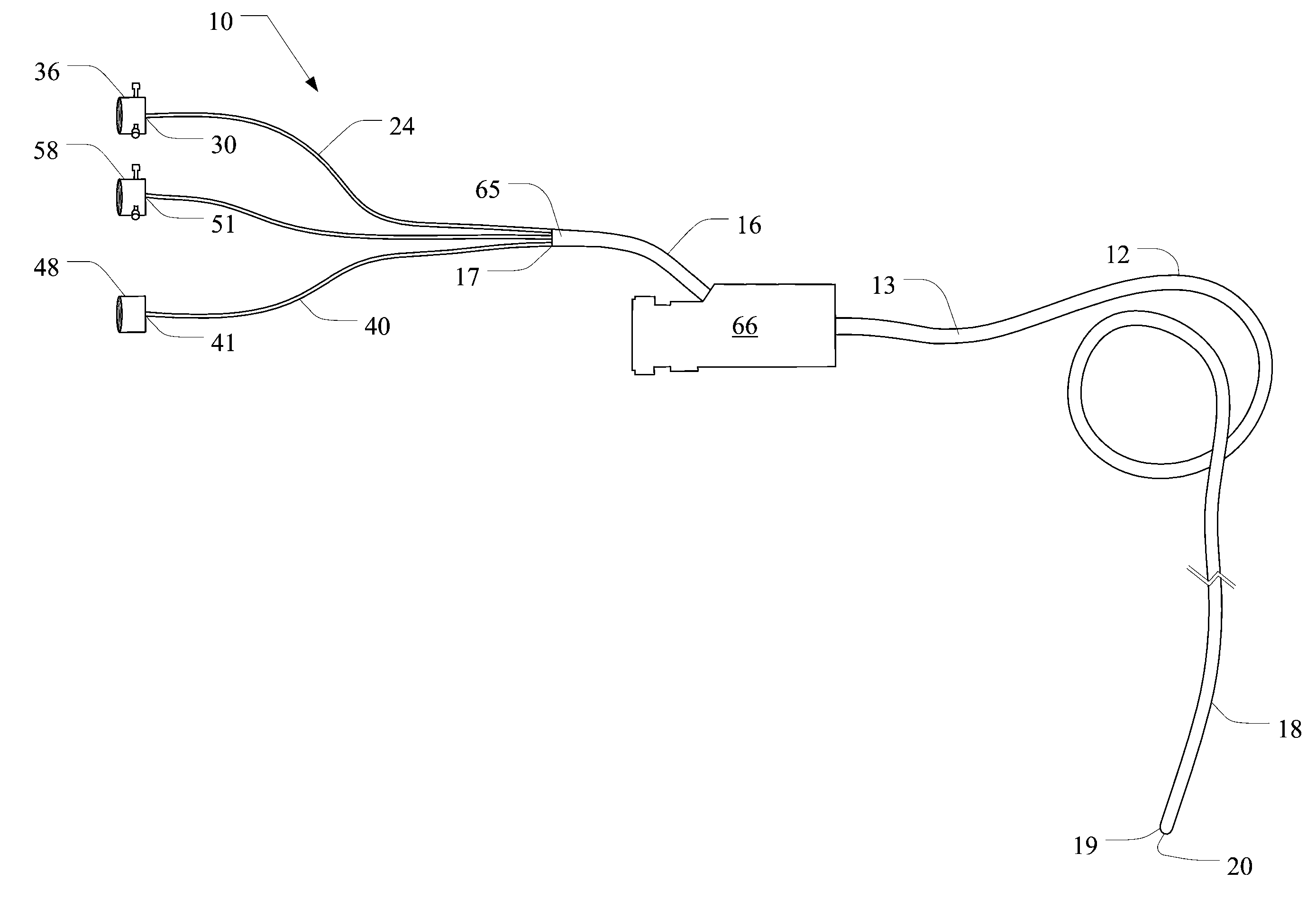
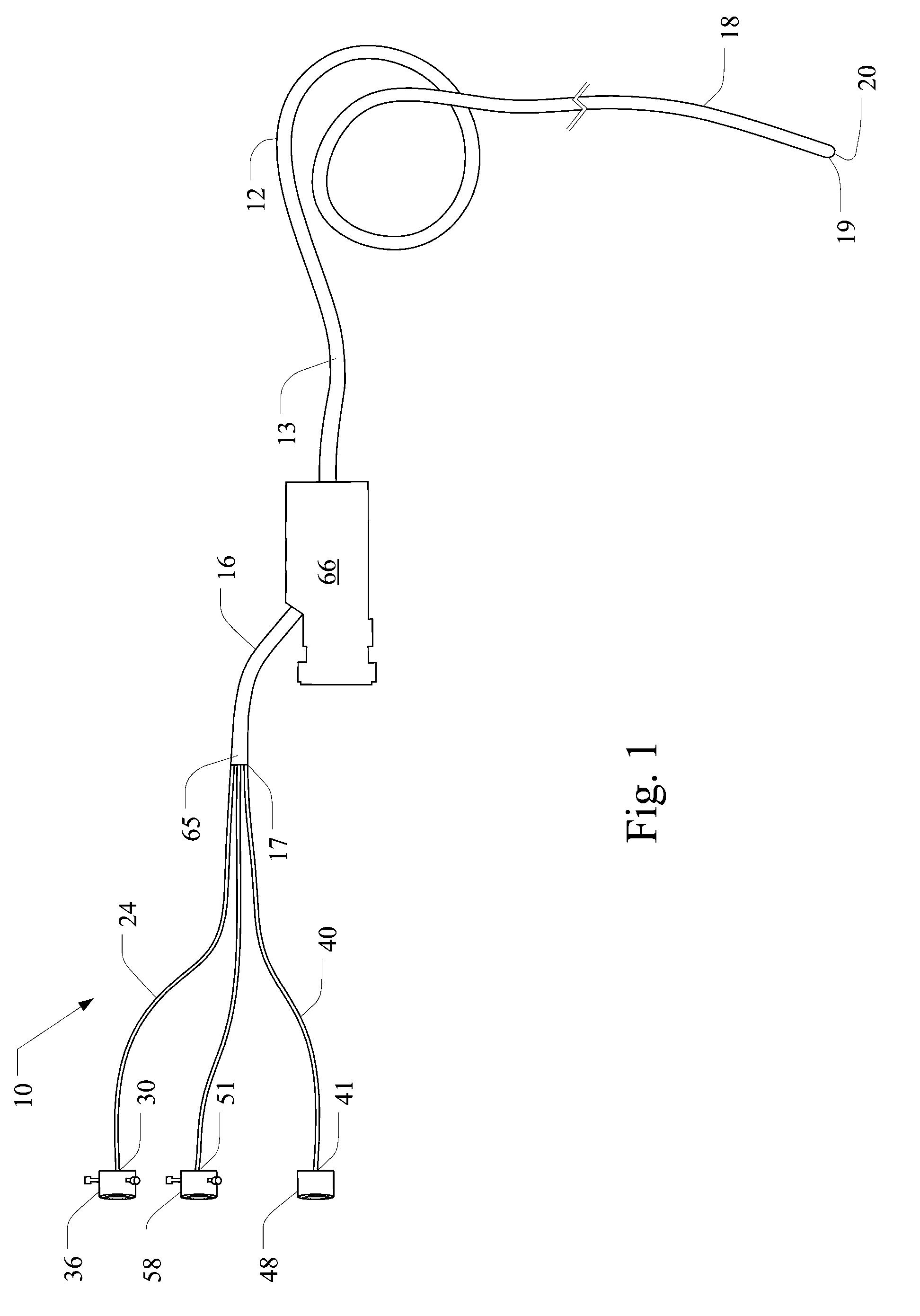
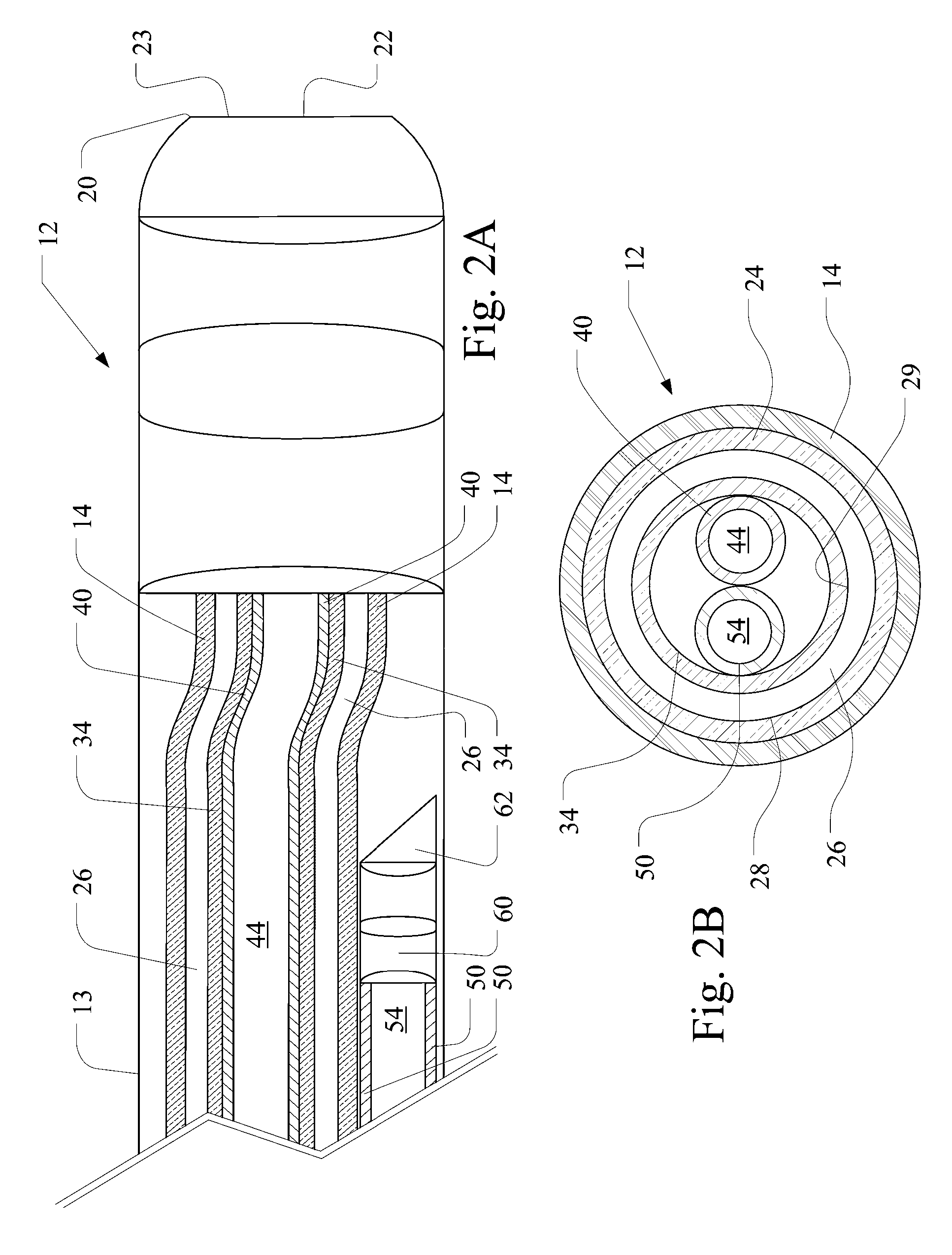
[0017]Embodiments of the present invention incorporate multiple single-mode imaging fibers, allowing for simultaneous imaging at both a gross level and a cellular level. One example of the present invention provides a trifurcated fiber design with two imaging fibers and one illumination fiber branching apart near the proximal end of the device. Distal to the trifurcation point, the annular illumination fiber encloses both of the imaging fibers. Each fiber is fitted with a specific adapter to allow coupling of the fiber to the desired video and imaging cameras or light source.

[0018]FIG. 1 illustrates an endoscopic imaging device 10 for endoscopy in a vessel, e.g., a body vessel of a patient. As shown, the device comprises an insertion tube 12 including and outer layer 13, preferably made of polytetraethylene, and an outer sheath 14 having a proximal portion 16 with a proximal end 17 and a distal portion 18 with a distal end 19. The distal end 19 has an open distal tip 20 that is pref...


PUM


Abstract

An endoscopic imaging device for endoscopy in a body vessel is disclosed. The device comprises an annular illumination tube comprising an annular illumination fiber for illuminating a body vessel during endoscopy. The device further includes a first imaging tube comprising a first imaging fiber for gross examination and navigation through the body vessel. The first imaging fiber is disposed within the annular illumination tube. The device further comprises a second imaging tube comprising a second imaging fiber for cellular imaging. The second illumination fiber is disposed adjacent the first imaging tube and within the annular illumination tube.

Description

CROSS-REFERENCE TO RELATED APPLICATIONS[0001]This application is a continuation of International Application No. PCT / US2006 / 038224 filed on Sep. 30, 2006, entitled “ENDOSCOPIC IMAGING DEVICE” and claims the benefit of U.S. Provisional Application Ser. No. 60 / 722,776 filed on Sep. 30, 2005, entitled “ENDOSCOPIC IMAGING DEVICE,” the entire contents of each are incorporated herein by reference.BACKGROUND OF THE INVENTION[0002]This invention relates to the field of endoscopy and more particularly to endoscopic devices that utilize fiber optic detection of fluorescence.[0003]Endoscopes are employed as a routine clinical macroscopic examination tool. One of the most common methods for producing high-quality, optically sectioned, microscopic images, confocal microscopy employs a pinhole or multiple pinholes to capture image data from a specific optical section. Moreover, a confocal raster scanning endoscope scans the tip of a fiber bundle to reconstruct a high-resolution image. With this m...

Claims


Application Information

Patent Timeline
Patent Type & Authority Applications(United States)
IPC IPC(8): A61B1/06
CPCA61B1/00009A61B1/00181A61B5/6852A61B1/3137A61B5/0068A61B1/0607A61B1/000094
Inventor ROBINSON, JOSEPH P.LEAVESLEY, SILAS J.XI, PENG
Owner PURDUE RES FOUND INC
Why Eureka
Try Eureka