Antibody inhibiting infection of papillomavirus

a technology of papillomavirus and anti-infectious antibodies, which is applied in the direction of antibody medical ingredients, dsdna viruses, peptide sources, etc., can solve the problems of loss of infection, ineffective use in underdeveloped countries, and inefficient packaging of dna, so as to inhibit the infection of mammalian cells

Inactive Publication Date: 2009-02-19
ROSALIND FRANKLIN UNIVERSITY OF MEDICINE AND SCIENCE
View PDF0 Cites 0 Cited by
  • Summary
  • Abstract
  • Description
  • Claims
  • Application Information

AI Technical Summary

Benefits of technology

[0015]In yet another further embodiment, the present invention discloses an isolated antibody, or antigen-binding fragment thereof, that binds specifically to residues 36-49 (SEQ ID NO:1) of bovine papillomavirus-1 (BPV-1) minor capsid L2. The antibody or the antigen-binding fragment is capable of inhibiting infection of a mammalian cell by bovine papillomavirus-1 (BPV-1) or human papillomavirus 16 (HPV-16).

Problems solved by technology

The Pap smear test is readily available in the US and other western countries, but is not effectively used in underdeveloped countries.
In contrast, blocking clathrin / endosome mediated entry results in the loss of infection.
Expression of viral L1 protein in the absence of other viral proteins results in the packaging of DNA at inefficient levels (7, 61).

Method used


Image

  • Antibody inhibiting infection of papillomavirus
  • Antibody inhibiting infection of papillomavirus
  • Antibody inhibiting infection of papillomavirus

Examples


example 1

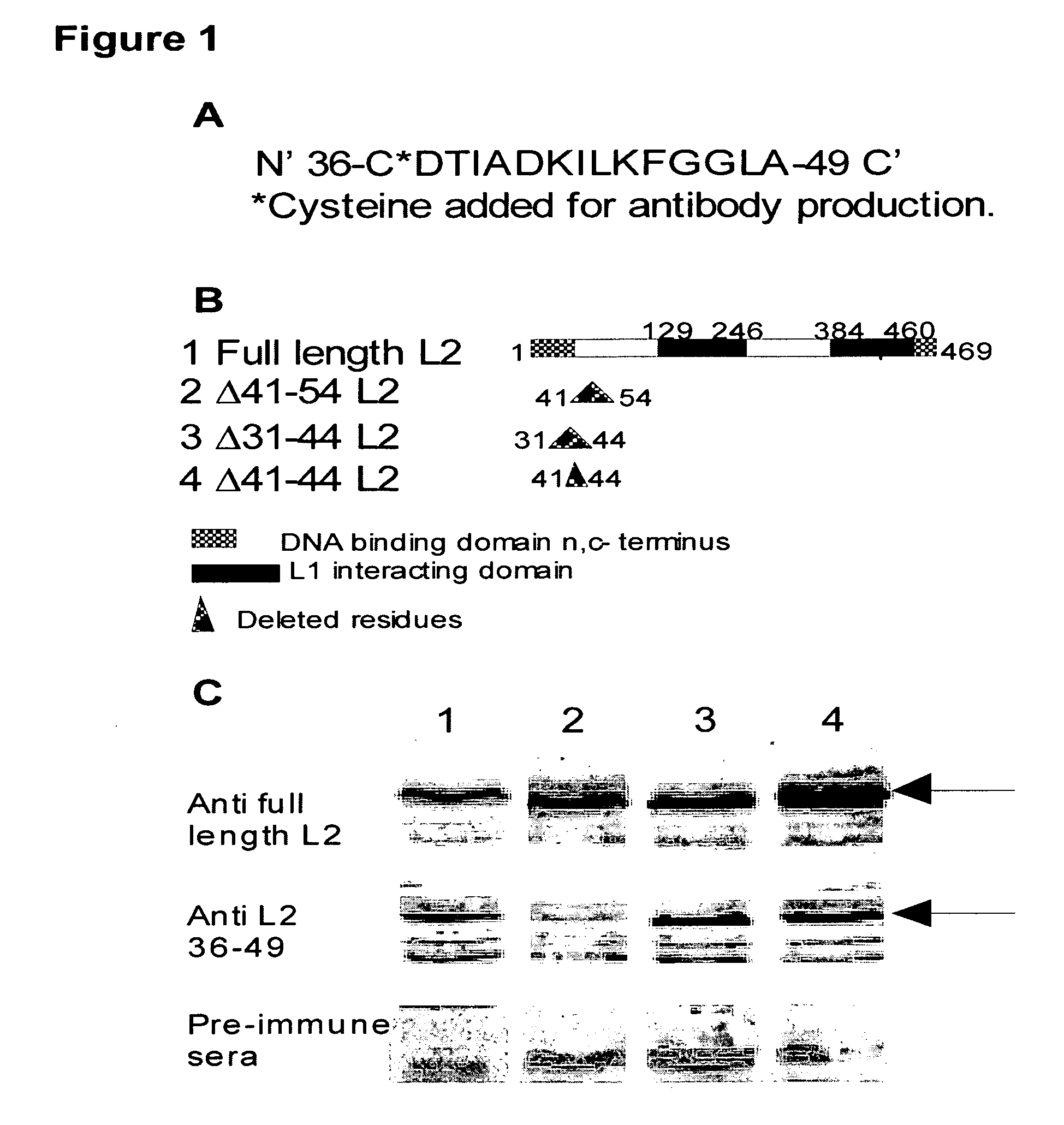
L2 Peptides and Antibodies

[0048]The following peptides were made by ADI (Dallas, Tex.): 1) WTP15L2-CDTIADKILKFGGLA corresponding to residues 36-49 of BPV-1 L2 (SEQ ID NO:1) with a cysteine moiety at the N-terminal, and 2) SCR15L2CIDGLGKLATIDAKF (SEQ ID NO:2) corresponding to the same BPV-1 L2 residues, with the cysteine moiety at the N-terminal, in random order. The peptides were resuspended in serum-free DMEM as per manufacturer's instruction at 10 mM concentration. The proper amount of peptide from stock was added to the cultures to attain the final concentrations in the neutralization blocking experiments. Antibodies to the L2 residues 36-49 were made by ADI after conjugation of the 36-49 amino acids peptide to keyhole-limpet hemocyanine (KLH). Two rabbits were immunized and bleeds were collected at 4 week intervals. Pre-immune serum was collected from both rabbits.

[0049]Antibodies were generated in the two rabbits using the peptide corresponding to BPV-1 L2 residues 36-49 conjug...

example 2

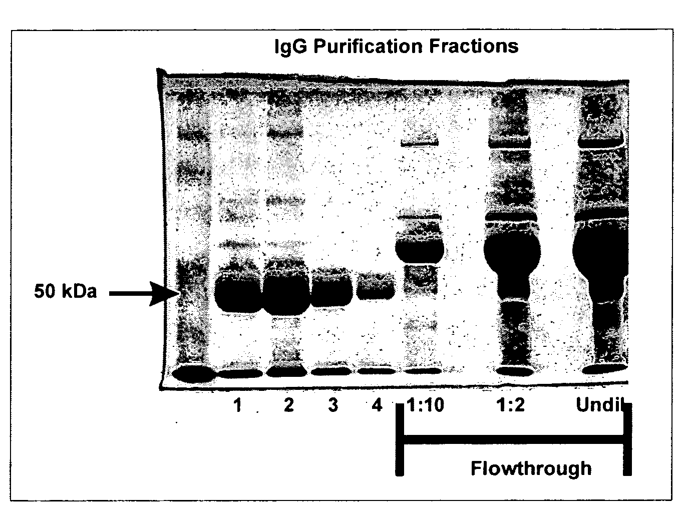
Pseudovirion Production and Purification

[0050]The bicistronic plasmid pShell carrying the BPV-1 L1 and L2 sequences, the GFP cDNA containing plasmid 8fwb, and the 293TT viral packaging cell line were a generous gift from Drs. Day and Schiller (6). Pseudovirus production was performed as described (6). In brief: 293TT cells at 70-80% confluence in a T175 cm2 flask were co-transfected with 15 ug of pShell and 15 ug of 8fwb, in 65 ul of lipofectamine 2000 (Invitrogen, Carlsbad, Calif.) as per manufacturer's instructions. After 24 hours, the cells were trypsinized and split into two new T175 cm2 flasks. After 24 hours, the cells were harvested by centrifugation and re-suspended in 500 ul of cell lysis buffer (Brij, benzonase, DNAsafe, in PBS) and incubated at 37° C. with gentle rotation for 16 hours. The virus preparation was put on ice for 5 minutes before the addition of 85 ul of 5M NaCl. After a 20 minute incubation on ice with inversion every 5-10 minutes, the preparations were spun...

example 3

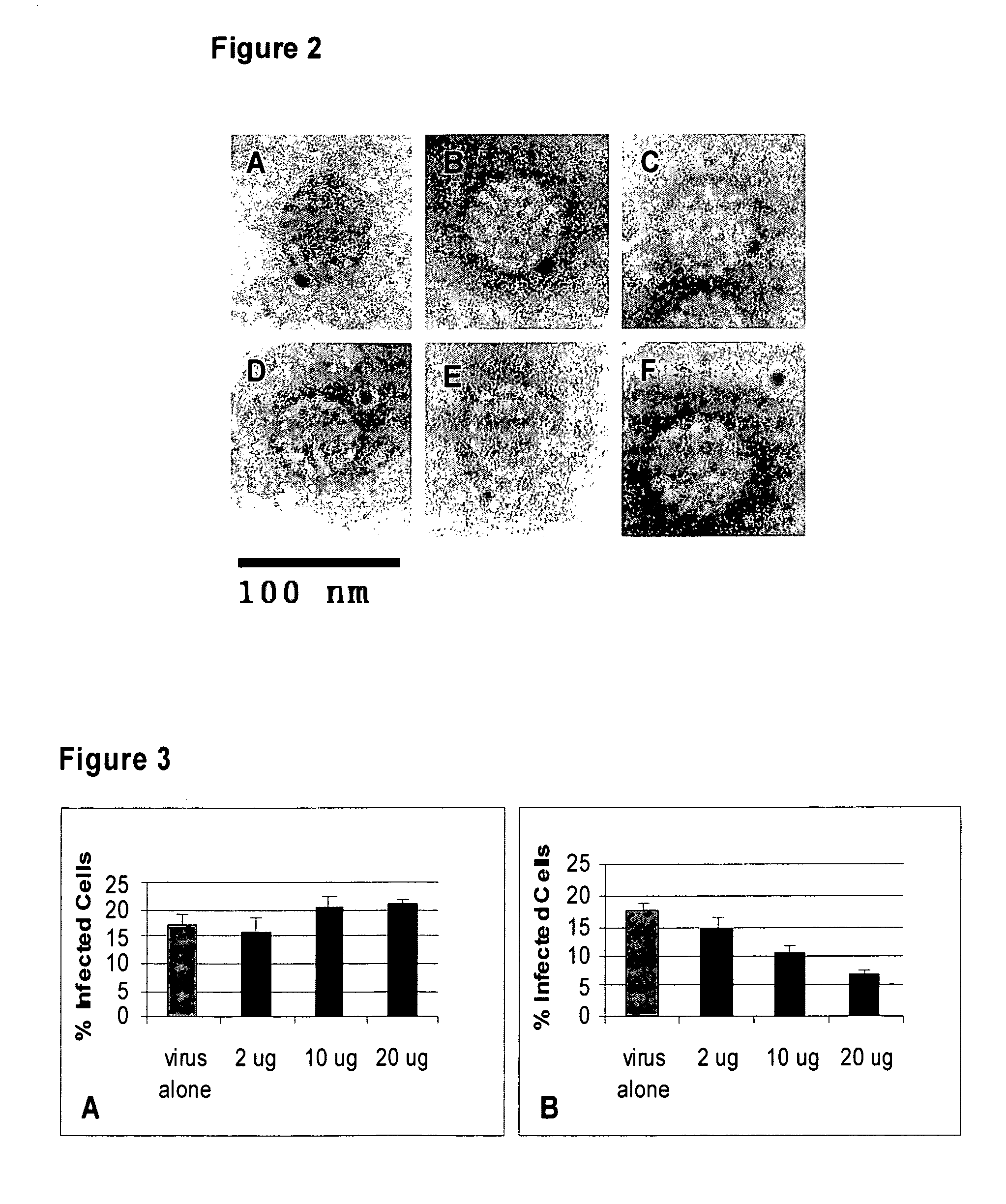
Determining Viral Titer

[0051]Viral titer was performed on Cos-7 cells. In brief, 50,000 Cos-7 cells / well were plated on a 24 well plate for 24 hours. At 24 hours, cells were washed in cold media (DME-10% FBS) and incubated on ice for 10 minutes in cold media. After 10 minutes cold media was added with 2, 5, or 8 ul of virus to the wells and the plate was incubated on ice for 2 hours. After two hours, the cells were washed ×3 in DME-10% FBS at room temperature, and incubated at 37° C. in 500 ul DME-10% FBS for 48 hours. Cells were then harvested and analyzed for GFP fluorescence by FACS analysis. 10,000 cells were counted and the percentage of green cells was used to determine the titer per ml of virus. Real time-PCR was performed on the DNA extracted from the virus preparation as described previously using GFP primers (6). The number of viral particles needed to obtain one infectious viral particle, i.e., a green cell, was determined by dividing the total number of viral particles o...


PUM

PropertyMeasurementUnit
diameteraaaaaaaaaa
diameteraaaaaaaaaa
diameteraaaaaaaaaa

Abstract

The present invention relates generally to an antibody that binds specifically to a neutralization epitope at the surface of papillomavirus (residues 36-49 of the minor capsid L2) that does not interfere with virus entry into endosomes and lysosomes. This antibody neutralizes infection of the virus in a dose dependent manner.

Description

CROSS-REFERENCE TO RELATED APPLICATION[0001]The present application claims priority to and the benefit of U.S. Provisional Application No. 60 / 879,163 filed on Jan. 8, 2007, and is incorporated by reference and made a part hereof.FEDERALLY SPONSORED RESEARCH OR DEVELOPMENT[0002]This invention was made with government support under National Institutes of Health (NIH) / National Cancer Institute (NCI) grant number K22:CA117971. The government has certain rights in the invention.TECHNICAL FIELD[0003]The present invention relates generally to an antibody that binds specifically to a neutralization epitope at the surface of papillomavirus (residues 36-49 of the minor capsid L2) that does not interfere with virus entry into endosomes and lysosomes. This antibody neutralizes infection of the virus in a dose dependent manner.BACKGROUND OF THE INVENTION[0004]Papillomavirus (PV) infection is the most common sexually transmitted disease in the U.S. (approximately 5.5 million per annum) (40). PVs ...

Claims


Application Information

Patent Timeline
Patent Type & Authority Applications(United States)
IPC IPC(8): A61K39/395C07K16/18C12N5/06A61P31/00
CPCA61K39/00A61K39/12C07K14/005C12N2710/20034C07K2316/96C07K2317/77C12N2710/20022C07K16/084A61P31/00C07K2317/76
Inventor MENESES, PATRICIO I.ROBERTSON, ERLE S.NGUYEN, KHALANIOSZ, VALERIE
Owner ROSALIND FRANKLIN UNIVERSITY OF MEDICINE AND SCIENCE
Why Eureka
Try Eureka