Advanced bio-compatible polymer surface coatings for implants and tissue engineering scaffolds

a biocompatible and scaffold technology, applied in the field of advanced biocompatible polymer surface coatings for implants and tissue engineering scaffolds, can solve the problems of many implants being lost, poor integration of the implant surface with the bone/tissue structure, and limited implant biocompatibility, so as to promote cell adhesion and proliferation, enhance the hydrophilic nature of the surface, and enhance the binding of bioactive agents

Inactive Publication Date: 2010-10-07
THE BOARD OF TRUSTEES OF THE UNIV OF ARKANSAS
View PDF16 Cites 70 Cited by
  • Summary
  • Abstract
  • Description
  • Claims
  • Application Information

AI Technical Summary

Benefits of technology

[0008]In another embodiment, at least one surface of the coating may undergo plasma/ion treatment to induce the formation of surface charges that enhance th

Problems solved by technology

Because most implants are made of metals, metal oxides, polymeric materials or tissue components obtained from animals or humans, implant bio-compatibility poses a limitation in many applications as implants need to perform complex functions in the human body and their binding to the host tissue is crucial.
Still a relatively large number of such mat

Method used


Image

  • Advanced bio-compatible polymer surface coatings for implants and tissue engineering scaffolds
  • Advanced bio-compatible polymer surface coatings for implants and tissue engineering scaffolds
  • Advanced bio-compatible polymer surface coatings for implants and tissue engineering scaffolds

Examples


example 1

Preparation of Polyurethane-Hydroxyapatite Composite Materials

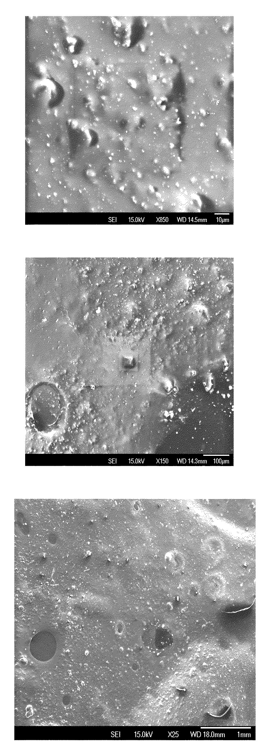
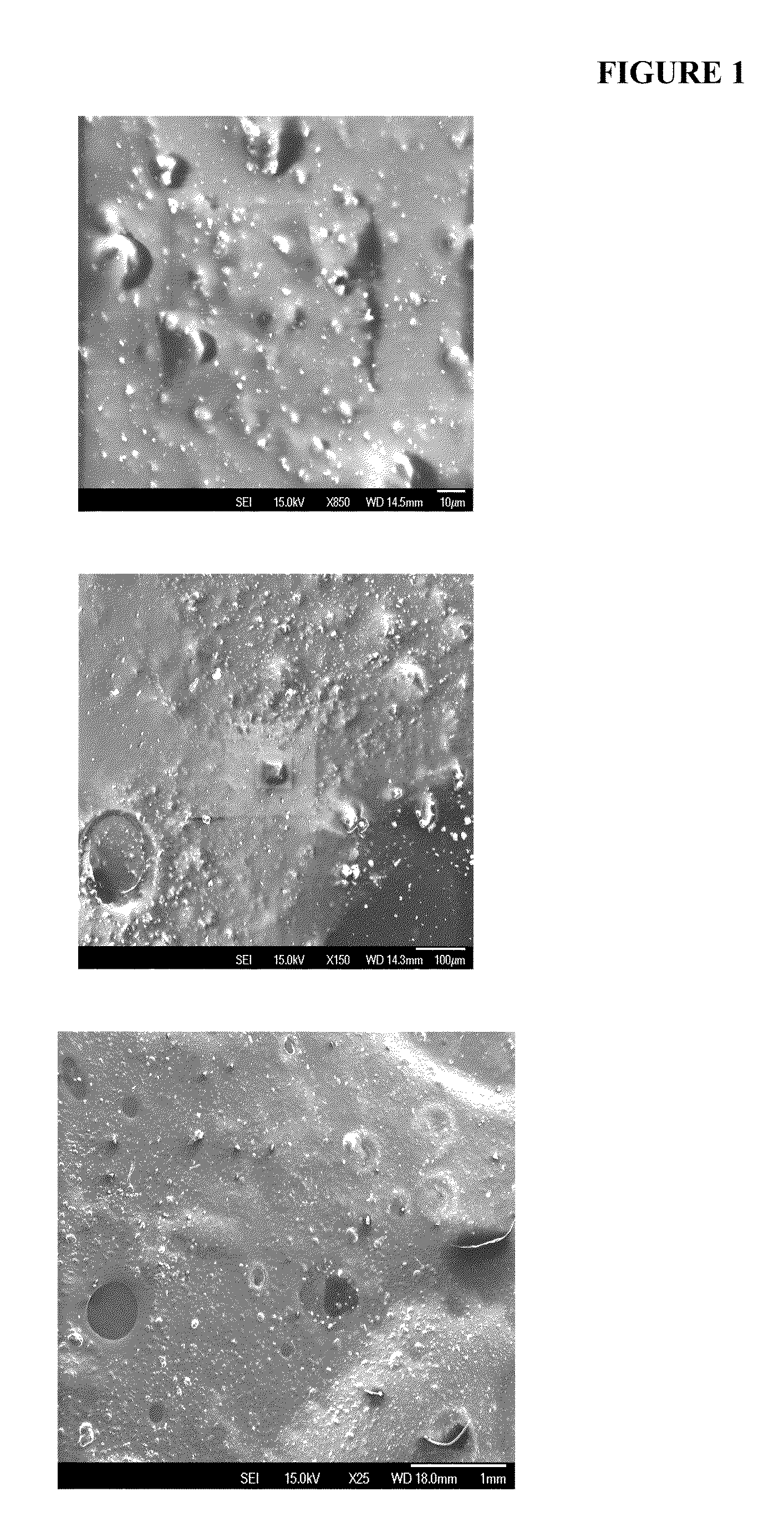
[0044]Polyurethane-hydroxyapatite (HA) composite materials are prepared by in-situ polymerization. The nanomaterials are mixed in the solvent of the polymeric scaffold and deposited by air spraying.

[0045]Similarly, for industrial applications, the composite films can be deposited by e-beam deposition, ion beam deposition, pulsed laser deposition, electrospray, or any other method known in the art, on the implant surfaces.

[0046]The coating can be exposed to any type of plasma discharge and covered with growth factors, proteins, amino acids, drugs, hydroxyapatite, or any other bioactive agent.

[0047]FIG. 1 presents a surface created by the deposition of the polymeric / nano-hydroxyapatite nanocomposites.

example 2

Cell Growth Assay on Coated and Non-Coated Surfaces

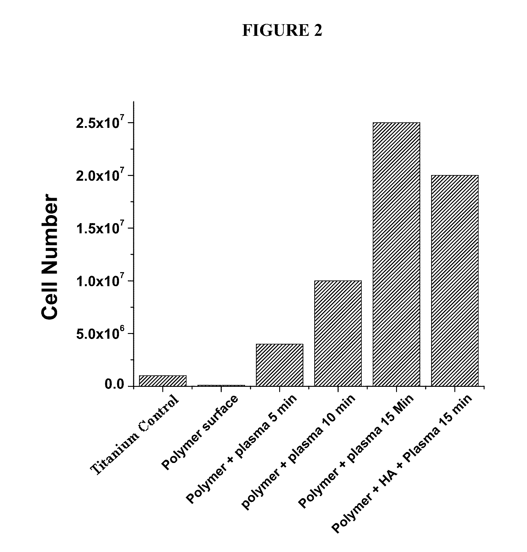
[0048]Osteoblast cells were incubated in tissue culture on top of the coatings and controls for 7 days at 37 deg. C. At day 0, at the start of the experiment, the number of cells was 105. The control surfaces were a roughened titanium surface and an untreated polymer surface. After 7 days in tissue culture, osteoblasts were counted using standardized cell counting methods. The number of cells on the untreated polymer surface was 105. The number of cells on the second control (titanium surface) was 106. The results for the polyurethane coatings and HA are shown in FIG. 2.

[0049]As show in FIG. 2, there is a significant increase in the number of cells on the plasma treated polymer coatings, as well as the polymer treated and HA coatings. The polymer surfaces which were plasma treated between 10 and 15 minutes demonstrated approximately 20 times more cells per unit area versus the controls. The plasma treated polymer surface with hydrox...

example 3

Visualization of Bone Cells on Various Surfaces

[0050]FIG. 3 shows enhanced growth of bone cells on the polymeric surfaces exposed to plasma and doped with HA nanoparticles (b) as compared to the regular Ti surfaces (a). The corrugated polymeric surfaces (doped with HA nanoparticles and plasma treated) (c) due to various modifications, have also been found to enhance significantly the proliferation of osteoblast cells. The visualization was done with optical microscopy and cellular staining.


PUM

PropertyMeasurementUnit
Sizeaaaaaaaaaa
Sizeaaaaaaaaaa
Crystallinityaaaaaaaaaa

Abstract

Disclosed herein are methodologies and compositions for coating materials, which can be used in a variety of biological applications.

Description

CROSS-REFERENCE TO RELATED APPLICATION[0001]This application claims the benefit of U.S. Provisional Application No. 61 / 212,110, filed Apr. 7, 2009.INTRODUCTION[0002]Annually, millions of implants are placed inside of organisms, including humans and animals. Most of these implants serve complex roles including but not limited to tissue replacement, mechanical support, tissue generation, cosmetic enhancement, complete or partial limb replacement, joint replacement, tooth replacement, spine reconstruction, defibrillators / pacemakers, in addition to electrodes and wires.[0003]Because most implants are made of metals, metal oxides, polymeric materials or tissue components obtained from animals or humans, implant bio-compatibility poses a limitation in many applications as implants need to perform complex functions in the human body and their binding to the host tissue is crucial. For example, dental implants need to adhere very strongly to the jaw bone. It is also important for implant su...

Claims


Application Information

Patent Timeline
IPC IPC(8): A61C8/00A61B19/02A61F2/02A61F2/28C04B35/00C09D1/00C09D189/00C03C3/00B32B5/16B05D3/00C23C14/34C23F1/00A61L2/08A01N43/20C12N5/00
CPCA61C8/0013Y10T428/25A61F2/3094A61F2002/3084A61F2310/00023A61F2310/00634A61F2310/00796A61L27/306A61L29/106A61L31/088C08K3/04C08K3/08C08K3/22C08K9/02C08L2203/02C09D5/14C09D7/1208C09D7/1266C09D189/00C12N5/0654C12N2533/18C23C14/083C23C14/28C23C26/00C23C30/00A61F2/30767C09D7/67
Inventor BIRIS, ALEXANDRU S.JENSEN, PEDERKANNARPADY, GANESH
Owner THE BOARD OF TRUSTEES OF THE UNIV OF ARKANSAS
Why Eureka
Try Eureka