[0010]In some embodiments, the present invention provides for specific locus probes designed for use with interphase stem cells, to detect chromosomal aberrations. Probes of the present invention are created, for example, from BAC cloning, yeast artificial chromosome (YAC) cloning, microdissection of metaphase chromosomes, PCR amplified DNA sequences, synthetic oligonucleotides and synthesized peptide nucleic acids (PNAs), etc. In some embodiments, probes used in assays of the present invention are directly or indirectly labeled with a fluorescent moiety for single fluorescence measurement. As well, multiple probes, each labeled with different fluorescent moieties of different wavelengths, may be used such that fluorescent signal multiplexing can occur in one area, targeting one or more chromosomes, thereby maximizing a stem cell sample being assayed and space on the assay substrate. In some embodiments, probes used are indirectly labeled with non-fluorescent moieties, such as digoxigenin, peroxidase or biotin, which can be conjugated to an antibody, avidin, etc., for detection using light microscopy. In one embodiment, the probes are made from synthesized peptide nucleic acids and labeled with digoxigenin. Fluorescent moieties useful in systems and methods of the present invention include, but are not limited to, cascade blue, coumarin, cyanine dyes (e.g., 2, 3, 5, 7), BODIPY dyes (e.g., FL, TMR, TR, 650 / 665) Alexa dyes (e.g., 488, 532, 546, 594), fluorescein isothiocyanate (FITC), spectrum green, tetramethylrhodamine (TMR), rhodamine B, spectrum orange, texas red, and spectrum red. Commerically available FISH probes, such as painting probes, only identify anolomies on metaphase chromosomes and not interphase chromosomes. Some embodiments of the present invention are directed to a rapid method for interphase detection of chromosomal anomolies, which does not require, for example, tissue culture, etc. and other time steps necessary for maintaining actively dividing cells.
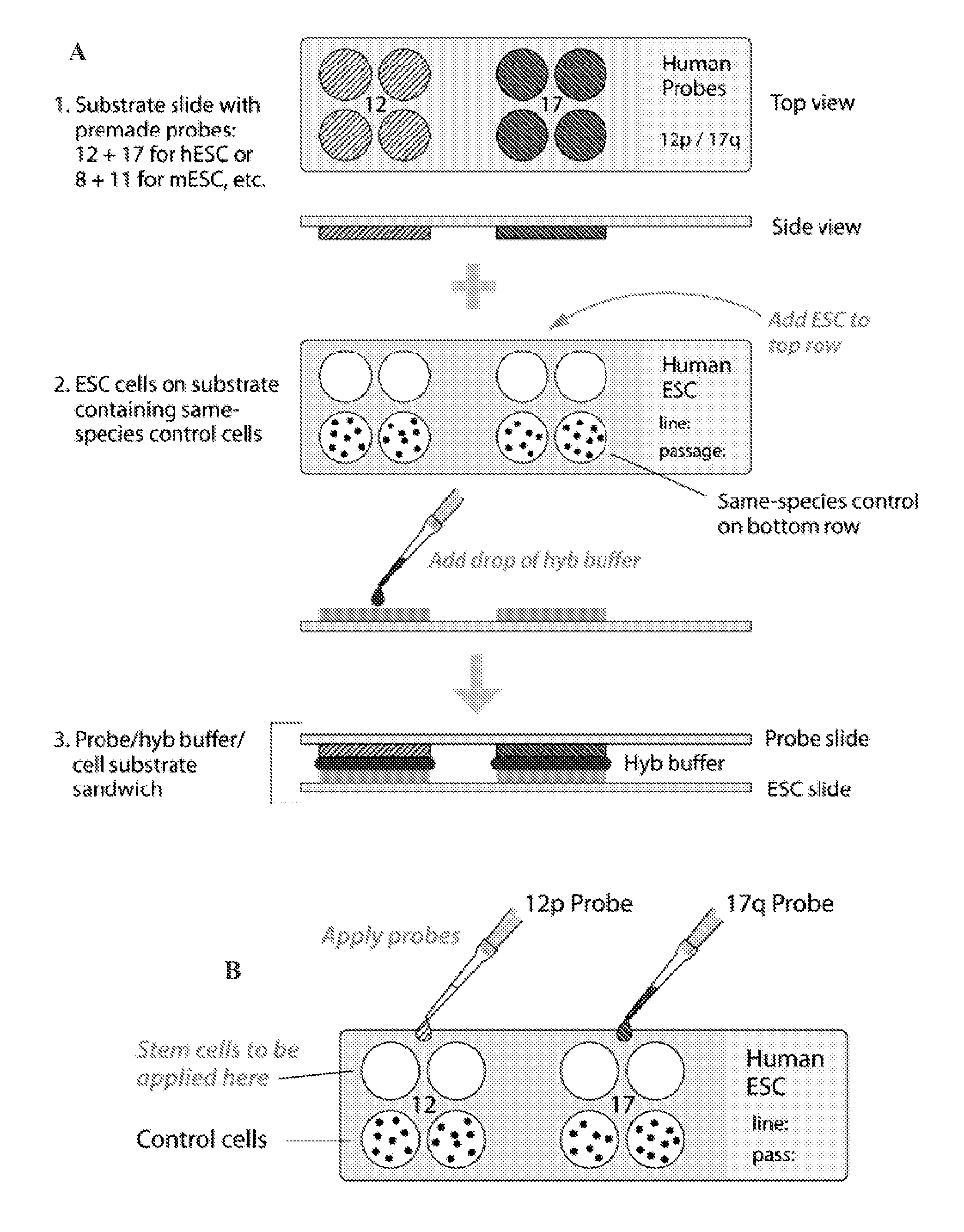
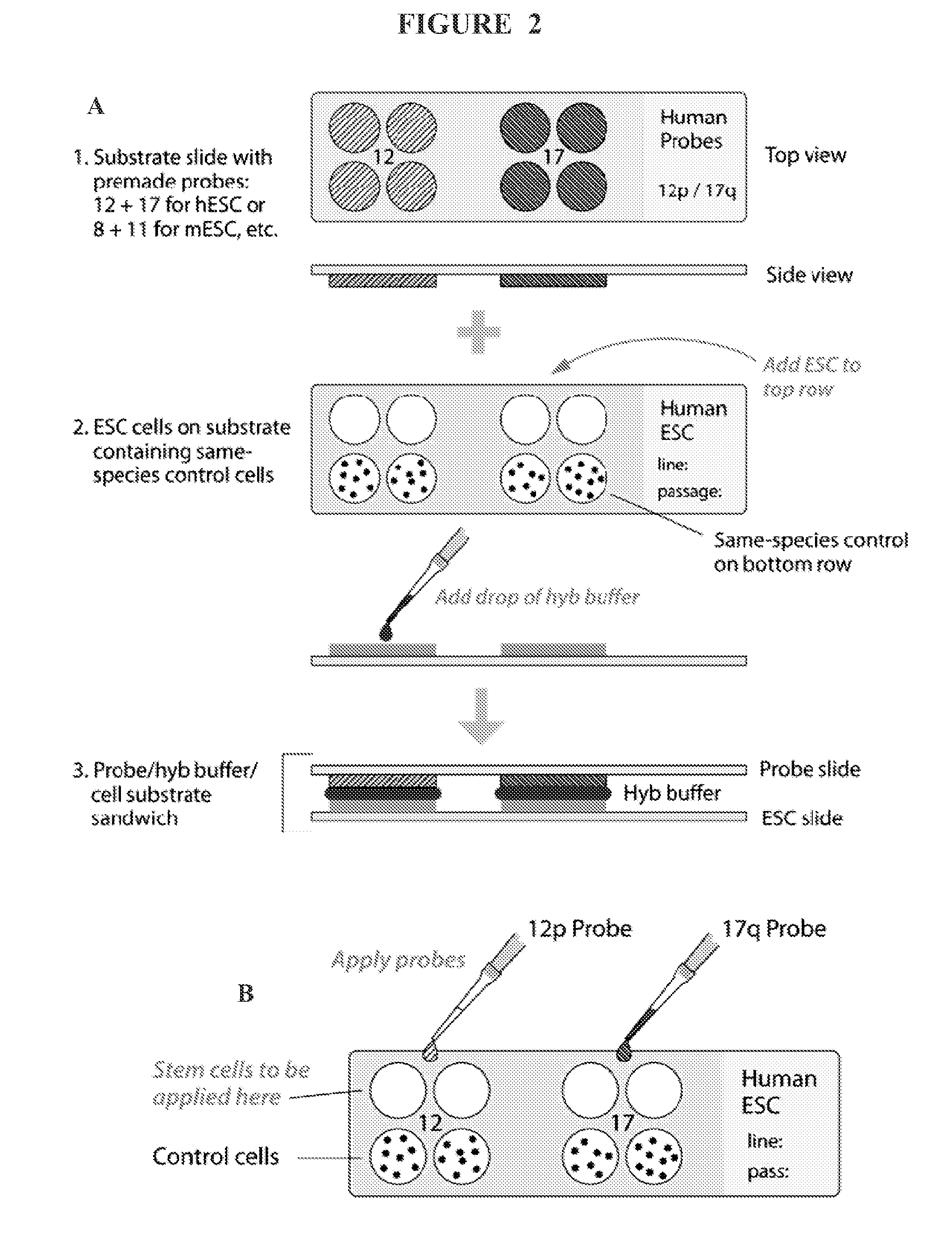
[0011]In some embodiments, an assay of the present invention comprises a substrate such as a slide (e.g., glass, polypropylene, polyethylene, etc,), a membrane surface, and the like. The assay substrate comprises multiple discrete locations for the application of different stem cell species or control cells on each discrete location. FIG. 2 depicts an exemplary substrate (slide) for use in an assay of the present invention. In some embodiments, samples from stem cell species for assay and control cells are taken from aliquots of passaged cells and placed each on a discrete location on an assay substrate. The labeled probes are subsequently added to each location on the substrate, and in situ hybridization is performed. Hybridization results are detected by any detection means, such as fluorescence (fluorescently labeled probe) microscopy and light microscopy (non-fluorescently labeled probe), either system being adaptable to an automatic reader.
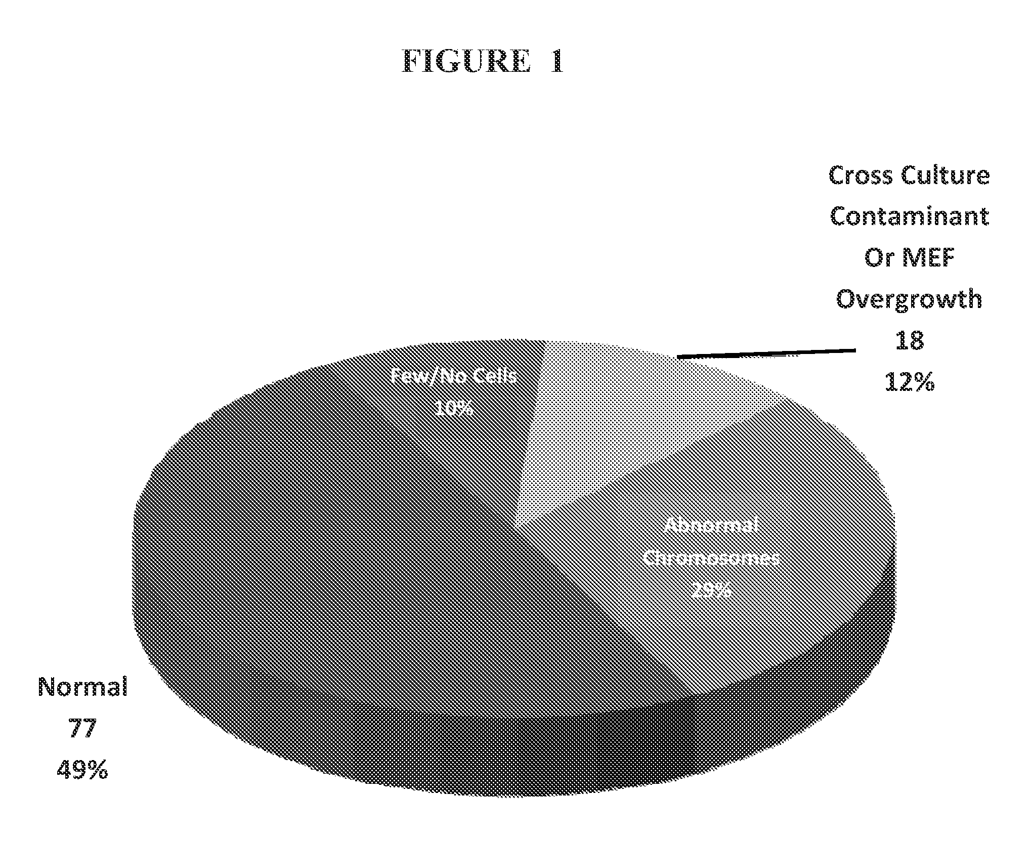
[0012]In some embodiments, an assay of the present invention comprises two substrates, or slides. One slide comprises, for example, discrete locations with samples from aliquoted control cells and stem cell species to be tested, and the second slide comprises labeled probes capable of hybridizing to one or more chromosomal aberrations. In this embodiment, hybridization buffer is applied to the probe slide that is laid atop the cell-containing slide, followed by hybridization and detection methods known to those skilled in the art (FIG. 2A). In some embodiments controls are used for comparison. For example, if the specimen to be tested has clear signals, with minimal cell overlapping, reliability of the results is possible by comparing the signal counts to those of the normal control cells that undergo the same hybridization conditions. As long as the normal control demonstrates two signals for the probe being assayed, finding signals greater than two in any of the stem cells being tested is considered evidence for low level trisomy. Scoring may also be based on cut-off values that are developed after scoring at least ten cultures with a specific probe. Also, when both probes show the same frequency of cells with four signals, it is suggestive of the presence of a low level of cells with teteraploidy. If low or high level trisomy of either probe is detected, or tetrasomy 12 due to formation of an isochromosome (two copies of the short-arm with loss of the long-arm) leading to duplication of the critical region of 12 to which the probe is directed, this would prompt the investigator performing the assays to go to an earlier normal passaged aliquot. A potential for misinterpretation, for example, is in the case of low level tetraploidy of both probes on the slide array, in which case a few cells would show four signals for chromosome 12 while a similar number would show four signals for 17. When both probes show the same frequency of cells with four signals strongly suggesting the presence of a low level of cells with tetraploidy, a slide array using a control probe for a genetic sequence unlikely to undergo in vitro selection would be provided for either quantifying, or ruling out, tetraploidy.
[0013]In some embodiments, the present invention provides assay kits. Assay kits of the present invention comprise a substrate, such as a slide, and pre-measured aliquots of reagents needed for hybridization and post-hybridization reactions. Reagents needed for hybridization and post-hybridization reactions include, but are not limited to, directly or indirectly labeled probes with or without blocking DNA (e.g., human placental DNA, salmon sperm DNA, etc.) or similar reagents (e.g., pepsin, protease, etc.), hybridization wash solutions (e.g., SSC, etc.), post-hybridization solutions (e.g., SSC with detergents, etc.), counterstains (e.g., propidium iodide, DAPI, etc), and the like. An example of the use of blocking DNA in FISH for chromosomal staining can be found in U.S. Pat. No. 5,447,841 (incorporated herein by reference in its entirety). In some embodiments, the substrate of the kit comprises pre-deposited control cells, wherein an investigator adds the stem cell species for testing. In some embodiments, an assay kit further provides labeled probes for adding to the substrate upon which is located the cells for testing (e.g., test and control cells). In other embodiments, the substrate comprises pre-deposited labeled probes, wherein the investigator adds the stem cells for testing and control cells (which can be additionally furnished in the assay kit). In some embodiments, an assay kit of the present invention comprises two substrates, one of which comprises pre-deposited control cells that have already been added to the labeled probes, and the second of which comprises pre-deposited labeled probes to which the stem cells are to be added.
[0014]In some embodiments, additional buffers, solutions, specialized slides or substrates, detection reagents, and the like necessary, sufficient or useful to perform washes, hybridizations, counterstaining, etc. are furnished in an assay kit of the present invention. An assay kit further comprises instructions for performing the assay, as well as scoring criteria for determining chromosomal aberration presence or absence and amount thereof, if present. In some embodiments, software is provided that is configured to analyze, store, correlate, or otherwise manipulate data obtained from use of the assay. In some embodiments, the software associates information about the source of the stem cell with information pertaining to the presence, absence, nature of, or level of aberration. In some embodiments, data obtained is compared to data in a database so as to predict or characterize a test sample based on its similar properties to a stored database sample. In some embodiments, the kit comprises detection equipment. In some embodiments, the kit provides a desktop or handheld device that carries out one or more of the steps useful in utilizing the invention, including, but not limited to, sample preparation, sample processing / incubation, assay preparation, assay use, label detection, software, data collection, data storage / analysis, and the like.
[0015]The present invention further provides probes or sets of probes particularly useful in identifying chromosomal aberrations in stem cells, as well as compositions comprising such probes (e.g., kits, reaction mixtures, slides, beads, etc.). The probes may hybridize to and detect a critical region that identifies all of the translocations and duplications associated with the most common chromosomal aberrations found in stem cell lines. Aberrant cells identified by the probes may be discarded. Cells lacking aberrations may be used for any number of purposes, including, but not limited to, maintenance of a cell lines, research, drug screening, transplantation into an organism for research or therapy, and the like.

