System and method for providing enhanced background rejection in thick tissue with differential-aberration two-photon microscopy

Inactive Publication Date: 2009-04-02
TRUSTEES OF BOSTON UNIV
View PDF18 Cites 34 Cited by
  • Summary
  • Abstract
  • Description
  • Claims
  • Application Information

AI Technical Summary

Benefits of technology

[0011]The present invention can also be viewed as providing methods for modulating the in-focus signal in a laser scanning nonlinear microscope, thereby enabling a better discrimination of in-focus signal from out-of-focus background. In this regard, one embodiment of such a method, among others, can be broadly summarized by the following steps: acquiring a standard two-photon excited fluorescence of thick tissue without extraneous aberrations; introducing an extraneous aberration pattern in an excitation beam path; acquiring two-photon excited fluorescence of the thick tissue having the introduced extraneous aberra

Problems solved by technology

While it is well accepted that TPEF microscopy provides greater imaging depth penetration in thick tissue than more conventional fluorescence imaging techniques, such as confocal or widefield microscopy, the depth penetration of TPEF microscopy remains nonetheless limited.
1) An excitation beam can undergo scattering when it propagates through tissue. This scattering weakens the ballistic (un-scattered) excitation power that attains the beam focus and thereby reduces the TPEF signal generated at the focus. Since scattering scales roughly exponentially with propagation distance, by dint of the Lambert-Beer law, the reduction in TPEF signal becomes particularly severe at larger focal depths. One strategy to maintain adequate ballistic excitation power at relatively large focal depths has involved the use of non-standard laser sources based on regenerative amplifiers. Unfortunately, such a strategy can only go so far in compensating for an exponential loss in ballistic power, even though it has been the most successful to date in pushing the limits of depth penetration.
2) The required increase in excitation power necessary to maintain (or try to maintain) adequate ballistic power at the beam focus can lead to significant power densities near the tissue surface. If the tissue is fluorescent near its surface, as is the case for example if the fluorescent labeling is homogeneously distributed throughout the sample, or if the sample is autofluorescent either intrinsically o

Method used


Image

  • System and method for providing enhanced background rejection in thick tissue with differential-aberration two-photon microscopy
  • System and method for providing enhanced background rejection in thick tissue with differential-aberration two-photon microscopy
  • System and method for providing enhanced background rejection in thick tissue with differential-aberration two-photon microscopy

Examples


Example

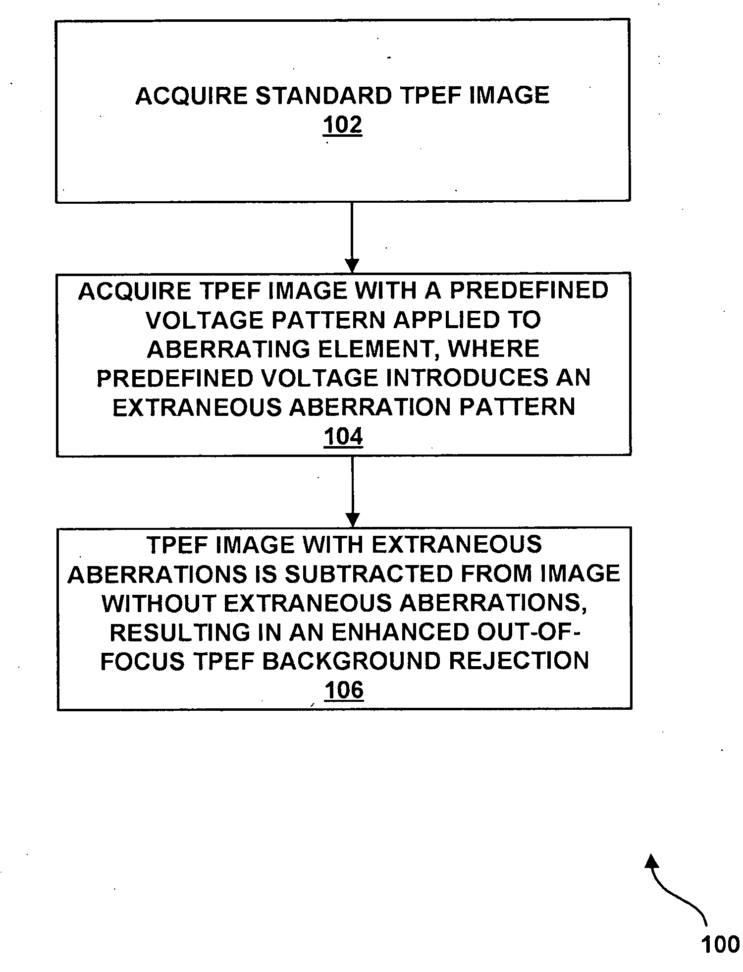
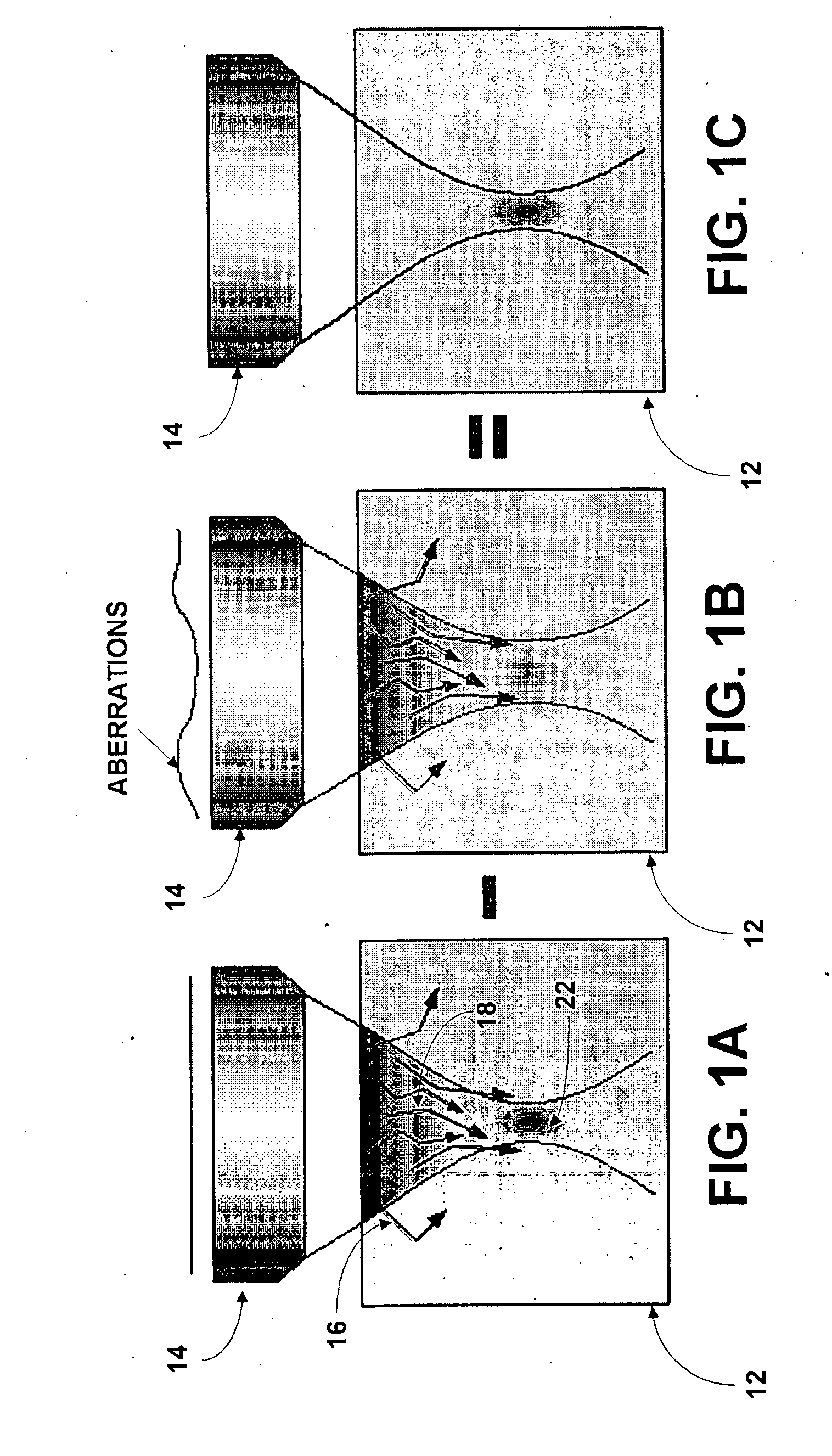
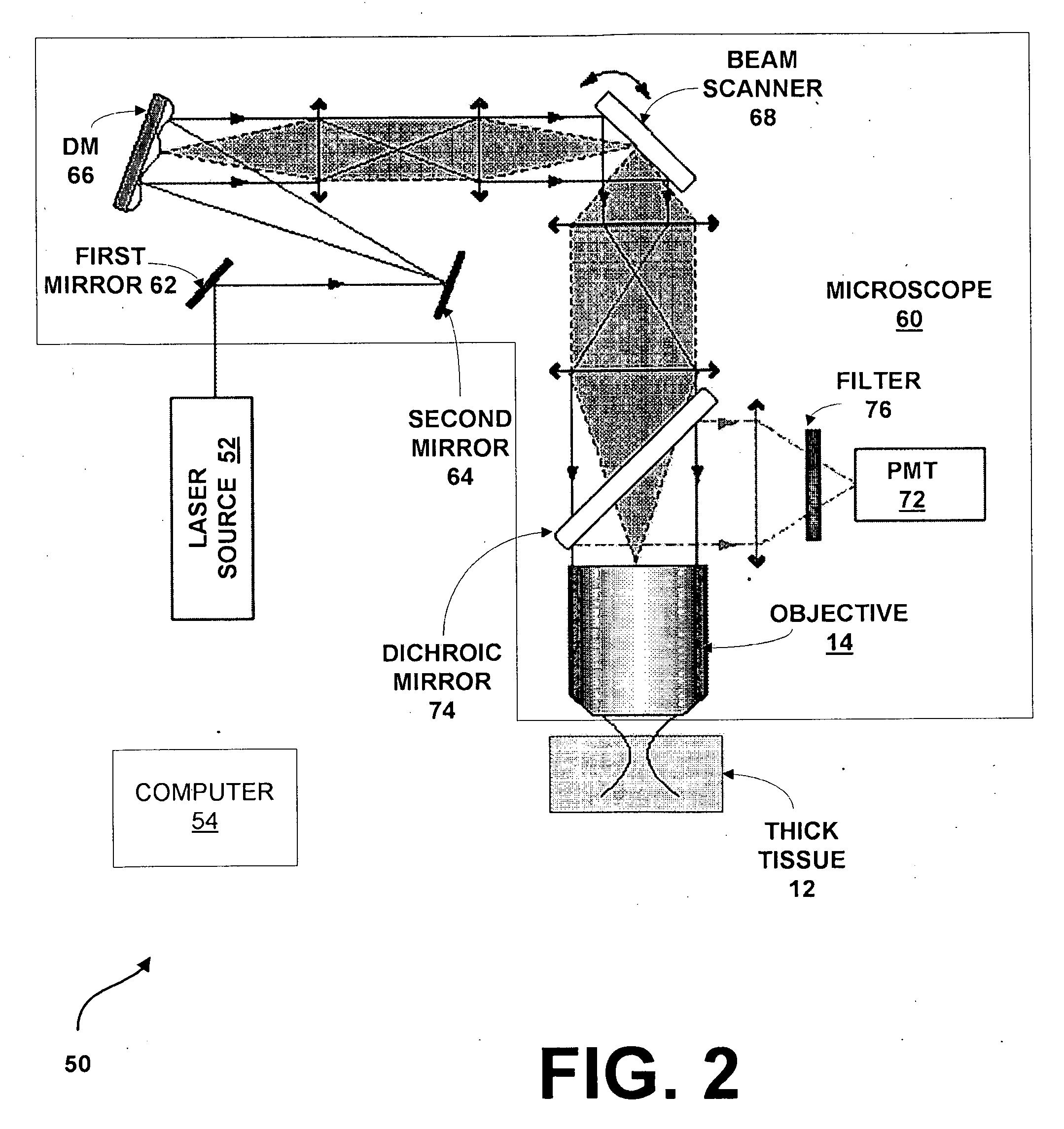
[0020]The present system and method provides a robust technique that significantly alleviates limitations to depth penetration of TPEF microscopy arising from out-of-focus background generated either by superficial ballistic excitation or by snakelike scattered excitation. The present system and method uses an aberrating element, such as, but not limited to, a deformable mirror, in an excitation light path. While in most applications involving a deformable mirror, the deformable mirror is meant to improve beam focus by compensating for sample-induced aberrations, in the present system and method the deformable mirror is meant to do just the opposite. Instead, the deformable mirror is used in the present system and method to introduce extraneous spatial aberrations in an excitation beam path so as to purposefully degrade the quality of the ballistic light focus, thereby quenching the TPEF signal and associated focal spot.

[0021]While the TPEF signal can be severely quenched with extra...


PUM


Abstract

A system for providing enhanced background rejection in thick tissue contains an aberrating element for introducing controllable extraneous spatial aberrations in an excitation beam path; at least one mirror capable of directing received laser pulses to the aberrating element; an objective; a beam scanner imaged onto a back aperture of the objective so that the beam scanner steers beam focus within the thick tissue; and a detector for recording signals produced by the tissue. An associated method comprises the steps of acquiring two-photon excited fluorescence of thick tissue without extraneous aberrations; introducing an extraneous aberration pattern in an excitation beam path; acquiring two-photon excited fluorescence of the thick tissue having the introduced extraneous aberration pattern; and subtracting the two-photon excited fluorescence with extraneous aberrations from the acquired standard two-photon excited fluorescence of the thick tissue without extraneous aberrations.

Description

CROSS-REFERENCE TO RELATED APPLICATION[0001]This application claims priority to copending U.S. Provisional Application entitled, “Enhanced Background Rejection In Thick Tissue With Differential-Aberration Two-Photon Microscopy,” having Ser. No. 60 / 975,855, filed Sep. 28, 2007, which is entirely incorporated herein by reference.FIELD OF THE INVENTION[0002]The present invention is generally related to imagine, and more particularly is related to deep tissue imaging.BACKGROUND OF THE INVENTION[0003]The statements in this section merely provide background information related to the present disclosure and may not constitute prior art.[0004]When a two-photon excited fluorescence (TPEF) microscope is used to image deep tissue, out-of-focus background can arise from both ballistic and non-ballistic excitation. TPEF microscopy has become a well-established tool for high-resolution imaging in scattering media such as thick tissue. While it is well accepted that TPEF microscopy provides greate...

Claims


Application Information

Patent Timeline
IPC IPC(8): G01N21/64
CPCG01N21/6458G02B26/06G02B21/365G02B21/16
Inventor MERTZ, JEROME
Owner TRUSTEES OF BOSTON UNIV
Why Eureka
Try Eureka