Spinal Cord's Epidural Space Detection By Using Fiber Optic Technology

a fiber optic technology and epidural space detection technology, applied in the field of hypodermic needles, can solve the problems of weak resistance of ligaments, headache or severe spinal injury after puncture of spinal cord, and tenacity of ligaments,

Inactive Publication Date: 2009-04-16
NATIONAL YANG MING UNIVERSITY
View PDF8 Cites 19 Cited by
  • Summary
  • Abstract
  • Description
  • Claims
  • Application Information

AI Technical Summary

Benefits of technology

[0007]Different tissues have different optical properties due to their different composition, such as absorbance and reflectivity, thus we can discriminate the tissue types where the needle reaches by analyzing their distinctive optical properties. Thus, the light analyzer of the present invention analyses the ratio of one reflective light to another, wherein the two reflective lights are produced by two incident lights on different wavelengths targeting to the same tissue, to provide the tissue type information. More specificity, the analyzer analyses the absorbency of the target tissue between two different lights. In one embodiment, if someone wishes to apply this device on epidural anesthesia, to the inventors' practical experience, the better light wavelengths for discrimination are one light wavelength from 620 nm to 680 nm and the other from 520 nm to 600 nm. In the best embodiment, one wavelength is 650 nm and the other is 580 nm. But in practical, there are some commercial products can emit 650 nm and 532 nm laser, so using these two wavelength light sources are more convenient. Besides, this light analyzer can also notify the user by sound or light when the distal tip of needle reaches the target tissue.

Problems solved by technology

For anesthesiologists, the most important issue in epidural anesthesia is whether the needle tip is accurately inserted into the correct position (epidural space).
This may in turn cause the post dural puncture headache (PDPH) or severe spinal injury.
For example, the tenacity of ligamentum flavum in old man is weak, and those in pregnant women and obese people also have weak resistance.

Method used


Image

  • Spinal Cord's Epidural Space Detection By Using Fiber Optic Technology
  • Spinal Cord's Epidural Space Detection By Using Fiber Optic Technology
  • Spinal Cord's Epidural Space Detection By Using Fiber Optic Technology

Examples


Embodiment Construction

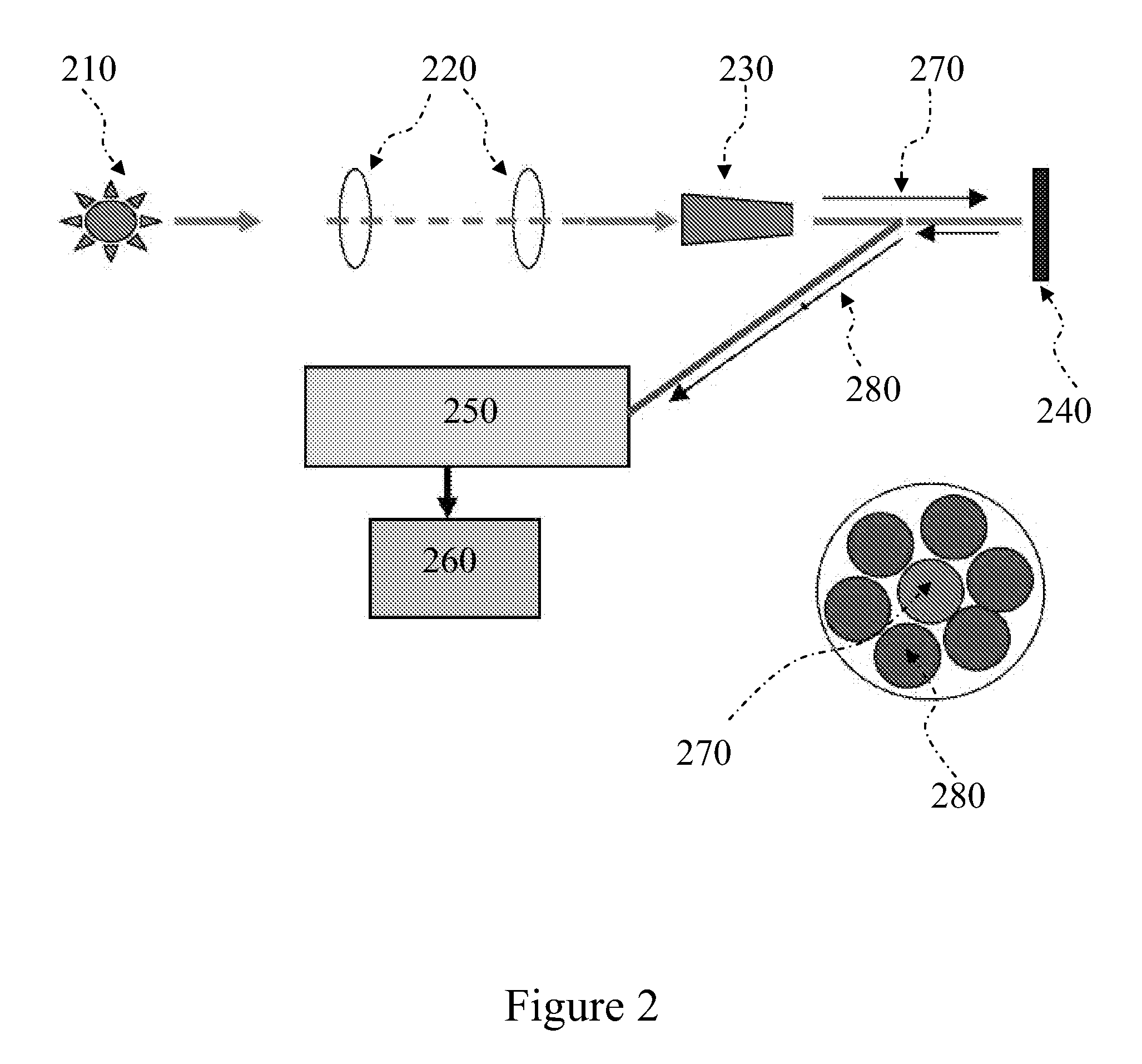
[0017]The idea of this invention is derived from the concept that different tissues due to their different composition have different optical properties, such as absorbance and reflection, so we can discriminate the tissue types where the needle reaches by their distinctive optical properties. Thus we provide a new device and a new method for tissue puncture, especially for epidural anesthesia, that can position a hypodermic needle in real time through optical technology to improve the accuracy of surgery.

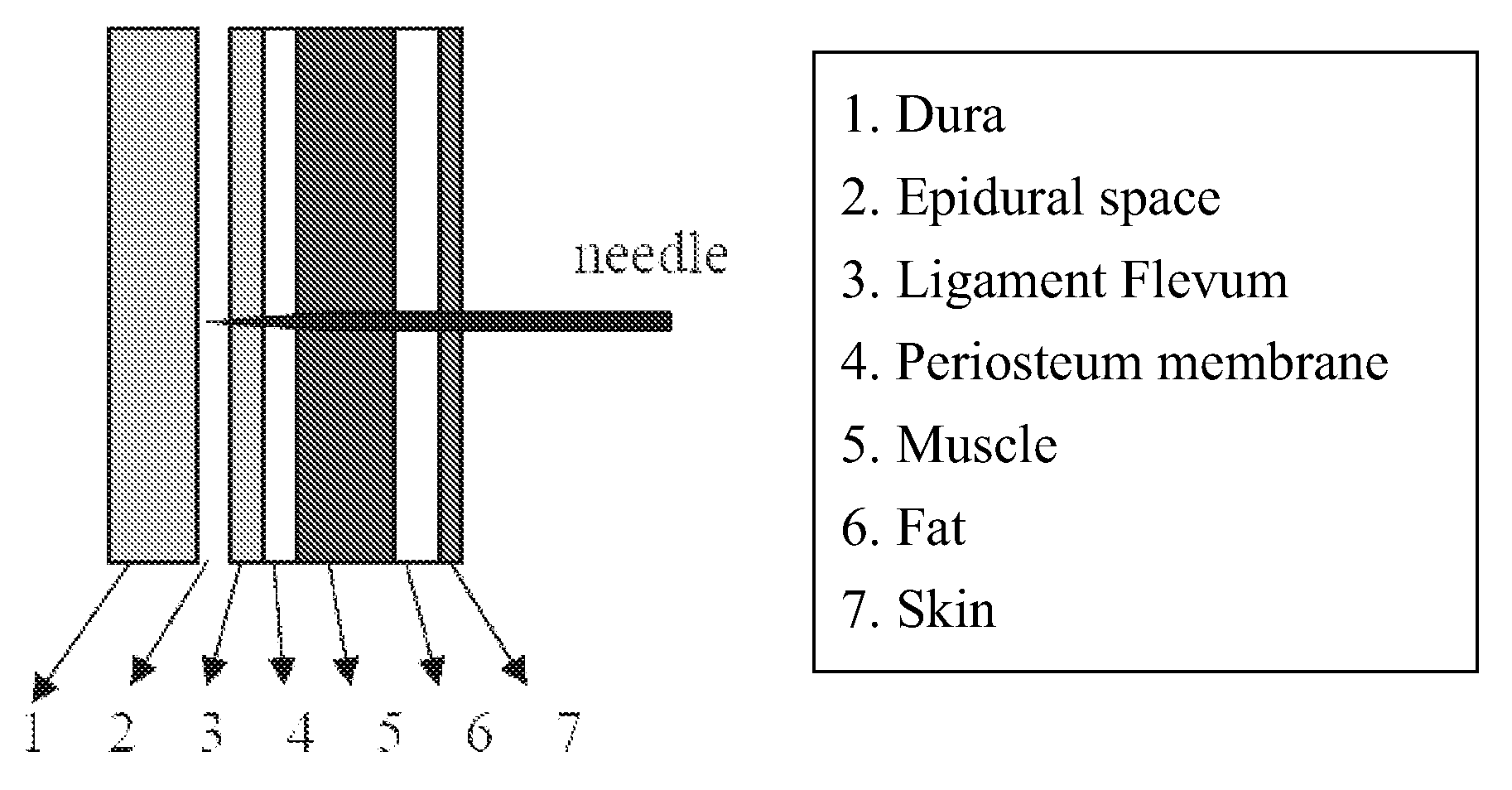
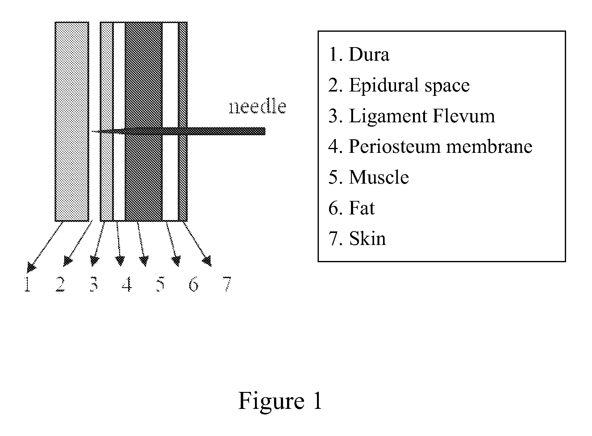
[0018]FIG. 1 shows a longitudinal profile of human posterior part and the tissue layers from outside to inside are skin, fat, muscle, periosteum membrane, ligament flavum, epidura space, and dura, but the spin is not indicated in this figure. During epidural anesthesia, the periosteum membrane is not necessary punctured.

[0019]In the present invention, the hypodermic needle used in the device is a normal clinical needle, which has a hollow inner bore to place an inner needle filled ...


PUM


Abstract

The present invention provides a device and a method for effectively processing epidural anesthesia by providing the needle-localization information in real time. The idea of this invention is derived from the concept that different tissues due to their different composition have different optical properties, such as absorbance and reflection, so we can discriminate the tissue types where the needle reaches by their distinctive optical properties. In this device, the inner solid needle of the puncture needle used before is replaced by a hollow needle which filled with optical fibers, and the distal end of those optical fibers are connected with two light sources of different wavelengths and a light analyzer. Thus, while qualifying and comparing the optical properties of those tissues, these device and method can be very helpful during epidural anesthesia and also reduce the risk of the surgery.

Description

FIELD OF THE INVENTION[0001]The present invention is related to the field of hypodermic needles and more specifically to hypodermic needles that are used to insert catheters or medication into the epidural space.DESCRIPTION OF RELATED ART[0002]Epidural anesthesia is a technique to reach the state of anesthetization by injecting some sedative into the epidural space. Those sedative blocks the spinal neural transmission temperately, so this surgery is also called “epidural block”, and is often applied on surgical operations. For anesthesiologists, the most important issue in epidural anesthesia is whether the needle tip is accurately inserted into the correct position (epidural space). As shown in FIG. 1, epidural space is a small lumen located between dural matter and ligamentum flavum that is hard to position and detect precisely, and its' lumen size varies with age and body shape. Normally, the epidural space in the adult lumbar spine is 3-6 mm deep, but in thorax it is only 1˜5 mm...

Claims


Application Information

Patent Timeline
Patent Type & Authority Applications(United States)
IPC IPC(8): A61N1/30A61M31/00
CPCA61M25/01A61M2025/0166A61M2025/0007
Inventor CHANG, YINTING, CHIEN-KUN
Owner NATIONAL YANG MING UNIVERSITY
Why Eureka
Try Eureka