Method and apparatus for remotely controlled navigation using diagnostically enhanced intra-operative three-dimensional image data

a three-dimensional image and remote control technology, applied in image enhancement, angiography, instruments, etc., can solve the problems of difficult registration of projection information provided, significant limitations associated with the use of x-ray projection imaging, and limited fluoroscopy, so as to achieve efficient diagnosis of conditions

Inactive Publication Date: 2009-04-23
STEREOTAXIS
View PDF100 Cites 146 Cited by
  • Summary
  • Abstract
  • Description
  • Claims
  • Application Information

AI Technical Summary

Benefits of technology

[0008]One object of the invention is to provide methods, devices and systems to perform a medical procedure utilizing diagnostically enhanced, intra-operative 3D image data set(s), the co-registered intra-operative data and diagnostic information being combined with a virtual or actual image of a remotely controlled navigation device into a real-time display. The 3D image data set can be acquired and reconstructed by various means including 3D X-ray rotational angiography, 3D / 4D ultrasound, MRI or other appropriate imaging modality. The 3D reconstructed image data set is registered to the navigation system by various means and approaches depending on the imaging source. For example, a 3D X-ray image can be inherently registered due to a known, fixed mechanical alignment of the X-ray and navigation system, while a 3D ultrasound data set could be registered using a localization system that tracks the position and orientation of the imaging device tip relative to the navigation system. The remotely navigated interventional device is visualized directly by the 3D imaging device (e.g. ultrasound) or indirectly by a localization means and associated device model to derive the virtual appearance of the device in the reconstructed 3D data set. The 3D reconstruction can be a fused representation of the anatomy whereby a static or periodically refreshed volumetric anatomical reconstruction is formed using a sweep of the external or internal imaging device and then fused with a real-time representation of a portion (e.g. a wedge) of the anatomy. The 3D reconstruction presents regions or targets based upon diagnostic and functional information related to the anatomy, the diagnostic information having been acquired through various internal and external methods. For example, the navigation device can be advanced to positions along a vessel or cardiac chamber wall to gather diagnostic information which when processed can then be displayed as regions of activity or therapy targets on the organ wall. The imaging device in this case could be a 3D ultrasound catheter, the catheter location being directly extracted from the image. There are many types of diagnostic information that could be collected including but not limited to voltage, electrical timing, impedance, tissue content and characterization, and blood pressure and velocity. By combining a diagnostically enhanced 3D or 4D reconstructed data set with a rendition of a remotely controlled navigation device that can be displayed directly or virtually co-registered to the 3D or 4D image data, the methods and systems of the present invention enable an operator to efficiently diagnose conditions and deliver correspondingly appropriate therapy to a plurality of targeted points within the patient anatomy.

Problems solved by technology

However significant limitations are associated with the use of x-ray projection imaging.
Besides subjecting the patient and potentially the operator to possibly large radiation dose, fluoroscopy is limited by the noisy nature of the acquired images, and by the superimposition of three-dimensional anatomy onto a single plane inherent to projection imaging.
While the pre-operative data provide an excellent 3D anatomical map of the region-of-interest at the time of the data acquisition, and therefore helps in planning the intervention, it is often difficult to register the projection information provided by the fluoroscopy to the pre-operative 3D reconstruction: the patient position with respect to the imaging chain might have changed; organs might have assumed a different shape or relative configuration as compared to the pre-operative acquisition; noise in the images renders the registration and registration evaluation difficult; and real-time demands put strict limits on the amount of computations that might be performed to bring two imaging modalities in registration.
Additionally, both the pre-operative 3D CT or ultrasound data and the fluoroscopy images present anatomical information from which diagnostic information might be difficult or impossible to extract; changes due to disease processes might not appear conspicuously on an anatomical map such as provided by x-ray attenuation coefficients that depend mostly on electron density at diagnostic energies.
However it does not disclose nor suggest the use of intra-operative 3D data, nor the use of ultrasound imaging, nor the use of two modalities of similar image quality; nor does it teach or suggest the use of magnetic navigation or the co-registration of diagnostic information onto image data.

Method used


Image

  • Method and apparatus for remotely controlled navigation using diagnostically enhanced intra-operative three-dimensional image data
  • Method and apparatus for remotely controlled navigation using diagnostically enhanced intra-operative three-dimensional image data
  • Method and apparatus for remotely controlled navigation using diagnostically enhanced intra-operative three-dimensional image data

Examples


Embodiment Construction

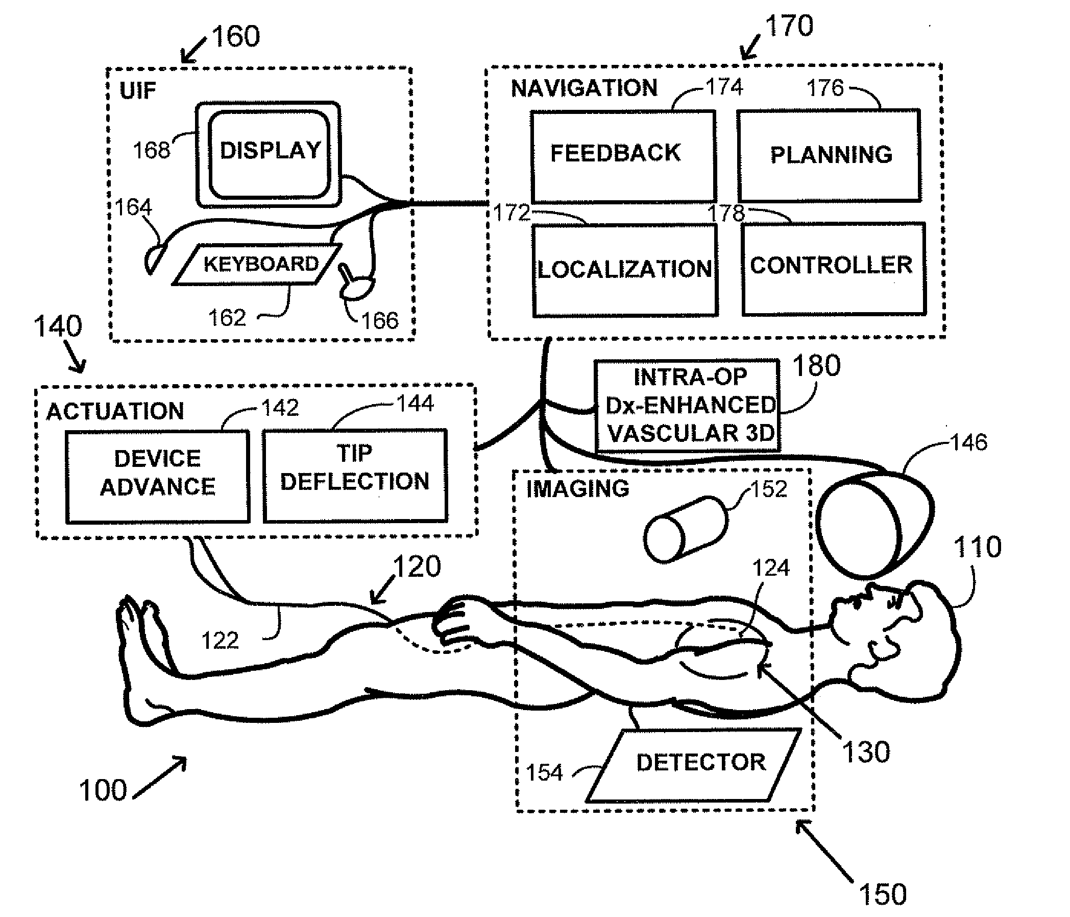
[0019]As illustrated in FIG. 1, a patient 110 is positioned within an interventional system, 100. An elongated navigable medical device 120 having a proximal end 122 and a distal end 124 is provided for use in the interventional system 100, FIG. 1-A, and the medical device is inserted into a blood vessel of the patient and navigated to an intervention volume 130. A means of applying force or torque to orient the device distal end 124 is provided, as illustrated by actuation block 140 comprising a device advance / retraction component 142 and a tip deflection component 144. The tip deflection means may be one of (i) a mechanical pull-wire system; (ii) a hydraulic or pneumatic system; (iii) an electrostrictive system; (iv) a magnetic system; or (v) other navigation system as known in the art. For illustration of a preferred embodiment, in magnetic navigation a magnetic field externally generated by magnet(s) assembly 146 orients a small magnet located at the device distal end (126, FIG....


PUM


Abstract

A method of performing intra-operative three-dimensional imaging and registering diagnostic functional information to the three-dimensional anatomical data is introduced. The availability of co-registered diagnostic information to intra-operative data enables fast and efficient navigation to pre-selected target areas, and allows automatic or semi-automatic treatment of cardiac cavity or vascular disease.

Description

CROSS-REFERENCE TO RELATED APPLICATIONS[0001]This application claims priority to U.S. Provisional Patent Application Ser. No. 60 / 981,472 filed Oct. 19, 2007. The disclosure of the above-referenced application is incorporated herein by reference.FIELD OF THE INVENTION[0002]This invention relates to methods, devices and systems for intra-operative three-dimensional image acquisition, the registration of a sequence of projection images to the three-dimensional reconstructed image data, and the display of diagnostic information registered to the three-dimensional reconstructed image data.BACKGROUND OF THE INVENTION[0003]Interventional medicine is the collection of medical procedures in which access to the site of treatment is made by navigation through one of the subject's blood vessels, body cavities or lumens. Interventional medicine technologies have been applied to the manipulation of medical instruments such as guide wires and catheters which contact tissues during surgical navigat...

Claims


Application Information

Patent Timeline
Patent Type & Authority Applications(United States)
IPC IPC(8): A61B5/05G06K9/00A61B19/00
CPCA61B1/00158G06T2207/30101A61B6/503A61B6/504A61B8/0891A61B8/12A61B8/4461A61B8/483A61B19/46A61B19/5244A61B2019/5289G06K2209/057G06T7/0038G06T2207/10072G06T2207/10121G06T2207/10136G06T2207/30048A61B5/7289A61B34/20A61B2090/364A61B90/06G06T7/38G06V2201/034
Inventor GARIBALDI, JEFFREY M.
Owner STEREOTAXIS
Why Eureka
Try Eureka