Extracellular histones as biomarkers for prognosis and molecular targets for therapy
- Summary
- Abstract
- Description
- Claims
- Application Information
AI Technical Summary
Benefits of technology
Problems solved by technology
Method used
Image
Examples
example 1
Materials & Methods
[0204]Materials. Mouse protein C, bovine factor V, X and thrombin, rat anti-mouse EPCR (MEPCR1560) and mouse TM (MTM1703) were produced in this laboratory according to the standard procedures. Human recombinant APC (Xigris) was purchased from Eli Lilly. Calf thymus histones (Sigma), calf thymus histone H4 (Roche), LPS from Salmonella typhimurium (Sigma), mouse recombinant IFN (Biosource), synthetic histone H4 peptides (GenScript), E. coli M15 strain (Qiagen) were also purchased. E. coli B7 strain was provided by Dr. F. B. Taylor.
[0205]Animals. Six to 10-week male C57BL / 6 mice (Jackson Lab) were used according to an animal protocol approved by Institutional Animal Care and Use Committees of the Oklahoma Medical Research Foundation.
[0206]Mouse peritoneal macrophage. Mice were injected intraperitoneally with 2 ml 3% thioglycollate medium. Four days later peritoneal exudate cells were harvested by lavage with 10 ml cold HBSS containing 10 U / ml heparin. Peritoneal cell...
example 2
Results
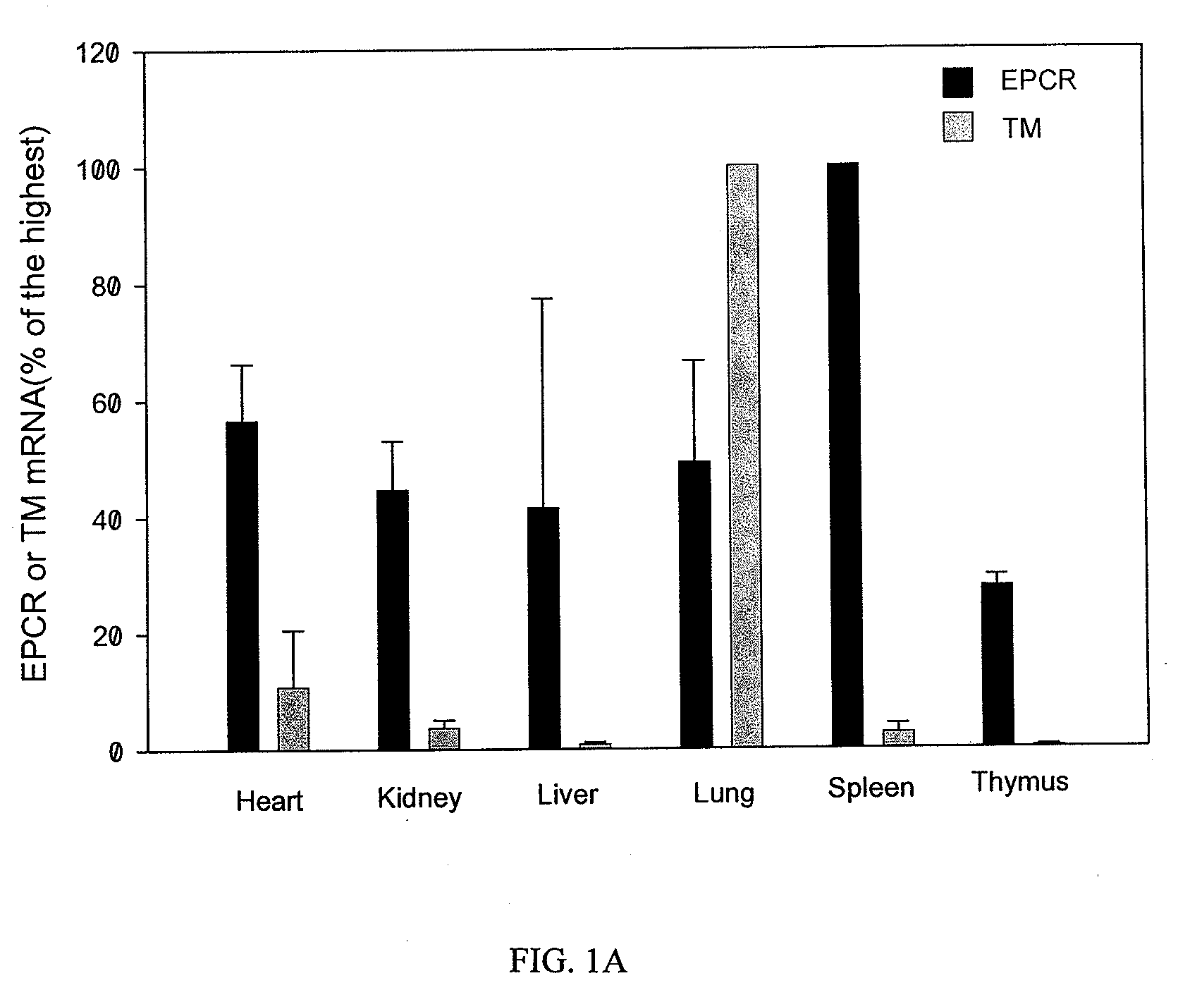
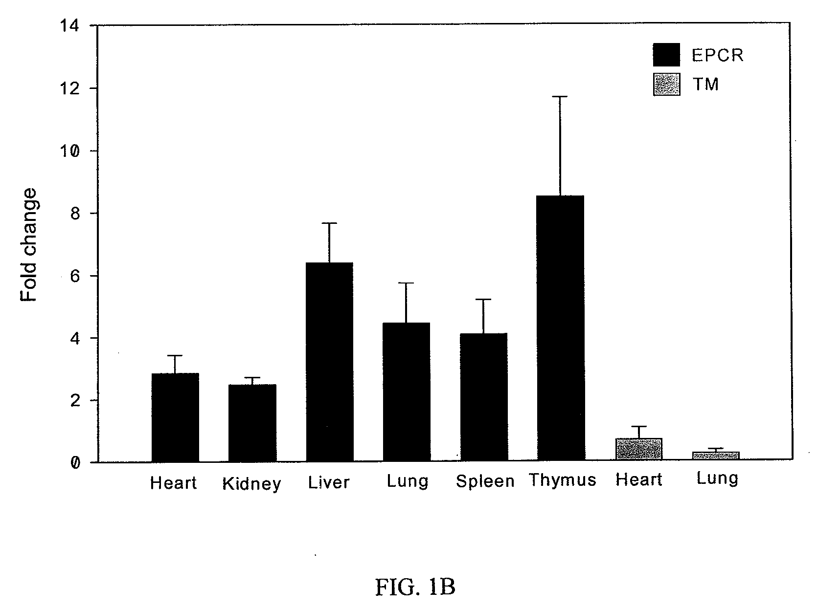
[0216]Upregulation of EPCR and protein C activation on macrophages activated by LPS and IFN. Unlike TM mRNA expression pattern which is highest in mouse lung tissue but low in heart, kidney, liver, spleen and thymus, EPCR mRNA is highly expressed in spleen and other organ tissues (FIG. 1A). In contrast to down regulation of TM mRNA in mouse lung and heart tissues, EPCR mRNA was up regulated by LPS challenge in all organ tissues we examined (FIG. 1B). Immunoprecipitation and Western blot confirmed that EPCR protein was highly expressed in spleen and other organ tissues (FIG. 1C). These results indicated that EPCR might be expressed on other cell types in addition to endothelial cells.
[0217]In the study of EPCR expression on various mouse immune cells, the inventors found that EPCR could be dramatically up regulated on peritoneal macrophages by LPS and IFN (FIG. 2A), in contrast to the down regulation of TM (FIG. 2B). EPCR mRNA was also greatly increased after LPS and IFN stimu...
example 3
Materials and Methods
[0222]Methods. Human protein C, bovine thrombin and rat anti-mouse protein C mAb (MPC1609) were produced in our laboratory according to standard procedures23. Human recombinant APC (Xigris) was purchased from Eli Lilly. Calf thymus histones (Sigma), calf thymus histone H1, H2A, H2B, H3 and H4 (Roche), phosphatidylcholine (PC), phosphatidylserine (PS), phosphatidylethanolamine (PE) (Avanti Polar Lipids), LPS from Salmonella typhimurium (Sigma), murine recombinant IFN (Biosource), goat anti-histone H3 (Santa Cruz) and PPACK (Calbiochem) were also purchased. PS / PC (20:80) and PE / PS / PC (40:20:40) liposomes were prepared by membrane extrusionil. Mouse anti-histone H2B (LG2-2) and anti-histone H4 (BWA-3) mAbs were generated from autoimmune mice as previously described24.
[0223]Animals. Six to 8 week male C57BL / 6 mice (Jackson Lab) were used according to an animal protocol approved by Institutional Animal Care and Use Committees of the Oklahoma Medical Research Foundati...
PUM
| Property | Measurement | Unit |
|---|---|---|
| Permeability | aaaaa | aaaaa |
| Content | aaaaa | aaaaa |
| Cytotoxicity | aaaaa | aaaaa |
Abstract
Description
Claims
Application Information
 - R&D
- Intellectual Property
- Life Sciences
- Materials
- Tech Scout
- Unparalleled Data Quality
- Higher Quality Content
- 60% Fewer Hallucinations
Browse by: Latest US Patents, China's latest patents, Technical Efficacy Thesaurus, Application Domain, Technology Topic, Popular Technical Reports.
© 2025 PatSnap. All rights reserved.Legal|Privacy policy|Modern Slavery Act Transparency Statement|Sitemap|About US| Contact US: help@patsnap.com


