Biodegradable nanoparticle having t-cell recognizable epitope peptide immobilized thereon or encapsulated therein

a biodegradable nanoparticle and epitope technology, applied in the direction of peptide/protein ingredients, immunological disorders, antibody medical ingredients, etc., can solve the problems of limited clinical use and likely side effects of treatmen

Inactive Publication Date: 2009-06-18
TAIHO PHARMA CO LTD
View PDF2 Cites 15 Cited by
  • Summary
  • Abstract
  • Description
  • Claims
  • Application Information

AI Technical Summary

Benefits of technology

[0047]The biodegradable nanoparticles for use in the present invention are not limited particularly in shape and are generally spherical and usually 80 nm to 100 μm, preferably 100 nm to 50 μm, in size. When so sized, the particles can be given, for example, an increased surface area per unit weight, thereby permitting the T cell recognizable epitope peptide to be immobilized in an increased amount, made retainable in tissues more effectively and to be taken into cells in controllable manner, hence advantageous effects. When otherwise shaped, nanoparticles are sized substantially the same as spherical nanoparticles.
[0048]The biodegradable nanoparticles for use in the present invention can be prepared by using known methods. Examples of preparation methods are submerged drying method, spray drying method, spherical crystallization method, solvent replacement method (precipitation / dialysis method), direct ultrasonic dispersion method, etc. For example, biodegradable nanoparticles comprising poly(γ-glutamic acid) and those comprising poly(ε-lysine) can be prepared by the solvent replacement method. Biodegradable polysaccharide nanoparticles can be prepared, for example, by the direct dispersion method. Biodegradable poly(organic acid) nanoparticles can be prepared, for example, emulsion-submerged drying method. Such method are suitably selected and used in combination to provide biodegradable nanoparticles which are adjusted or controlled in material, components, molecular weight, size, electric charge and other parameters in conformity with the purpose. When desired, nanoparticles may be joined by matrix cross-linking.
[0049]Various T cell recognizable epitope peptides are usable for immobilization on or encapsulation in biodegradable nanoparticles. Preferable as T cell recognizable epitope peptides are cedar pollen T cell recognizable epitope peptides. These peptides include, for example, P1: 277-290 (KQVTIRIGCKTSSS) (Yoshitomi T et al: Immunology 10)7: 517-520, 2002) which is BALB / c mouse T cell recognizable epitope peptide for Cryj1, P2: 70-83 (HFTFKVDGIIAAYQ) and P2: 246-259 (RAEVSYVHVNGAKF (Yoshitomi T at al: Immunology 107: 517-520, 2002, Hirahara S et al; J. Allery Clin. Immunol. 102: 961-967, 1998, Murasugi T et al; Eur. J. Pharmacol. 510: 143-148, 2005) which are BALB / c mouse T cell recognizable epitope peptidea for Cryj2, human T cell recognizable epitope peptides for Cryj1 reported in p16-30, p81-95, p91-105, p106-120, p111-125, p151-165, p156-170, p191-205, p211-225, p231-245, p301-315, p316-330, p331-345, etc, human T cell recognizable epitope peptides for Cryj2 reported in p66-80.p76-90, p81-95, p96-107, p141-155, p146-160, p181-195, p186-200, p236-250, p336-350, p346-360, p351-365 (Sone T et al; J. Immunol. 161:448-457, 1998, Hirahara K et al: J. Allergy Clin. Immunol. 108: 94-100. 2001), etc. More preferable are human or mouse cedar T cell recognizable epitope peptide. These peptides are used singly or in combination as T cell recognizable epitope peptides of the invention.
[0050]A suitable T cell recognizable epitope peptide can be selected for immobilization on or encapsulation in biodegradable nanoparticles, in accordance with the state of the subject of administration, for example, the kind, age, body weight and health condition of the animal, the kind of disease to be prevented, and / or already developed and to be treated, or the cause thereof. Biodegradable nanoparticles may have immobilized thereon or encapsulated therein a single kind of or at least two kinds of T cell recognizable epitope peptides.
[0051]The T cell biodegradable epitope peptide can be immobilized on or encapsulated in biodegradable nanoparticles by various known methods. The epitope peptide may be immobilized on or encapsulated in biodegradable nanoparticles directly or by means of a linker such as polyethylene glycol (PEG). Peptides are immobilized or encapsulated by known methods, such as the bonding method using covalent bonds, ionic bonds or intermolecular forces, adsorption method or inclusion method. For example, the functional group on the biodegradable nanoparticle may be linked to the functional group of the peptide by a covalent bond for immobilization or encapsulation. The immobilization or encapsulation may be effected by an ionic bond when the charge on the nanoparticle is opposite to that of the peptide. For example, the peptide can be immobilized on the biodegradable poly(γ-glutamic acid) nanoparticle by the inclusion method by introducing a hydrophobic amino acid into poly(γ-glutamic acid) by covalent bonds, dissolving the resulting acid in an organic solvent and subsequently adding an aqueous solution of the peptide dropwise to the solution. Alternatively, the peptide may be immobilized on or encapsulate in biodegradable nanoparticles by a suitable combination of the bonding method, adsorption method and / or inclusion method. Such a mode of immobilization or encapsulation can be suitably selected in accordance with the purpose of use (e.g., the kind of subject or disease).
[0052]The biodegradable nanoparticles of the invention having a T cell recognizable epitope peptide immobilized thereon or encapsulated therein have the advantage that the peptide remains unaffected in its stereostructure, such that the immobilized or encapsulated peptide is less likely to alter in amount or properties even after freeze-drying and can be preserved for a prolonged period of time.

Problems solved by technology

While this immunotherapy has been accepted as the sole therapy of which a complete cure can be expected unlike drug therapies, the treatment is likely to produce side effects such as anaphylaxis since the antigen used is a pollen extract.
Accordingly, the therapy has the problem that the extract can be administered only in very small amounts to suppress the development of the side effect, and the period of administration is as long as several years, therefore has found limited clinical use.
The administration of peptides to the living body generally appears to involve the following problems.(1) The peptide is decomposed by an enzyme and therefore can not be maintained at an effective concentration (problem of stability).(2) The digestive tract membrane is very low in permeability to the peptide, which therefore can not be administered orally without problems.(3) If attempted for the purpose of inducing a reaction through mucosal immunity, the submucous administration of the peptide involves problems with respect to tissue retentivity.

Method used


Image

  • Biodegradable nanoparticle having t-cell recognizable epitope peptide immobilized thereon or encapsulated therein
  • Biodegradable nanoparticle having t-cell recognizable epitope peptide immobilized thereon or encapsulated therein
  • Biodegradable nanoparticle having t-cell recognizable epitope peptide immobilized thereon or encapsulated therein

Examples


example 1

[0063](1) Preparation of Mouse Cedar Pollen T Cell Recognizable Epitope Peptides as Converted to PEG (Polyethylene Glycol)

[0064]P1: 277-290; KQVTIRIGCKTSSS (hereinafter referred to as “P1”) was selected as BALB / c mouse T cell recognizable epitope peptide for Cryj1, and P2: 246-259; RAEVSYVHVNGAKF (hereinafter referred to as “P2”) was selected as a BALB / c mouse T cell recognizable epitope peptide for Cryj2. The conversion of these peptides to PEG (polyethylene glycol) was made by Toray Research Center Co. Ltd. Mouse cedar pollen T cell recognizable epitope peptides as converted to PEG (polyethylene glycol) were prepared by the Fmoc solid-phase process.

[0065](2) Preparation of Biodegradable Nanoparticles Having Mouse Cedar Pollen T Cell Recognizable Epitope Peptide Immobilized Thereon

[0066]Poly(γ-glutamic acid) (γ-PGA, 300,000 in molecular weight) in an amount of 607 mg (4.7 unit mmols) was dissolved in 100 ml of 54 mM aqueous solution of sodium hydrogencarbonate (pH 8.5). Subsequentl...

example 2

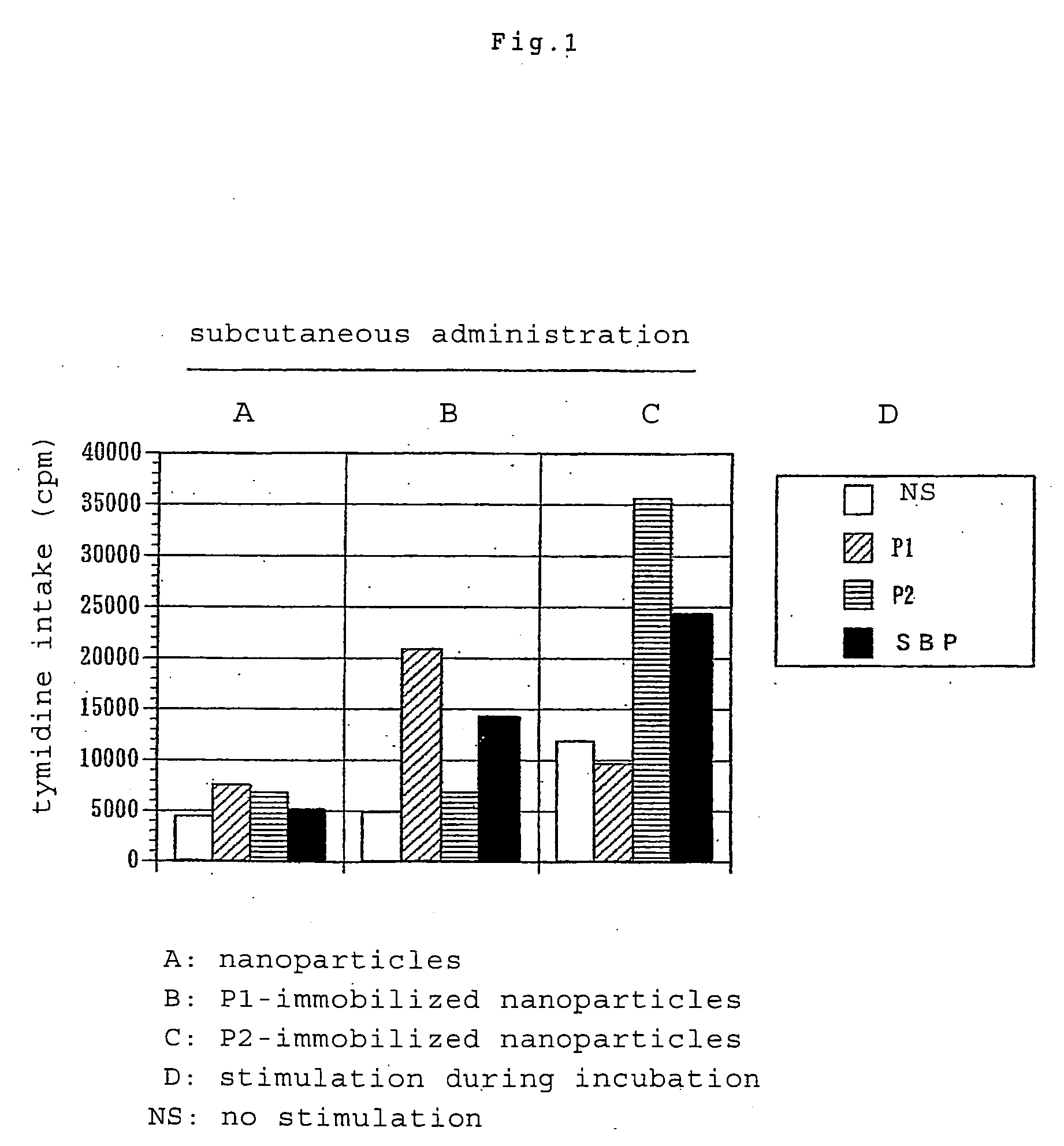
Immunization Potential of Biodegradable Nanoparticles Having Immobilized Thereon Mouse Cedar Pollen T Cell Recognizable Epitope Peptide

[0067]Subcutaneously injected into the footpads of BALB / c mice (6-week-old male) was 100 μl (50 μl for each footpad) of a suspension of biodegradable nanoparticles having immobilized thereon Cryj1 or Cryj2 T cell recognizable epitope peptide (P1 or P2) (the amount of nanoparticles corresponding to 20 μg of the peptide) or nanoparticles having no peptide immobilized thereon. On day 11, a draining lymph node was removed to collect lymph node cells, which were then suspended in a mixture of RPM 11640 medium and 10% fetal calf serum (FCS). Such cells from two mice were combined together for use in each group. The cells were placed onto a 96-well incubation plate in an amount of 5×10⁵ in each well. Further placed into each well as an antigen stimulus was P1 or P2 to a final concentration of 20 μg / ml or a cedar pollen roughly purified antigen (Sugi Basic P...

example 3

[0070]Immunomodulating Effect of Biodegradable Nanoparticles Having Immobilized Thereon Mouse Cedar Pollen T Cell Recognizable Epitope Peptide

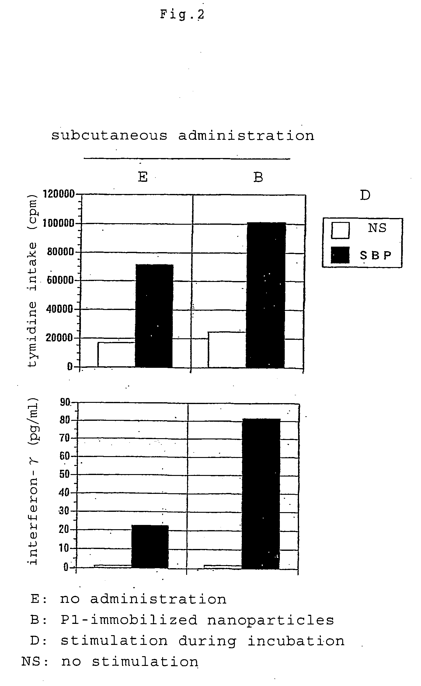
[0071](1) Th1 Cytokine Immunity Induction by Subcutaneous Administration

[0072]Subcutaneously injected into the footpads of BALB / c mice (10-week-old male) was 100 μl (50 μl for each sole) of a suspension of biodegradable nanoparticles having immobilized thereon Cryj1 T cell recognizable epitope peptide (P1) (the amount of nanoparticles corresponding to 50 μg of the peptide). On day 16, a suspension of 5 μg of cedar pollen roughly purified antigen SBP in Freund's incomplete adjuvant was injected into the right footpads to give rise to an immune response. Five days thereafter, draining lymph node cells were collected, placed onto a 96-well incubation plate in the same manner as in Example 2 and stimulated with cedar pollen roughly purified antigen SBP at a final concentration of 10 μg / ml. The proliferation of cells was measured by the same method...


PUM

PropertyMeasurementUnit
sizeaaaaaaaaaa
sizeaaaaaaaaaa
pHaaaaaaaaaa

Abstract

A biodegradable nanoparticle having a T cell recognizable epitope peptide immobilized thereon or encapsulated therein of the present invention is usable as a safe and effective immunotherapeutic agent, and is useful as an immunotherapeutic agent for treating, for example, pollinosis, year-round nasal allergic disease and seasonal nasal allergic disease.

Description

TECHNICAL FIELD[0001]The present invention relates to biodegradable nanoparticles having immobilized thereon or encapsulated therein a T cell recognizable epitope peptide, more particularly a T cell recognizable epitope peptide of pollinosis patients, and / or to an immunotherapeutic agent comprising the nanoparticle.BACKGROUND ART[0002]The immunotherapy for pollinosis is attributable to the finding in 1910's that injections of an extract of pollen to pollinosis patients in amounts gradually increasing from a small amount were effective. This method, which is termed also the desensitization therapy, has since been found empirically effective and practiced widely. While this immunotherapy has been accepted as the sole therapy of which a complete cure can be expected unlike drug therapies, the treatment is likely to produce side effects such as anaphylaxis since the antigen used is a pollen extract. Accordingly, the therapy has the problem that the extract can be administered only in ve...

Claims


Application Information

Patent Timeline
Patent Type & Authority Applications(United States)
IPC IPC(8): A61K38/16A61P37/08
CPCA61K9/0019A61K9/0048A61K9/5146B82Y5/00A61K47/48907A61K2039/57A61K39/35A61K47/6935A61P11/02A61P37/08A61K39/00A61K47/34A61K47/42A61K47/50
Inventor AKASHI, MITSURUIKIZAWA, KOICHI
Owner TAIHO PHARMA CO LTD
Why Eureka
Try Eureka