
Pro-clotting enzyme, and method for detection of endotoxin or (1-3)-beta-d-glucan using the same

a pro-clotting enzyme and endotoxin technology, applied in the field of nucleic acid fragment encoding a pro-clotting enzyme, can solve the problems of limited standard technique and difficult development of pro-ce activity, and achieve the effects of stable pro-ce activity and quality, low cost, and good stability

Inactive Publication Date: 2009-08-20
SEIKAGAKU KOGYO CO LTD
View PDF5 Cites 14 Cited by
  • Summary
  • Abstract
  • Description
  • Claims
  • Application Information

AI Technical Summary

Benefits of technology

[0055]The nucleic acid fragment of the present invention is very useful, since the virus of the present invention, which is useful for steady, low-cost, and large-scale production of Pro-CE of satisfactory quality, can be provided through employment of the nucleic acid fragment. The virus of the present invention is very useful, since the cell of the present invention, which is useful for steady, low-cost, and large-scale production of Pro-CE of satisfactory quality, can be provided through employment of the virus. The cell of the present invention is very useful, since the cell can produce a protein maintaining Pro-CE activity and satisfactory quality, steadily, at low cost, and on a large scale and can also provide the method and kit of the present invention. The method and kit of the present invention realize detection and measurement of Et and BG without preparing a lysate from horseshoe crabs, which are precious bio-resources, and can provide a very useful technique in terms of protection of lifeforms, provision of animal alternatives, cost, precision, reproducibility, etc.

Problems solved by technology

Although Pro-CE was previously cloned (Non-Patent Document 2), a protein (Pro-CE) maintaining an enzymatic activity is difficult to develop when the corresponding nucleic acid fragment is employed.
More specifically, the standard technique is limited to selection of 23 clones from a λgt11 cDNA library (1,500,000 clones) by use of an anti-CE antibody, subcloning to a pUC118 / 119 vector, and determination of the nucleotide sequence.

Method used


Image

  • Pro-clotting enzyme, and method for detection of endotoxin or (1-3)-beta-d-glucan using the same
  • Pro-clotting enzyme, and method for detection of endotoxin or (1-3)-beta-d-glucan using the same
  • Pro-clotting enzyme, and method for detection of endotoxin or (1-3)-beta-d-glucan using the same

Examples


example 1

Expression of Pro-CE by Use of Insect Cells

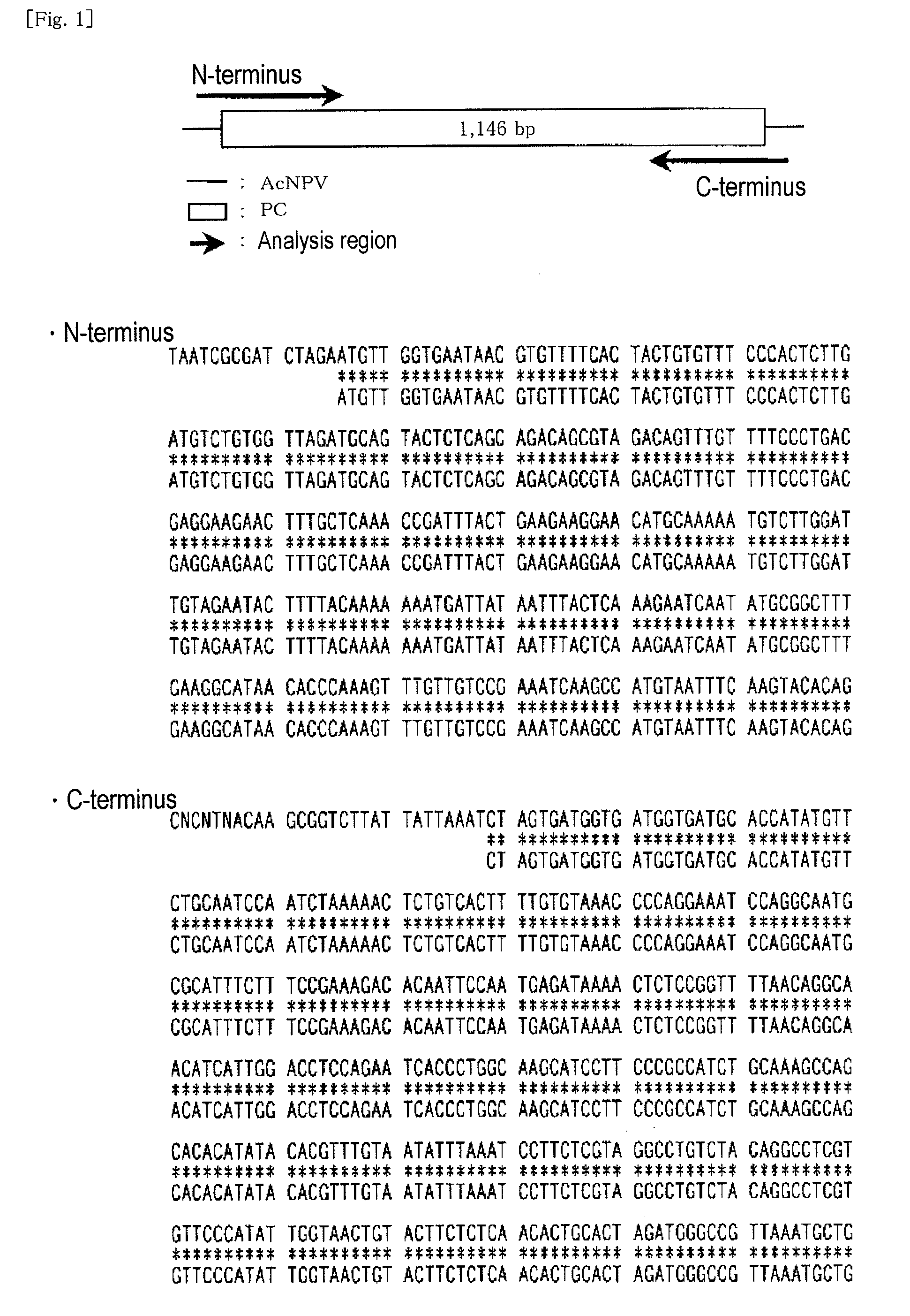
[0162]A cDNA fragment represented by SEQ ID NO: 3 (having a nucleotide sequence of nucleotides 1 to 1125 in SEQ ID NO: 3, a His-Tag sequence being attached to the C-terminus) was kindly offered by Dr. Tatsushi MUTA (Department of Molecular and Cellular Biochemistry, Kyushu University Graduate School of Medical Science; currently, Department of Bio-Science, Tohoku University Graduate School). The cDNA fragment had been prepared through a method disclosed in Non-Patent Document 2. The cDNA fragment was introduced into a transfer vector (pPSC8), and a clone having a predetermined nucleotide sequence was selected. The thus-selected expression vector (PC / pPSC8) DNA fragment and a baculovirus (AcNPV) DNA fragment were co-transfected into Sf9 cells. The virus fluid obtained from the culture supernatant was purified and amplified. The viral DNA fragment was extracted from the cells infected with the baculovirus, and sequenced. Insect cells (expresS...

example 2

Detection of Recombinant Pro-CE Activity

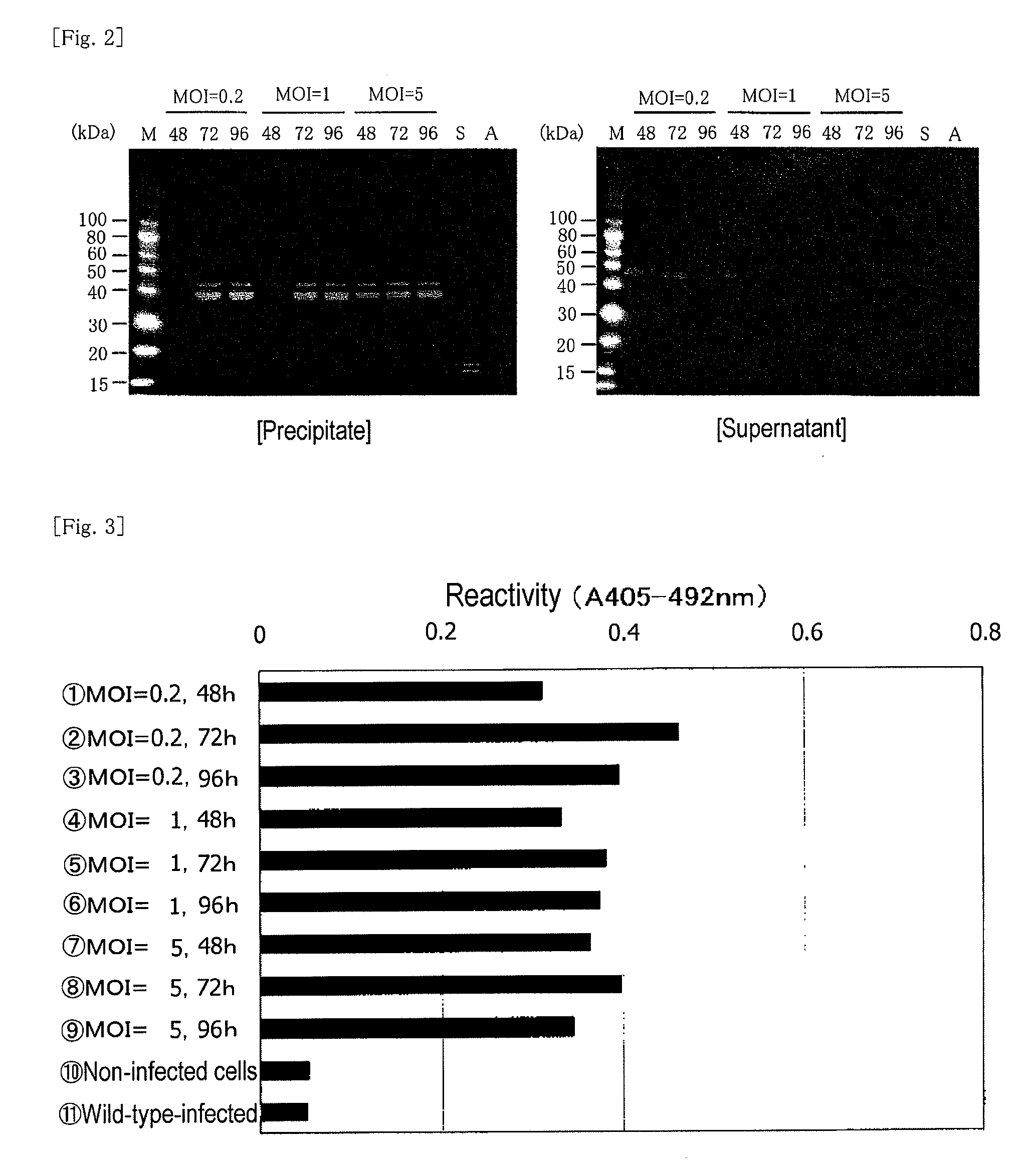
[0182]Insect cells (expresSF+) were diluted with a serum-free Sf-900 II medium so as to adjust the concentration to 1.5×10⁶ cells / mL, and the diluted product (100 mL / per flask) was placed in nine 250-mL Erlenmeyer flasks. The aforementioned third-generation virus fluid was added thereto so as to attain MOIs of 0.2, 1, and 5 (3 flasks each), respectively. Each mixture was subjected to shake cultivation at 130 rpm and 28° C. for 48, 72, and 96 hours. After completion of cultivation, each culture liquid was centrifuged at 3,000×g and 4° C. for 20 minutes, to thereby fractionate into the supernatant and the precipitate. The supernatant was preserved in a frozen state.

Sample 1: MOI=0.2, 48 hours

Sample 2: MOI=0.2, 72 hours

Sample 3: MOI=0.2, 96 hours

Sample 4: MOI=1, 48 hours

Sample 5: MOI=1, 72 hours

Sample 6: MOI=1, 96 hours

Sample 7: MOI=5, 48 hours

Sample 8: MOI=5, 72 hours

Sample 9: MOI=5, 96 hours

Sample 10: non-virus-infected cells

Sample 11: wild-typ...

example 3

Detection of Expression of Activity in a Complete Reconstitution System Employing Recombinant Pro-CE and Recombinant Factor G

Reagents

[0191](1) recombinant factor G (culture supernatant; α-subunit:β-subunit=2:1)

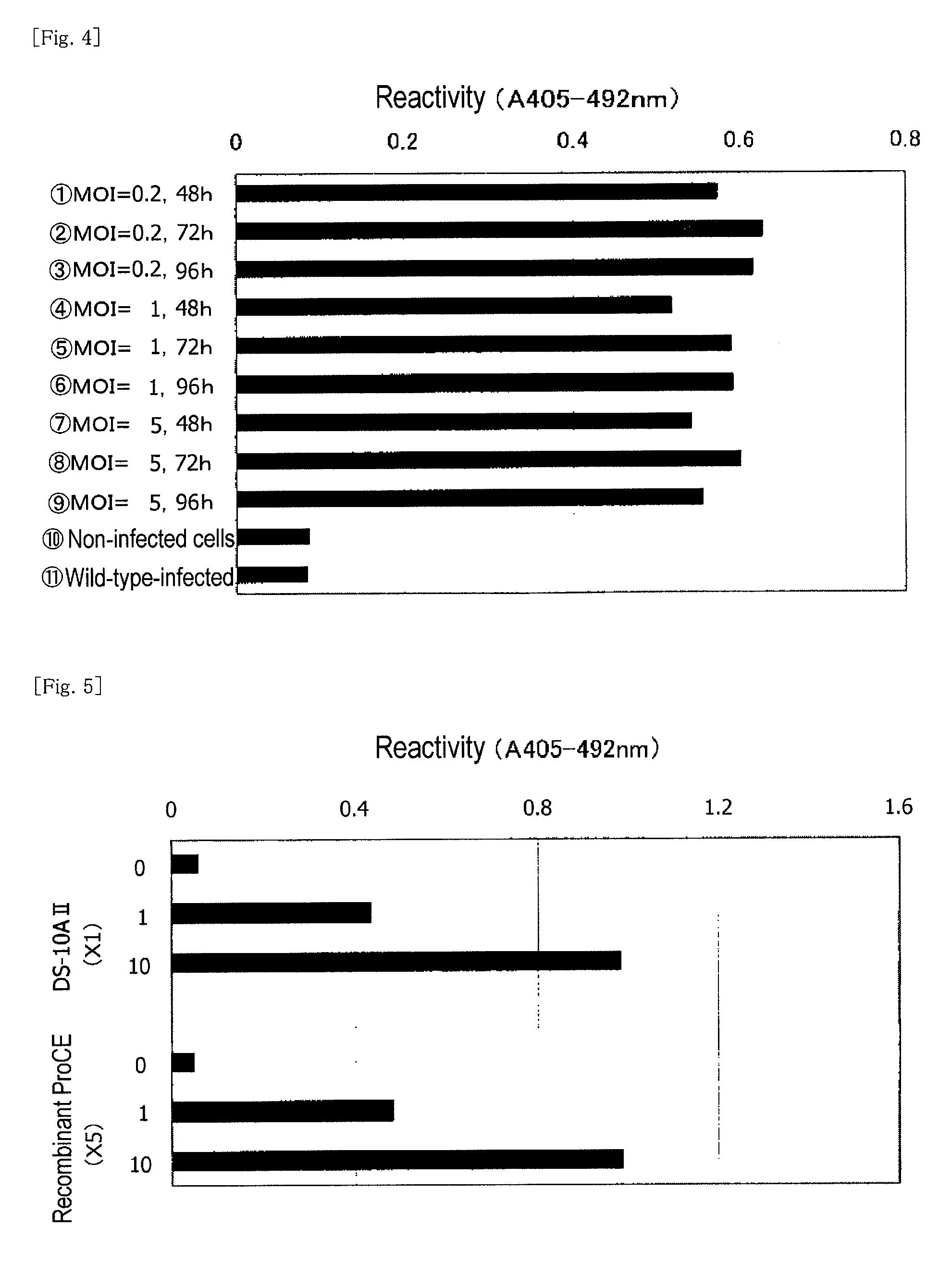
(2) recombinant Pro-CE (culture supernatant of sample 2 in Example 2)

(3) BG:CSBG (1,495 ng / vial) dissolved in distilled water (1.495 mL), followed by ×10-stepwise dilution

[0192]Each of the culture supernatants (1) and (2), which had been produced through the method disclosed in Japanese Patent Application Laid-Open (kokai) No. 2006-271384 (Patent Document 2), was five-fold diluted with Tris-HCl buffer (pH: 7.5) (50 mL) containing 150 mM NaCl. In this experiment, a nucleotide sequence represented by SEQ ID NO: 21 was employed as a DNA fragment encoding α-subunit of factor G. To the thus-diluted recombinant factor G (25 μL), recombinant Pro-CE (25 μL), Tris-HCl buffer (pH: 8.0) (final concentration: 80 mM), MgSO4 (final concentration: 64 mm), CaCl2 (final concentration: 0.4 mM),...


PUM

PropertyMeasurementUnit
Volumeaaaaaaaaaa
Volumeaaaaaaaaaa
Volumeaaaaaaaaaa

Abstract

Objects of the present invention are to provide a DNA fragment encoding a limulus-derived pro-clotting enzyme, a virus harboring the DNA fragment, a cell harboring the virus, a method of producing the pro-clotting enzyme by use of the cell, and means for assaying an endotoxin or (1→3)-β-D-glucan employing the enzyme, wherein these elements are capable of producing an endotoxin or (1→3)-β-D-glucan assay reagent of satisfactory quality, steadily, at low cost, and on a large scale. In the present invention, for example, a DNA fragment encoding a protein having an amino acid sequence defined by SEQ ID NO: 4 is selected as a nucleic acid fragment encoding a limulus-derived pro-clotting enzyme, and the corresponding recombinant pro-clotting enzyme. Use of the enzyme can provide a high-sensitivity method and kit for detecting (1→3)-β-D-glucan and an endotoxin, utilizing a cascade reaction system in a horseshoe crab lysate.

Description

TECHNICAL FIELD[0001]The present invention relates to a nucleic acid fragment encoding a pro-clotting enzyme derived from a horseshoe crab (hereinafter may be referred to as a limulus-derived pro-clotting enzyme), to a virus harboring the nucleic acid fragment, to a cell harboring the virus, to a method of producing the pro-clotting enzyme by use of the cell, and to a method and a kit for detecting Et or BG employing the pro-clotting enzyme produced through the production method.BACKGROUND ART[0002]There are disclosed methods for determining Et or BG by use of an amebocyte lysate of a horseshoe crab (i.e., a horseshoe crab hematocyte extract, hereinafter referred to simply as a lysate). These methods are based on coagulation of the lysate by Et or BG. The coagulation reaction occurs through cascade reaction of several coagulation factors (Patent Document 1 and Non-Patent Document 1).[0003]For example, when BG is brought into contact with the lysate, factor G contained in the lysate ...

Claims


Application Information

Patent Timeline
IPC IPC(8): C12Q1/26C12P21/06C12N9/00C12N7/00C12N5/06C07H21/04
CPCC07K14/43509C12N9/6402G01N33/579C12Q1/56C12N2799/026Y02P20/582C12N9/6408
Inventor TAMURA, HIROSHITAKAHASHI, SHOJI
Owner SEIKAGAKU KOGYO CO LTD
Learn More